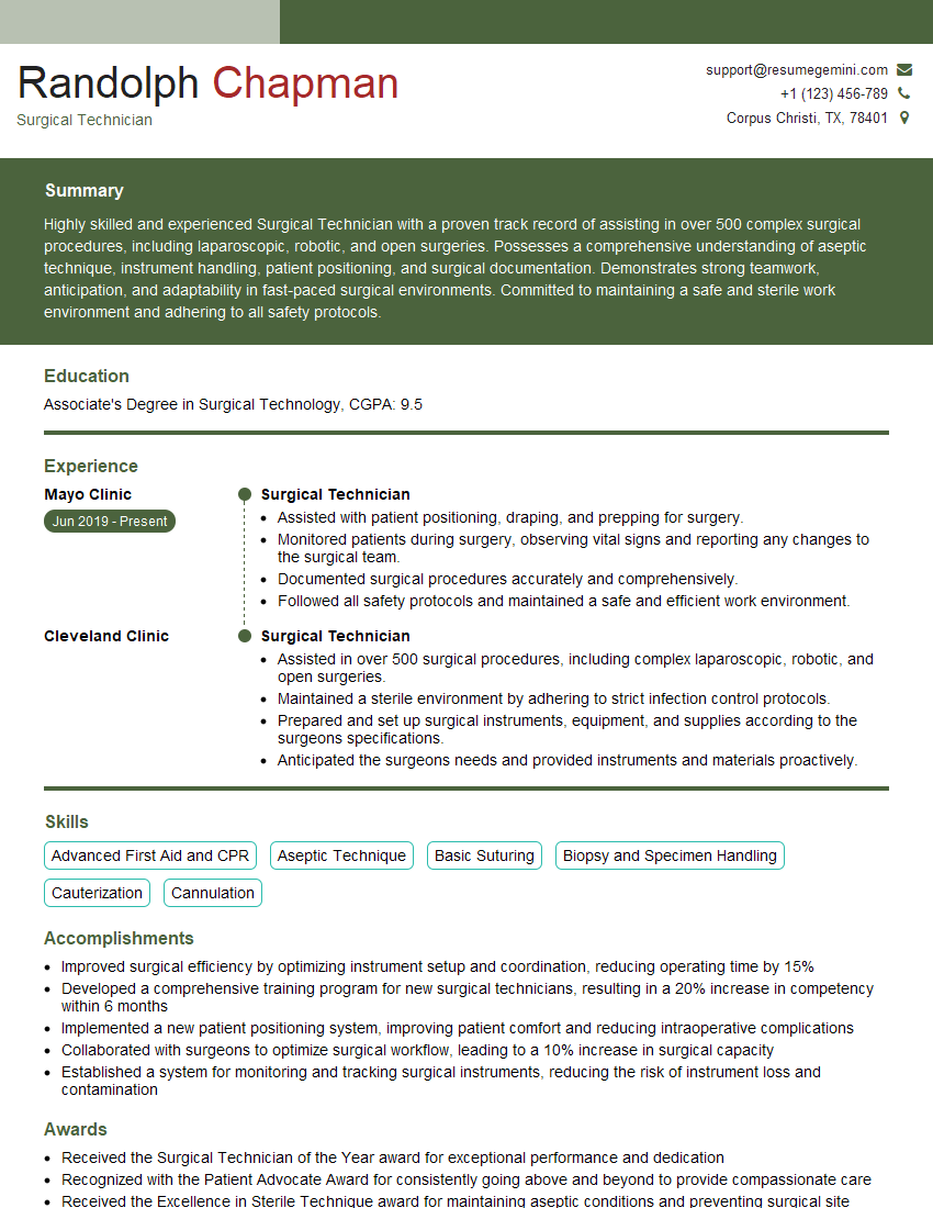
Feeling uncertain about what to expect in your upcoming interview? We’ve got you covered! This blog highlights the most important Pancreatic and Biliary Stone Removal interview questions and provides actionable advice to help you stand out as the ideal candidate. Let’s pave the way for your success.
Questions Asked in Pancreatic and Biliary Stone Removal Interview
Q 1. Describe the different endoscopic techniques used for biliary stone removal.
Endoscopic techniques for biliary stone removal primarily revolve around Endoscopic Retrograde Cholangiopancreatography (ERCP). Within ERCP, several methods exist to extract stones. The most common involves cannulating the bile duct (the tube carrying bile from the liver to the small intestine) with a catheter and then using various tools to retrieve stones. These tools include:
- Ballon catheters: These inflate to sweep stones into the duodenum (the first part of the small intestine).
- Dorsey baskets: These wire baskets are used to snag and retrieve stones.
- Stone extraction forceps: These are small, delicate forceps specifically designed for grasping and removing stones.
- Lithotripsy: This technique uses ultrasonic or laser energy to break up larger stones into smaller, more manageable fragments, which can then be extracted more easily.
In some cases, especially with very difficult-to-access stones or those impacted in the bile duct, additional techniques like sphincterotomy (cutting the muscle controlling the bile duct opening) may be necessary to facilitate stone removal. The choice of method depends on the size, location, number, and hardness of the stones, as well as the patient’s overall condition.
Q 2. Explain the indications and contraindications for ERCP.
ERCP, or Endoscopic Retrograde Cholangiopancreatography, is indicated when there’s biliary obstruction—a blockage in the bile duct—typically caused by gallstones, tumors, or strictures (narrowing of the duct). Specific indications include:
- Biliary stones causing jaundice (yellowing of the skin and eyes), abdominal pain, or cholangitis (infection of the bile duct).
- Suspected bile duct strictures needing dilation or stent placement.
- Diagnosis and treatment of pancreatic disorders such as pancreatitis or tumors.
Contraindications, or reasons to avoid ERCP, include:
- Severe coagulation disorders (problems with blood clotting), which increase bleeding risk during the procedure.
- Uncontrolled infections, which could worsen following the procedure.
- Recent major abdominal surgery, as it might hinder access and increase complications.
- Severe cardiopulmonary disease, putting the patient at higher risk under sedation and anesthesia.
It’s important to note that even with contraindications, the benefits of ERCP might outweigh the risks in certain life-threatening situations.
Q 3. What are the complications associated with ERCP and how are they managed?
ERCP, while a minimally invasive procedure, carries potential complications. These can range from minor to life-threatening. Common complications include:
- Pancreatitis: Inflammation of the pancreas, a serious complication that can be mild or severe and potentially fatal. It is usually managed with supportive care, including intravenous fluids, pain management, and bowel rest.
- Bleeding: Bleeding can occur at the site of cannulation or sphincterotomy, and can usually be managed endoscopically. In severe cases, interventional radiology techniques or surgery may be necessary.
- Infection (Cholangitis): This complication requires intravenous antibiotics and supportive measures. In some cases, drainage procedures are necessary.
- Perforation: A rare but serious complication involving a hole in the bile duct or duodenum, typically requiring surgical repair.
- Post-ERCP pain: Abdominal pain is relatively common post-procedure, usually managed with analgesics.
The risk of complications is influenced by the operator’s experience, the complexity of the case, and the patient’s overall health. Prevention involves careful patient selection, meticulous technique, and prompt recognition and management of early warning signs.
Q 4. Discuss the role of laparoscopic cholecystectomy in biliary stone management.
Laparoscopic cholecystectomy, or removal of the gallbladder through small incisions, plays a crucial role in managing biliary stones, especially when stones are primarily located within the gallbladder itself (cholelithiasis). While ERCP focuses on stones within the bile duct, laparoscopic cholecystectomy addresses the gallbladder, the main reservoir of stones. Removing the gallbladder eliminates the source of many biliary stones. This is particularly important to prevent future episodes of biliary colic (severe pain from gallstones obstructing the cystic duct), cholecystitis (inflammation of the gallbladder), and cholangitis. In cases of choledocholithiasis (stones in the common bile duct), a laparoscopic cholecystectomy is often combined with ERCP to remove stones from both the gallbladder and bile duct.
Q 5. Compare and contrast endoscopic ultrasound (EUS) and ERCP.
Both EUS (Endoscopic Ultrasound) and ERCP are endoscopic procedures used in the management of pancreaticobiliary diseases, but they serve different purposes:
- EUS is a diagnostic technique using ultrasound waves to obtain high-resolution images of the pancreas, bile ducts, and surrounding structures. It excels in detecting small lesions, assessing the extent of tumors, and guiding fine-needle aspiration for tissue sampling. EUS does not directly treat stones.
- ERCP is a therapeutic procedure used to diagnose and treat problems in the bile and pancreatic ducts. It’s primarily used to remove stones, stent strictures, or collect samples. ERCP offers less detailed imaging than EUS.
In practice, EUS and ERCP are often used complementarily. EUS may be used pre-ERCP to assess the location and size of stones, guiding the ERCP procedure and optimizing treatment strategies. For example, EUS can help identify small stones that might be missed on other imaging techniques, or assess the feasibility of stone removal through ERCP.
Q 6. How do you diagnose pancreatic stones?
Diagnosing pancreatic stones involves a combination of imaging and clinical assessment. Symptoms can include abdominal pain, weight loss, and signs of pancreatic insufficiency (malabsorption of nutrients). Imaging techniques used include:
- Endoscopic Ultrasound (EUS): Provides high-resolution images of the pancreas and allows for direct visualization of stones.
- Computed Tomography (CT) scan: Can identify stones, but the details might be less clear than EUS.
- Magnetic Resonance Cholangiopancreatography (MRCP): A non-invasive technique that produces detailed images of the pancreatic and bile ducts.
Laboratory tests, such as amylase and lipase levels (enzymes produced by the pancreas), may help identify pancreatic inflammation, indicating the presence of stones or other issues.
Q 7. What are the surgical approaches for pancreatic stone removal?
Surgical approaches for pancreatic stone removal are generally reserved for cases where endoscopic methods fail or are not feasible. These approaches include:
- Pancreaticoduodenectomy (Whipple procedure): This is a major surgery that involves removing the head of the pancreas, the duodenum (first part of the small intestine), a portion of the bile duct, and sometimes the gallbladder. It’s used for large stones or stones associated with tumors.
- Distal pancreatectomy: This procedure removes the tail of the pancreas, typically indicated when stones are located in the tail. It’s less extensive than the Whipple procedure.
- Puestow procedure: A less invasive approach involving opening the pancreatic duct and removing stones. It may be done with or without the assistance of laparoscopic techniques.
The choice of surgical approach is highly individualized and depends on the location, size, and number of stones, as well as the patient’s overall health and the presence of any other medical conditions. The Whipple procedure is considered the gold standard for many complex cases.
Q 8. Describe the management of post-ERCP pancreatitis.
Post-ERCP pancreatitis is a dreaded complication following endoscopic retrograde cholangiopancreatography (ERCP), a procedure used to diagnose and treat biliary and pancreatic diseases. Management focuses on supportive care and aims to prevent severe complications. Severity dictates the approach.
Mild Pancreatitis: This typically presents with mild abdominal pain, slightly elevated amylase and lipase levels, and no organ dysfunction. Management involves bowel rest (nil by mouth), intravenous fluids to maintain hydration, and analgesics for pain control. Patients are closely monitored for worsening symptoms. They are usually discharged once symptoms improve and labs normalize.
Moderate to Severe Pancreatitis: This involves more significant abdominal pain, substantial enzyme elevation, and potential organ failure (e.g., respiratory distress, kidney failure). Management escalates to include intensive care unit (ICU) admission, intravenous fluids with close monitoring of fluid balance, nutritional support (parenteral nutrition), and analgesics. Antibiotics may be considered if infection is suspected. In severe cases, interventions such as endoscopic necrosectomy or surgical debridement might be necessary to manage pancreatic necrosis. Close monitoring of vital signs, organ function, and inflammatory markers is crucial.
Example: I recently managed a patient who developed mild post-ERCP pancreatitis. After a day of bowel rest, intravenous fluids, and pain medication, their symptoms resolved, and they were discharged with follow-up instructions. In contrast, a patient with severe post-ERCP pancreatitis required ICU admission, aggressive fluid resuscitation, and close monitoring, emphasizing the importance of individualized management.
Q 9. Explain the role of sphincterotomy in biliary stone removal.
Sphincterotomy plays a crucial role in biliary stone removal during ERCP. The sphincter of Oddi, a muscular valve controlling the flow of bile and pancreatic juice into the duodenum, can be a significant obstacle to stone removal.
The procedure: A sphincterotomy involves using a specialized electrosurgical knife (or sometimes a laser) passed through the endoscope to make a small incision in the sphincter of Oddi. This incision allows easier access to the common bile duct, facilitating the retrieval of stones using balloon catheters or baskets.
Benefits: Sphincterotomy effectively widens the opening, allowing for complete stone extraction and preventing recurrence. It improves bile flow, preventing complications like cholangitis (infection of the bile duct).
Example: A patient presented with multiple large stones obstructing the common bile duct. After careful cannulation, we performed a sphincterotomy to facilitate easier passage and removal of the stones using a balloon catheter. Post-procedure, their jaundice resolved, and the patient recovered well.
Q 10. What are the different types of biliary stones and their composition?
Biliary stones, or gallstones, are classified based on their composition and location. Most are found in the gallbladder (cholelithiasis), but some can lodge in the bile ducts (choledocholithiasis).
- Cholesterol Stones: These are the most common type, largely composed of cholesterol. They often appear yellowish and are usually radiolucent (not visible on plain X-rays).
- Pigment Stones: Primarily composed of bilirubin (a byproduct of hemoglobin breakdown), these stones are darker in color, often black or brown. They are usually associated with conditions causing increased bilirubin levels such as hemolysis.
- Mixed Stones: These stones contain a mixture of cholesterol and pigment.
Compositional differences impact treatment: For example, larger cholesterol stones might be amenable to ESWL, whereas smaller pigment stones might be easier to remove endoscopically.
Q 11. Describe the use of extracorporeal shock wave lithotripsy (ESWL) in biliary stones.
Extracorporeal shock wave lithotripsy (ESWL) uses focused shock waves to break down stones into smaller fragments. While effective for urinary stones, its role in biliary stone management is limited.
Limitations: ESWL for biliary stones has shown inconsistent results. The stones need to be relatively radiopaque and of a certain size to be effectively targeted. Furthermore, the fragments produced may not readily pass through the bile ducts, potentially leading to complications such as ductal obstruction or pancreatitis.
Practical Considerations: ESWL is rarely the first-line treatment for biliary stones. ERCP with sphincterotomy remains the preferred method. ESWL might be considered for specific cases, such as patients who are poor surgical candidates or have stones that are difficult to access endoscopically, but it’s crucial to assess each case individually and weigh the potential benefits and risks.
Q 12. How do you differentiate between choledocholithiasis and cholelithiasis?
The key difference lies in the location of the stones:
- Cholelithiasis: Refers to gallstones located within the gallbladder.
- Choledocholithiasis: Indicates gallstones present in the common bile duct (the main duct carrying bile from the liver to the small intestine).
Clinical Significance: Choledocholithiasis usually presents with more severe symptoms because it causes direct biliary obstruction, potentially leading to jaundice, cholangitis, or pancreatitis. Cholelithiasis, while potentially symptomatic, might be asymptomatic for extended periods.
Q 13. What are the imaging modalities used in evaluating biliary and pancreatic stones?
Several imaging modalities are used to evaluate biliary and pancreatic stones:
- Ultrasound (US): This is usually the initial imaging technique. It can effectively visualize gallstones in the gallbladder and sometimes in the bile ducts.
- Computed Tomography (CT): Provides detailed images of the abdomen and can identify stones in the biliary and pancreatic ducts with greater accuracy than ultrasound, though with the use of contrast agents.
- Magnetic Resonance Cholangiopancreatography (MRCP): A non-invasive technique offering excellent visualization of the biliary and pancreatic ducts without the need for contrast agents. This makes it a great choice for patients with impaired renal function.
- Endoscopic Ultrasound (EUS): Provides high-resolution images and can be used to visualize stones and assess the surrounding structures.
The choice of imaging modality depends on the clinical suspicion, patient factors, and the information needed to guide management.
Q 14. Describe your experience with managing patients with complex biliary anatomy.
Managing patients with complex biliary anatomy poses significant challenges, requiring expertise and careful planning. Complex anatomy can include variations in the bile duct structure, presence of strictures (narrowing), or aberrant vessels.
My Approach: I typically begin with a thorough review of the patient’s medical history, imaging studies, and any prior interventions. Pre-procedural planning might involve 3D imaging reconstruction to better understand the anatomy and potential challenges. During the procedure, I use advanced endoscopic techniques, such as intraoperative cholangiography (injecting contrast into the bile ducts to visualize their anatomy during the procedure), to navigate the complex anatomy safely and effectively. In cases of severe strictures, balloon dilation or stenting might be necessary before stone extraction. Sometimes, a multidisciplinary approach with surgical colleagues is required, especially if complex anatomical variations make endoscopic management impossible.
Example: I recall a case involving a patient with a horseshoe-shaped bile duct and multiple stones. Careful planning and use of intraoperative cholangiography guided the stone extraction, ensuring complete clearance without causing injury to adjacent structures. Collaboration with a surgeon was important to ensure a successful outcome. Post-procedural follow-up is also crucial to monitor for potential complications and the long-term success of the intervention.
Q 15. Discuss the challenges in managing patients with pancreatic cancer and stones.
Managing patients with pancreatic cancer and stones presents unique challenges due to the intricate anatomy of the pancreas and biliary system, and the often aggressive nature of the cancer. The presence of stones can exacerbate existing pancreatic inflammation (pancreatitis) or even obstruct the bile duct, leading to jaundice and liver damage. Treatment decisions must carefully balance the need for cancer control with the risks of surgery, particularly in patients who may be weakened by the disease. For example, a patient with a pancreatic tumor obstructing the bile duct might require a complex procedure like a Whipple resection, which carries significant risks of post-operative complications like pancreatic fistula or infection. The delicate balance between aggressive cancer treatment and minimizing surgical trauma is paramount.
- Challenges include:
- High risk of post-operative complications: Pancreatic surgery is inherently complex and carries a higher risk of complications compared to other abdominal procedures.
- Advanced age and comorbidities: Many patients with pancreatic cancer are older and have other health issues that increase surgical risk.
- Tumor location and extent: The location and size of the tumor significantly impact surgical feasibility and outcome.
- Nutritional deficiencies: Pancreatic cancer often leads to malabsorption and weight loss, making patients more susceptible to complications.
Career Expert Tips:
- Ace those interviews! Prepare effectively by reviewing the Top 50 Most Common Interview Questions on ResumeGemini.
- Navigate your job search with confidence! Explore a wide range of Career Tips on ResumeGemini. Learn about common challenges and recommendations to overcome them.
- Craft the perfect resume! Master the Art of Resume Writing with ResumeGemini’s guide. Showcase your unique qualifications and achievements effectively.
- Don’t miss out on holiday savings! Build your dream resume with ResumeGemini’s ATS optimized templates.
Q 16. How do you assess a patient’s risk for post-operative complications?
Assessing a patient’s risk for post-operative complications after pancreatic or biliary surgery requires a thorough pre-operative evaluation. This includes a detailed medical history, physical examination, and a battery of tests. We use validated risk stratification tools such as the P-POSSUM (Physiological and Operative Severity Score for enUmerating Mortality and morbidity) or the Clavien-Dindo classification to objectively quantify risk. Factors influencing risk include age, overall health, nutritional status (albumin levels), presence of diabetes or chronic obstructive pulmonary disease (COPD), the extent of the surgery, and the presence of infection or inflammation. For instance, a patient with a history of diabetes, low albumin levels, and a large, obstructing pancreatic tumor will have a higher risk of post-operative complications compared to a patient with a smaller stone and good overall health. This risk assessment guides decision-making about the type of surgery, the need for intensive care support, and the optimal timing of the procedure.
Q 17. What are the key factors in determining the appropriate treatment modality for biliary stones?
Choosing the right treatment for biliary stones depends on several factors. The size, number, and location of stones are crucial, as are the patient’s overall health and the presence of any complications like cholangitis (infection of the bile duct) or jaundice. For small stones in the common bile duct, endoscopic retrograde cholangiopancreatography (ERCP) with sphincterotomy and stone extraction might be the preferred method. This minimally invasive procedure involves inserting a scope through the mouth to access and remove the stones. Larger stones or those that are difficult to access endoscopically might require surgery, such as laparoscopic cholecystectomy (removal of the gallbladder) and common bile duct exploration. In cases of severe complications or anatomical abnormalities, open surgery might be necessary. The patient’s age, comorbidities, and preference also play a role in decision-making. We always strive for the least invasive approach that addresses the problem effectively.
Q 18. Explain the role of pre-operative optimization in pancreatic surgery.
Pre-operative optimization is critical to improving outcomes in pancreatic surgery. It focuses on addressing any underlying medical conditions that could increase the risk of complications. This includes managing diabetes, optimizing nutritional status (often requiring nutritional support), treating infections, and ensuring the patient’s lungs and heart are functioning well. For example, a diabetic patient may need their blood sugar carefully controlled for several weeks before surgery. A patient with malnutrition might require intravenous nutrition to improve their overall health and reduce the risk of post-operative complications such as wound healing problems. These efforts are crucial because patients undergoing pancreatic surgery are already at high risk, and good pre-operative health substantially increases their chances of a successful outcome and a faster recovery.
Q 19. How do you counsel patients about the risks and benefits of various treatment options?
Counseling patients about the risks and benefits of different treatment options is a crucial part of my practice. I use a patient-centered approach, explaining the procedures in clear, simple language, avoiding medical jargon whenever possible. I present the potential benefits, such as relief of symptoms, improved quality of life, and increased life expectancy (especially in cancer cases), alongside the potential risks, which can include infection, bleeding, pancreatic fistula, and the need for further procedures. I encourage patients to ask questions and provide them with written materials to reinforce the discussion. I also involve family members or caregivers as appropriate. I believe shared decision-making is essential; patients should be fully informed and empowered to make choices that align with their values and preferences. A realistic understanding of the risks and rewards is key to informed consent.
Q 20. Describe your approach to managing patients with recurrent biliary stones.
Recurrent biliary stones are a significant concern. The approach depends on the cause of recurrence. If the recurrence is due to incomplete stone removal, repeat ERCP or surgery may be necessary. If it’s related to retained stones in the gallbladder, cholecystectomy is indicated. However, in some cases, the cause might be sludge, which are tiny stones that can repeatedly form. This may be managed with medication and lifestyle adjustments. Careful investigation is crucial to determine the underlying cause of recurrence before choosing a treatment strategy. Long-term surveillance, sometimes involving regular imaging, might be necessary to prevent future episodes. The ultimate goal is to prevent further complications like cholangitis or pancreatitis.
Q 21. What are the latest advancements in the field of pancreatic and biliary stone removal?
The field of pancreatic and biliary stone removal is constantly evolving. Advancements include minimally invasive techniques, such as NOTES (Natural Orifice Transluminal Endoscopic Surgery), which reduces surgical trauma. New endoscopic devices allow for more precise and efficient stone extraction. There’s also ongoing research in using less invasive methods for treating pancreatic cancer, such as targeted therapy and immunotherapy, that could reduce the need for extensive surgery. Improved imaging techniques, including advanced endoscopy and magnetic resonance cholangiopancreatography (MRCP), enhance diagnostic accuracy and allow for better planning of minimally invasive procedures. Furthermore, the development of newer, safer stents and drainage techniques minimizes complications and improves patient outcomes.
Q 22. Explain the role of minimally invasive surgery in the management of biliary stones.
Minimally invasive surgery, such as laparoscopic cholecystectomy (removal of the gallbladder) and endoscopic retrograde cholangiopancreatography (ERCP), has revolutionized biliary stone management. These techniques offer several advantages over open surgery, including smaller incisions, reduced pain, shorter hospital stays, faster recovery times, and decreased risk of infection.
Laparoscopic cholecystectomy is the gold standard for gallstones lodged in the gallbladder. In this procedure, small incisions are made, and a camera and specialized instruments are used to remove the gallbladder. ERCP, on the other hand, is an endoscopic procedure used to remove stones located in the bile ducts. A thin, flexible tube is passed through the mouth, down the esophagus, and into the bile duct, where stones are removed or fragmented. Think of it like using a tiny camera and tools to navigate the plumbing system of your digestive tract to remove blockages. The minimally invasive nature reduces patient trauma and enhances recovery.
Q 23. Discuss the management of cholangitis.
Cholangitis is a serious infection of the bile duct system, often caused by a blockage from gallstones or tumors. Management requires prompt and aggressive treatment to prevent life-threatening complications. The first step involves stabilizing the patient, which includes intravenous fluids, antibiotics, and pain relief. Next, the biliary drainage is crucial. This is typically achieved using ERCP to remove the obstruction or placing a stent to restore bile flow. In severe cases, emergency surgery may be necessary to relieve the obstruction and drain the infected bile. Imagine it as unclogging a severely blocked drain – the infection needs to be addressed, and the blockage removed to restore function. Delay in treatment can lead to sepsis and organ failure. Early diagnosis and intervention are key to successful outcomes.
Q 24. Describe the different types of stents used in biliary drainage.
Several types of stents are used in biliary drainage, each with its unique properties. These stents are temporarily placed to maintain bile flow, often after stone removal or in cases of malignant strictures. Common types include:
- Plastic stents: These are relatively inexpensive and easy to place, but they have a higher risk of blockage and require removal after a few weeks.
- Metallic stents: More durable than plastic stents, they offer longer patency (remain open), but they can migrate or cause complications like perforation. These are often preferred for patients with malignant biliary obstructions.
- Self-expandable metallic stents (SEMS): These stents expand within the bile duct, offering excellent patency and requiring less frequent replacement.
- Covered metallic stents: These stents have a coating that prevents tissue ingrowth, reducing the risk of complications. They are often used in situations where tumor infiltration of the bile duct is a significant concern.
The choice of stent depends on several factors, including the location and nature of the obstruction, the patient’s overall health, and the expected duration of drainage. Careful consideration is vital to optimize the benefits and minimize potential adverse effects.
Q 25. What are the criteria for admission and discharge after ERCP?
Admission criteria for ERCP usually include the presence of biliary stones, cholangitis, or other biliary obstructions confirmed by imaging studies. Patients may also be admitted for suspected pancreaticobiliary pathology needing diagnostic and therapeutic intervention. Discharge criteria are typically met after the procedure is completed successfully, and the patient is stable, with no significant complications. This usually includes showing normal bowel sounds, minimal abdominal pain, and being able to tolerate oral intake. Close monitoring of vital signs and lab results also influences discharge decisions. The patient should also be educated about potential complications and post-procedure care. Each case is individual, and the physician will make the final determination based on the patient’s specific condition.
Q 26. How do you manage bleeding complications after ERCP?
Bleeding after ERCP is a potential, albeit infrequent, complication. Management depends on the severity of bleeding. Minor bleeding often resolves spontaneously with close monitoring. In more significant cases, endoscopic techniques, such as injection of epinephrine or clipping of the bleeding site, are employed. If endoscopic hemostasis is unsuccessful, angiographic embolization (blocking the bleeding vessel) or even surgery may be necessary. Early recognition and prompt management are crucial in preventing serious complications. Think of it as a plumbing leak – various methods exist to stop the flow, from simple tightening to more significant repairs.
Q 27. Explain the importance of postoperative pain management in pancreatic surgery.
Postoperative pain management in pancreatic surgery is paramount due to the highly sensitive nature of the pancreas and surrounding structures. A multimodal approach is generally adopted, combining different analgesics to minimize pain, reduce opioid requirements, and prevent adverse effects. This includes regional anesthesia (nerve blocks), local infiltration of analgesics, and the use of opioid and non-opioid analgesics. Careful monitoring of pain levels, adjusting medication as needed, and employing non-pharmacological techniques (e.g., physiotherapy) are essential for optimal pain control and patient comfort. Managing pain effectively promotes faster recovery and improves patient satisfaction. Think of it as building a multi-layered defense against pain, using various strategies to minimize discomfort and provide relief. Effective pain management is a significant aspect of achieving successful surgical outcomes.
Q 28. Discuss the use of prophylactic antibiotics in pancreatic and biliary surgery.
Prophylactic antibiotics in pancreatic and biliary surgery aim to reduce the risk of surgical site infection and other infectious complications. The choice of antibiotic, dosage, and duration are guided by local infection patterns and the specific surgical procedure. Broad-spectrum antibiotics are often used perioperatively to cover various bacteria. Selective decontamination of the bowel is sometimes employed to reduce bacterial load. However, overuse of antibiotics contributes to antibiotic resistance, so a judicious approach, guided by infection risk stratification, is crucial. The decision to use prophylactic antibiotics involves a careful balancing act between infection prevention and the potential harms of antibiotic usage. Proper antibiotic stewardship is critical in this scenario.
Key Topics to Learn for Pancreatic and Biliary Stone Removal Interview
- Anatomy and Physiology: Thorough understanding of the pancreaticobiliary system, including the anatomy of the pancreas, bile ducts, gallbladder, and surrounding structures. Focus on relevant blood supply and lymphatic drainage.
- Pathophysiology of Stones: Mechanism of stone formation (cholelithiasis, choledocholithiasis, pancreatitis), risk factors, and clinical presentations. Understand the differences in stone composition and their implications.
- Diagnostic Modalities: Mastery of imaging techniques used in diagnosis (ultrasound, CT, MRCP, ERCP) and their interpretation. Be prepared to discuss the strengths and limitations of each modality.
- Endoscopic Techniques: Detailed knowledge of ERCP (Endoscopic Retrograde Cholangiopancreatography) including cannulation techniques, sphincterotomy, stone extraction methods (basket, balloon, electrohydraulic lithotripsy), and management of complications.
- Surgical Techniques: Understanding of different surgical approaches to stone removal (e.g., cholecystectomy, choledochotomy), including minimally invasive techniques and their indications and contraindications. Be familiar with postoperative care and potential complications.
- Medical Management: Knowledge of pharmacological interventions used in managing pancreatitis, cholangitis, and other related complications. Understand the role of analgesics, antibiotics, and supportive care.
- Complications and Management: Thorough understanding of potential complications associated with pancreatic and biliary stone removal procedures (e.g., perforation, bleeding, pancreatitis, infection) and their management strategies.
- Case Studies and Problem Solving: Practice analyzing clinical scenarios, interpreting diagnostic images, and formulating appropriate treatment plans. Be ready to discuss your decision-making process and justify your choices.
Next Steps
Mastering Pancreatic and Biliary Stone Removal demonstrates a high level of surgical skill and clinical judgment, significantly enhancing your career prospects in gastroenterology, surgery, or related specialties. To stand out to potential employers, create an ATS-friendly resume that showcases your expertise effectively. ResumeGemini is a trusted resource to help you build a professional resume that highlights your skills and experience. We provide examples of resumes tailored to Pancreatic and Biliary Stone Removal to guide you in crafting your perfect application.
Explore more articles
Users Rating of Our Blogs
Share Your Experience
We value your feedback! Please rate our content and share your thoughts (optional).
What Readers Say About Our Blog
Hi, I have something for you and recorded a quick Loom video to show the kind of value I can bring to you.
Even if we don’t work together, I’m confident you’ll take away something valuable and learn a few new ideas.
Here’s the link: https://bit.ly/loom-video-daniel
Would love your thoughts after watching!
– Daniel
This was kind of a unique content I found around the specialized skills. Very helpful questions and good detailed answers.
Very Helpful blog, thank you Interviewgemini team.
