Cracking a skill-specific interview, like one for Awake Craniotomy, requires understanding the nuances of the role. In this blog, we present the questions you’re most likely to encounter, along with insights into how to answer them effectively. Let’s ensure you’re ready to make a strong impression.
Questions Asked in Awake Craniotomy Interview
Q 1. Describe the patient selection criteria for awake craniotomy.
Patient selection for awake craniotomy is crucial for success and patient safety. It hinges on several factors, primarily the location of the tumor or lesion relative to eloquent cortical areas responsible for speech, movement, sensation, or vision. Awake craniotomy is indicated when removing a lesion near these areas risks significant neurological deficits if the procedure is performed under general anesthesia.
Specifically, we consider:
- Tumor location: Lesions close to eloquent cortex are prime candidates.
- Patient’s cognitive ability: The patient must be able to understand and follow instructions, cooperate during the procedure, and provide feedback. This often involves pre-operative cognitive testing.
- Medical stability: The patient needs to be medically fit enough to tolerate the prolonged procedure and potential complications.
- Psychological suitability: The patient should be psychologically prepared for the unique experience of an awake craniotomy and able to manage anxiety. We often involve neuropsychology to assess this.
- Tumor type and characteristics: The surgical approach and the feasibility of awake surgery are influenced by the nature of the tumor.
For example, a patient with a tumor encroaching on Broca’s area (responsible for speech production) would be a suitable candidate for awake craniotomy to allow for real-time monitoring and preservation of speech function during resection.
Q 2. Explain the different types of neuromonitoring used during awake craniotomy.
Neuromonitoring during awake craniotomy is vital for ensuring the safety and preservation of neurological function. Several modalities are used, often in combination, to provide comprehensive monitoring:
- Electroencephalography (EEG): Provides continuous monitoring of brain electrical activity. Changes in EEG patterns can signal cortical injury.
- Direct Electrical Cortical Stimulation (ECS): This involves directly stimulating the brain’s surface with small electrical currents to map eloquent cortical areas. It’s crucial for identifying functional areas to avoid damaging them.
- Transcranial magnetic stimulation (TMS): Non-invasive technique for mapping cortical areas prior to incision.
- Motor evoked potentials (MEPs): Monitor the integrity of motor pathways by stimulating the motor cortex and measuring muscle responses. Changes can indicate damage to motor pathways.
- Somatosensory evoked potentials (SSEPs): Monitor the integrity of sensory pathways. Changes can indicate damage to sensory pathways.
- Language mapping techniques: These techniques use various methods, including verbal fluency tasks, picture naming, and sentence repetition, to monitor language function during surgery.
The specific types of monitoring used depend on the location of the tumor and the potential risk to eloquent areas. For example, during resection near Broca’s area, language mapping (using tasks like naming pictures or repeating sentences) and EEG are very important. If the motor cortex is near the target area, then MEPs would be used.
Q 3. How do you manage intraoperative complications such as bleeding or swelling during an awake craniotomy?
Intraoperative complications during awake craniotomy, such as bleeding or swelling, require immediate and decisive action. The surgeon’s experience and the quality of the neuromonitoring team are essential here.
Bleeding: Small bleeds are often managed with meticulous hemostasis (stopping the bleeding) using techniques like bipolar cautery or small absorbable sponges. Larger bleeds might require temporary packing or the use of fibrin sealant to control bleeding. We always prioritize minimizing manipulation of the brain to prevent further trauma.
Swelling: Swelling is managed by optimizing the patient’s cerebral perfusion pressure (CPP), which depends on blood pressure, intracranial pressure, and other factors. This involves careful management of fluids, blood pressure, and sometimes the use of osmotic agents to reduce brain swelling. In severe cases, a temporary opening to the subarachnoid space (craniotomy) might be considered to reduce pressure.
Constant communication between the surgical team, anesthesiologist, and neurophysiologist is crucial in addressing these complications promptly and effectively. Monitoring parameters, like blood pressure, pulse rate, and EEG, are constantly watched.
Q 4. What are the key steps in performing a cortical mapping procedure?
Cortical mapping involves identifying and mapping eloquent cortical areas. The precise steps can vary depending on the techniques used but generally involve:
- Preoperative planning: Identifying the location of the tumor and potential eloquent areas using imaging techniques.
- Anesthesia: Typically involves sedation, not general anesthesia, to maintain patient responsiveness for feedback.
- Craniotomy: A portion of the skull is removed to access the brain.
- Dural opening: The protective covering of the brain (dura mater) is opened.
- Cortical stimulation: The surgeon uses a small probe to stimulate different cortical areas. The patient is asked to perform tasks like moving limbs or repeating words, allowing the surgeon to identify the location of these functions.
- Mapping the functional areas: The functional areas are carefully marked on the brain’s surface to guide the resection.
- Tumor resection: The surgeon meticulously removes the tumor while avoiding the mapped functional areas.
- Closure: The dura, skull, and scalp are closed in layers.
A skilled neurosurgeon uses their knowledge of neuroanatomy and sophisticated monitoring tools to maximize safety during this delicate procedure. It’s crucial to remember that this is a highly specialized procedure that demands precise technique and a multidisciplinary team approach.
Q 5. Discuss the role of language mapping and motor mapping in awake craniotomy.
Language mapping and motor mapping are critical components of awake craniotomy. They directly help the surgeon protect essential brain functions during tumor resection.
Language mapping: Identifies the cortical areas responsible for speech production (Broca’s area) and comprehension (Wernicke’s area). During surgery, language tasks are used to monitor function. If stimulation of an area interferes with language, it indicates proximity to a critical region that must be preserved.
Motor mapping: Locates the areas of the motor cortex that control specific body movements. Stimulation of these areas during surgery will cause movements in the corresponding body parts. This allows the surgeon to avoid damaging these areas.
For example, if a tumor is near Broca’s area, the surgeon might use a simple naming test or sentence repetition task to monitor the patient’s speech abilities while stimulating the nearby cortex. Any disruptions in speech would signal a need to avoid that area. Similarly, if a lesion is near the motor cortex, MEPs would guide resection to avoid disrupting motor pathways.
Q 6. Explain the anesthetic considerations during awake craniotomy procedures.
Anesthesia for awake craniotomy is unique. The goal is to balance the need for patient responsiveness and cooperation with comfort and minimal anxiety. General anesthesia is avoided as it renders the patient incapable of providing feedback about neurological function.
Instead, we use a combination of techniques, often described as ‘monitored anesthesia care’ (MAC):
- Sedation: Sedative medications, like propofol or dexmedetomidine, are titrated to maintain patient comfort and reduce anxiety. The patient remains awake and communicative but is relaxed.
- Analgesia: Pain medication is given to manage any discomfort.
- Local anesthesia: Local anesthetic is injected into the scalp and skull to reduce pain from the surgical incision.
The anesthesiologist’s role is critical in maintaining the optimal level of sedation, managing vital signs, and addressing any complications that might arise. The level of sedation needs to be carefully adjusted throughout the surgery to maintain the balance between patient cooperation and comfort.
Q 7. How do you communicate with the patient during an awake craniotomy?
Communication with the patient during an awake craniotomy is paramount to its success. It requires careful planning, a calm and reassuring environment, and a well-coordinated team.
We use a variety of communication techniques:
- Preoperative briefing: The patient receives detailed explanations of the procedure, the role of neuromonitoring, and what to expect. This helps alleviate anxiety and promotes cooperation.
- Simple instructions: During the procedure, instructions are kept clear, concise, and easily understandable. This could involve a simple request like ‘point to your nose’ or ‘repeat this word.’
- Nonverbal cues: Observation of subtle changes in facial expressions or gestures can also provide useful feedback.
- Communication board: A communication board with simple images or words can be used for patients who have difficulty verbalizing their responses.
- Frequent communication with the patient: Throughout the procedure, the neurosurgeon or a dedicated member of the team will maintain ongoing dialogue with the patient to ensure their cooperation and comfort.
A good rapport between the surgical team and the patient is critical to maintaining a successful awake craniotomy. The trust and open communication fosters the patient’s ability to provide effective feedback during the critical process of mapping and resection.
Q 8. What are the potential risks and complications associated with awake craniotomy?
Awake craniotomy, while offering significant advantages, carries inherent risks. These risks can be broadly categorized into those related to the surgery itself, the awake state, and potential complications from the underlying condition requiring surgery.
- Surgical Risks: These include bleeding (hemorrhage), infection, cerebrospinal fluid (CSF) leak, damage to surrounding brain tissue, stroke, and seizures. The risk of these complications is generally higher than in a traditional asleep craniotomy, though this is often balanced by the improved precision achieved.
- Risks Related to the Awake State: Maintaining a patient’s awake state throughout the procedure requires meticulous monitoring. Movement, anxiety, and unexpected responses can complicate the surgery. Rarely, patients may experience psychological distress during the procedure.
- Complications from Underlying Condition: The underlying condition requiring the awake craniotomy (often a brain tumor near eloquent cortical areas) itself poses risks, such as tumor progression or related neurological deficits. The surgery is designed to minimize these, but they remain a consideration.
- Anesthesia Risks: Even though the patient is awake, general anesthesia is usually utilized to induce sleep for the initial incision and closure to minimize discomfort and potential movement. Associated risks include those related to general anesthesia such as nausea, vomiting and allergic reactions to medications.
It’s crucial to remember that the severity of these risks varies depending on factors like the patient’s overall health, the location and size of the surgical area, and the surgeon’s experience. A thorough pre-operative assessment is essential to minimize potential complications.
Q 9. Describe the postoperative management of patients undergoing awake craniotomy.
Postoperative management after an awake craniotomy focuses on monitoring for complications and supporting the patient’s recovery. This involves a multidisciplinary approach.
- Neurological Monitoring: Continuous monitoring of neurological status is crucial. This involves regular neurological examinations to assess level of consciousness, motor strength, speech, and sensory function.
- Pain Management: Pain control is a priority. This may involve a combination of oral and intravenous analgesics, tailored to the patient’s needs and tolerability.
- Wound Care: Meticulous wound care is essential to prevent infection. This includes regular wound inspections and dressing changes.
- Rehabilitation: Depending on the location and extent of the surgery, patients may require physical therapy, occupational therapy, and speech therapy to regain lost function.
- Imaging Follow-up: Postoperative imaging, such as CT or MRI scans, helps to assess the surgical site and rule out any complications.
- Cognitive Monitoring and Support: Postoperative cognitive dysfunction can occur. Neuropsychological testing may be necessary, along with supportive care and education.
The length of hospital stay varies depending on the individual case, but patients generally require close monitoring for several days after surgery. Discharge planning involves providing detailed instructions to the patient and family regarding medication, wound care, and follow-up appointments.
Q 10. How do you ensure patient safety and comfort during the procedure?
Ensuring patient safety and comfort during an awake craniotomy is paramount. This involves a meticulous approach to every aspect of the procedure.
- Detailed Pre-operative Assessment: A thorough assessment evaluates the patient’s cognitive function, ability to cooperate, and tolerance for the awake state. This may include neuropsychological testing.
- Comprehensive Anesthesia Plan: Anesthesiologists play a vital role, managing sedation levels to ensure the patient’s comfort and cooperation while maintaining neurological monitoring. They must allow sufficient alertness for participation but manage potential anxiety.
- Intraoperative Monitoring: Continuous monitoring of vital signs, brain activity (EEG), and neurological function is crucial. This helps to detect any complications early.
- Communication and Feedback: Open and consistent communication between the patient, surgical team, and anesthesiologists is crucial. The patient is given clear instructions on what to expect and is encouraged to communicate any discomfort or changes in sensation.
- Minimally Invasive Techniques: The adoption of minimally invasive techniques whenever possible can reduce the invasiveness of the procedure, leading to less trauma and improved recovery.
- Supportive Care: Providing constant reassurance, and employing distraction techniques, such as music or conversation, can significantly reduce anxiety.
For instance, if a patient starts feeling discomfort or experiences anxiety, the team uses strategies such as adjusting sedation levels or employing psychological calming techniques to ensure comfort and safety.
Q 11. What are the latest advancements in awake craniotomy techniques and technology?
Awake craniotomy has benefited from several recent advancements, focusing on enhancing precision, minimizing invasiveness, and improving patient experience.
- Intraoperative Neurophysiological Monitoring (IONM): Advanced IONM techniques, such as direct cortical stimulation (DCS) and electrocorticography (ECoG), provide real-time feedback on brain function, allowing for greater precision during tumor resection.
- Fluorescence-Guided Surgery: The use of fluorescent dyes that target specific tumor cells allows surgeons to visualize and remove cancerous tissue with greater accuracy.
- Advanced Imaging Techniques: Real-time imaging techniques like intraoperative MRI and CT scanning provide surgeons with detailed views of the surgical site, increasing precision and minimizing damage to surrounding healthy tissue.
- Minimally Invasive Approaches: Smaller incisions and the use of advanced tools allow for less trauma, faster recovery times, and improved cosmetic outcomes.
- Virtual Reality (VR) and Augmented Reality (AR): These technologies can be used to create 3D models of the brain and to guide the surgical procedure, improving planning and accuracy.
These advancements contribute to safer and more effective awake craniotomies, leading to better patient outcomes.
Q 12. Compare and contrast awake craniotomy with traditional craniotomy procedures.
Awake craniotomy and traditional craniotomy differ primarily in the patient’s state during the procedure. The choice depends heavily on the location of the pathology and its proximity to eloquent cortical areas.
| Feature | Awake Craniotomy | Traditional Craniotomy |
|---|---|---|
| Patient State | Awake and responsive (often with sedation) | Asleep under general anesthesia |
| Tumor Location | Near eloquent cortical areas | Anywhere in the brain |
| Surgical Precision | Higher, due to real-time feedback | Lower, relying on pre-operative imaging |
| Risk of Neurological Deficit | Lower, if successful mapping | Potentially higher, as mapping is limited |
| Recovery Time | Variable but potentially faster for certain procedures | Generally longer recovery |
| Patient Cooperation | Essential, requires patient education and preparation | Not required |
In essence, awake craniotomy is a more specialized technique reserved for cases where preserving neurological function is paramount. Traditional craniotomy remains a valuable approach for many brain surgeries.
Q 13. Discuss the ethical considerations related to awake craniotomy.
Awake craniotomy raises several ethical considerations, particularly regarding patient autonomy, informed consent, and the potential for psychological distress.
- Informed Consent: Patients must be fully informed about the risks and benefits of the procedure, including the potential for discomfort, anxiety, and unexpected responses during the surgery. This requires careful explanation and shared decision making.
- Autonomy: Maintaining patient autonomy is crucial. Patients must be empowered to participate actively in decisions regarding their care. This may include choosing to forgo awake craniotomy if the risks outweigh the benefits in their specific case.
- Psychological Well-being: The potential for psychological distress during the procedure must be carefully considered and addressed through appropriate support and monitoring. If a patient becomes significantly distressed, modifications to the approach are often necessary.
- Resource Allocation: Awake craniotomies often demand specialized personnel and equipment, leading to ethical considerations about resource allocation and equitable access to this advanced surgical technique.
Ethical oversight and robust communication between the surgical team and the patient are vital in ensuring that the procedure is conducted ethically and respectfully.
Q 14. How do you address patient anxiety and concerns before an awake craniotomy?
Addressing patient anxiety and concerns before an awake craniotomy requires a compassionate and thorough approach.
- Detailed Explanation: The surgical team should clearly explain the procedure, including the reasons for choosing an awake craniotomy, the potential risks and benefits, and what the patient can expect during the procedure.
- Reassurance and Support: Offering reassurance and emotional support helps reduce anxiety. Patients should be encouraged to express their concerns and receive honest answers.
- Pre-operative Preparation: This includes providing information about the environment, the monitoring equipment, and the role of the patient during the procedure. Simulation or rehearsal techniques can also reduce anxiety.
- Meeting with the Anesthesiologist: A separate meeting with the anesthesiologist allows them to address any concerns regarding anesthesia and pain management. The anesthesiologist is key in managing the level of comfort and alertness during surgery.
- Psychological Support: Offering access to psychological support, such as counseling or relaxation techniques, can significantly benefit patients who experience high levels of anxiety.
A calm, reassuring approach that actively involves patients in the process enhances their sense of control and diminishes their anxiety.
Q 15. What are the different types of brain tumors that are suitable for awake craniotomy?
Awake craniotomy is best suited for brain tumors located in eloquent brain areas – regions responsible for critical functions like speech, movement, or sensation. Removing these tumors without causing permanent neurological deficits requires the patient to be awake and able to participate in testing. Several tumor types benefit from this approach:
- Low-grade gliomas: These slow-growing tumors often infiltrate eloquent areas, making precise surgical removal crucial. Awake craniotomy allows real-time functional mapping to preserve healthy tissue.
- Meningiomas: Depending on their location and proximity to vital brain areas, meningiomas can benefit from awake craniotomy to minimize functional deficits.
- Brain metastases: In certain situations, awake craniotomy might be considered for strategically located metastases near eloquent areas, particularly if the tumor location necessitates a high degree of precision during resection.
- Cavernous malformations (CMs): Awake surgery helps to map the eloquent cortex that is surrounding and at risk of being injured.
The suitability of awake craniotomy is determined on a case-by-case basis, considering the tumor’s location, size, growth rate, patient’s overall health, and their cognitive abilities.
Career Expert Tips:
- Ace those interviews! Prepare effectively by reviewing the Top 50 Most Common Interview Questions on ResumeGemini.
- Navigate your job search with confidence! Explore a wide range of Career Tips on ResumeGemini. Learn about common challenges and recommendations to overcome them.
- Craft the perfect resume! Master the Art of Resume Writing with ResumeGemini’s guide. Showcase your unique qualifications and achievements effectively.
- Don’t miss out on holiday savings! Build your dream resume with ResumeGemini’s ATS optimized templates.
Q 16. Explain the role of neuropsychology in the assessment and rehabilitation of patients undergoing awake craniotomy.
Neuropsychology plays a vital role throughout the awake craniotomy process. Before surgery, neuropsychological testing helps establish a baseline cognitive function, identifying areas of strength and weakness. This helps the surgical team understand the potential impact of the tumor and the surgery on the patient’s cognitive abilities. During surgery, the neuropsychologist collaborates with the surgical team, assessing the patient’s cognitive function in real-time, providing continuous feedback on the impact of the surgery. This may involve tasks like naming pictures, following instructions, or repeating sentences. Post-surgery, neuropsychological rehabilitation focuses on addressing any cognitive deficits resulting from the procedure. This might include speech therapy, occupational therapy, and cognitive retraining exercises to help the patient regain their pre-operative cognitive abilities and improve their quality of life.
For instance, if a tumor is near the language center, pre-operative testing can pinpoint specific language functions that might be affected. During surgery, continuous language testing ensures that the resection remains within safe limits. Post-operatively, neuropsychological rehabilitation can address any language deficits the patient experiences.
Q 17. Describe your experience with specific awake craniotomy cases.
I’ve been involved in numerous awake craniotomies, each case presenting unique challenges and rewards. One particularly memorable case involved a young musician with a low-grade glioma near his motor cortex controlling his left hand. Pre-operative neuropsychological testing showed excellent dexterity and fine motor skills crucial for his profession. During surgery, real-time motor mapping allowed us to precisely resect the tumor while maintaining his hand function. Post-operatively, he showed minimal deficits and fully regained his pre-operative dexterity, returning to his musical career. Another case involved a patient with a tumor near the language center. Continuous language monitoring during surgery was crucial for avoiding aphasia (language loss). While there were some minor temporary speech difficulties, intensive speech therapy after surgery completely resolved them.
Q 18. How do you manage unexpected neurological deficits during the procedure?
Unexpected neurological deficits during an awake craniotomy require immediate and decisive action. The first step is to immediately stop the surgical manipulation and assess the patient’s condition. This involves collaboration among the neurosurgeon, anesthesiologist, neuropsychologist, and the entire surgical team. We utilize advanced neuro-monitoring techniques like electrocorticography (ECoG) to pinpoint the location and nature of the deficit. Depending on the severity and type of deficit, the surgical strategy may be modified. This might involve adjusting the resection plan, halting the procedure temporarily, or even abandoning the resection in rare situations. Post-operative management involves close monitoring of the patient’s condition and implementing appropriate rehabilitation strategies. The patient is thoroughly monitored to observe any temporary or permanent effects.
Q 19. What are the challenges and limitations of awake craniotomy?
Awake craniotomy, despite its advantages, has limitations. Patient cooperation is paramount. Patients with anxiety, cognitive impairment, or communication difficulties may not be suitable candidates. The procedure is time-consuming and requires a highly specialized multidisciplinary team. The process is also psychologically demanding for both the patient and surgical team. Potential complications can include bleeding, infection, and neurological deficits, although these risks are generally minimized through meticulous planning and execution. Furthermore, the technical challenges associated with maintaining patient comfort and ensuring precise localization of eloquent areas during the surgery add to the complexities of awake craniotomies.
Q 20. Discuss the importance of teamwork and communication in awake craniotomy.
Teamwork and communication are absolutely essential for successful awake craniotomy. It requires seamless collaboration between the neurosurgeon, anesthesiologist, neuropsychologist, nurses, and surgical technicians. Clear and consistent communication during the procedure is crucial. The team must work as a cohesive unit, making real-time decisions based on the patient’s response and neurophysiological data. Regular briefings before and during the surgery, alongside clear role definitions, ensure everyone is aware of the plan, potential challenges, and their respective responsibilities. Post-operative care also necessitates close communication among team members and with the patient and their family to ensure optimal recovery.
Q 21. Describe your approach to patient education and counseling before and after awake craniotomy.
Patient education and counseling are crucial before and after awake craniotomy. Pre-operatively, I take the time to explain the procedure in detail, including the benefits, risks, and potential complications. I address the patient’s concerns and anxieties, emphasizing the importance of their active participation throughout the surgery. I also detail what to expect during the surgery and what is involved with the awake portion of the surgery. This process usually involves several consultations. Post-operatively, I provide clear instructions for recovery, including medication management, physical therapy, and cognitive rehabilitation. I remain available to address their questions and concerns, providing emotional support during their recovery process. Open communication and empathy build trust and help patients adjust to their new reality after surgery.
Q 22. How do you utilize advanced imaging techniques to plan and guide awake craniotomy procedures?
Advanced imaging is crucial for meticulous planning and precise execution in awake craniotomies. We utilize high-resolution MRI, including functional MRI (fMRI) and diffusion tensor imaging (DTI), to precisely map eloquent brain areas like speech and motor cortices. This allows us to create a detailed 3D model of the patient’s brain, identifying the tumor’s location relative to these critical structures. Preoperative planning software then allows us to simulate the surgical approach, optimizing trajectory and minimizing the risk of damage. Intraoperative navigation systems, using real-time image guidance, further refine our surgical plan, ensuring accuracy throughout the procedure. For instance, fMRI helps identify areas crucial for language function, guiding us to avoid these regions when removing a tumor near the language centers.
For example, a patient with a tumor near Broca’s area (responsible for speech production) will undergo fMRI to identify the precise location of Broca’s area. This precise mapping helps us determine the safest surgical trajectory, maximizing tumor removal while preserving speech function.
Q 23. Explain your familiarity with different types of surgical instruments used in awake craniotomy.
The instruments used in awake craniotomy are specialized to ensure precision and minimize trauma. We utilize microsurgical instruments, including various sizes of retractors, dissectors, and forceps, offering excellent control and visualization during the procedure. Specialized suction devices allow for gentle removal of tumor tissue while minimizing bleeding. High-speed drills and ultrasonic aspirators are used for bone removal, creating access to the brain while maintaining structural integrity. Electrocorticography (ECoG) grids and electrodes are essential tools for continuous monitoring of brain activity. Furthermore, we have a wide array of instruments for closing the wound, including specialized sutures and clips, carefully chosen based on the tissue type and the specific surgical need. The selection of instruments always depends on the tumor location and size, and each procedure requires a tailored set.
Q 24. How do you ensure accurate and reliable neuromonitoring data during the surgery?
Accurate and reliable neuromonitoring is paramount in awake craniotomy. We use a multi-modal approach, combining different monitoring techniques to provide a comprehensive assessment of the patient’s neurological status. This includes continuous EEG monitoring to detect any changes in brain activity, somatosensory evoked potentials (SSEPs) to monitor the integrity of sensory pathways, motor evoked potentials (MEPs) to assess motor function, and electromyography (EMG) to monitor muscle activity. Data is meticulously recorded and analyzed by a dedicated neurophysiologist throughout the surgery. Regular calibration and rigorous quality control measures are in place to ensure data accuracy and reliability. Any significant changes are immediately reported to the surgical team, enabling prompt adjustments to the surgical strategy.
Think of it as having multiple safety nets in place. If one monitoring method shows a subtle change, others can provide confirmation and offer a fuller picture of the patient’s condition.
Q 25. Describe your experience with the use of intraoperative neurophysiological monitoring (IONM) during awake craniotomy.
Intraoperative neurophysiological monitoring (IONM) is absolutely central to the success of awake craniotomies. It allows us to continuously monitor the patient’s neurological function in real-time, providing crucial feedback during the surgical manipulation of brain tissue near eloquent areas. Changes in IONM signals during surgery, such as decreased amplitude or increased latency in evoked potentials, can immediately alert us to potential injury, allowing us to alter our approach before permanent damage occurs. The neurophysiologist’s real-time interpretation of IONM data is critical for decision-making during the surgery. We have established protocols for handling various IONM findings; for instance, a drop in MEP amplitude might signify a possible disruption of motor pathways, prompting us to immediately adjust the surgical technique or reposition the retractors. The integration of IONM is a cornerstone of our safe and effective awake craniotomy practice.
Q 26. What are the key performance indicators (KPIs) you use to evaluate the success of an awake craniotomy?
Key performance indicators (KPIs) for evaluating the success of an awake craniotomy include complete resection of the tumor, preservation of neurological function (measured by postoperative neurological examination and cognitive assessments), minimal blood loss, and short hospital stay. We also assess the extent of postoperative complications such as infection or cerebrospinal fluid leaks. Patient-reported outcomes, including quality of life surveys, are crucial long-term measures of success. Achieving a high rate of gross total resection with minimal morbidity demonstrates the skill and effectiveness of the surgical approach. We track all these parameters meticulously to ensure optimal patient outcomes and continuously improve our surgical techniques.
Q 27. How do you handle unexpected situations or complications during an awake craniotomy procedure?
Unexpected situations during awake craniotomy demand swift and decisive action. Our team is highly trained to handle various complications, ranging from unexpected bleeding to changes in the patient’s neurological status. The immediate response involves careful assessment of the situation, utilizing IONM data and clinical observation. We may adjust the surgical approach, employ additional monitoring techniques, or temporarily suspend the procedure if necessary. If severe bleeding occurs, we implement damage control strategies. Open communication between the entire surgical team, including the anesthesiologist and neurophysiologist, is crucial in coordinating our response and ensuring the patient’s safety. We have established protocols for managing different emergencies and regularly conduct simulation drills to prepare for unexpected scenarios. Our goal is to always prioritize patient safety and minimize the impact of any complications.
Q 28. Describe a time when you had to adapt your surgical approach during an awake craniotomy due to unexpected findings.
In one case, during an awake craniotomy for a patient with a glioma near the motor cortex, we encountered an unexpected vascular malformation adhering to the tumor. Preoperative imaging hadn’t fully revealed the extent of this vascular network. Real-time IONM data indicated an increased risk of neurological damage if we proceeded with our initial surgical plan. We immediately adapted our approach, carefully dissecting around the malformation to protect the blood vessels while still ensuring optimal tumor removal. This involved utilizing specialized microsurgical instruments and adjusting the surgical trajectory to minimize risk. The patient’s neurological function was meticulously monitored throughout this modification, resulting in a successful outcome with complete tumor resection and preservation of motor function. This experience reinforced the importance of flexibility and adaptability in awake craniotomy and the critical role of intraoperative neuromonitoring in guiding our decision-making.
Key Topics to Learn for Awake Craniotomy Interview
- Patient Selection & Assessment: Understanding the criteria for selecting appropriate candidates for awake craniotomy, including neurological examination techniques and pre-operative imaging interpretation.
- Intraoperative Monitoring Techniques: Deep dive into the various monitoring modalities used during awake craniotomy, such as EEG, EMG, and evoked potentials. Focus on understanding their limitations and how to interpret the data effectively.
- Surgical Techniques & Approaches: Familiarize yourself with different surgical approaches used in awake craniotomy, including craniotomies and cortical mapping strategies. Consider the benefits and drawbacks of each.
- Anesthesia and Sedation Management: Grasp the nuances of anesthesia management during awake craniotomy, focusing on maintaining optimal patient awareness and minimizing discomfort.
- Communication and Collaboration: Understand the importance of clear and effective communication with the patient, anesthesiologist, and surgical team throughout the procedure.
- Post-operative Care and Complications: Learn about potential post-operative complications and the management strategies employed to mitigate risks and ensure optimal patient recovery.
- Ethical Considerations: Explore the ethical implications of awake craniotomy, focusing on informed consent, patient autonomy, and risk management.
- Functional Neurosurgery Principles: Understand the fundamental principles of functional neurosurgery and its applications in awake craniotomy procedures.
- Advanced Imaging Techniques: Explore the role of advanced neuroimaging techniques in pre-operative planning and intraoperative guidance during awake craniotomy.
- Problem-Solving Scenarios: Practice analyzing hypothetical scenarios involving unexpected intraoperative events or complications during awake craniotomy and how to react effectively.
Next Steps
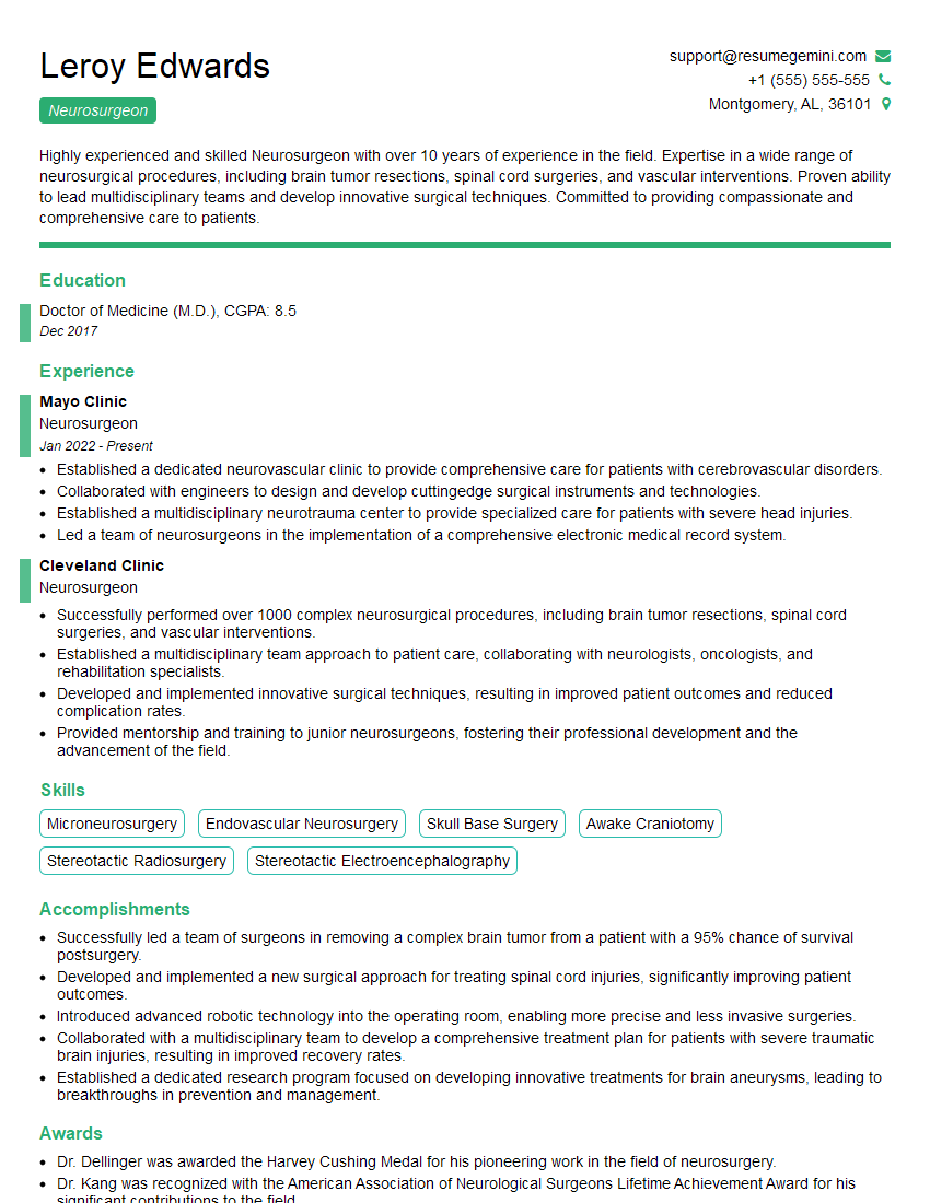
Mastering the intricacies of awake craniotomy significantly enhances your expertise in neurosurgery and opens doors to exciting career opportunities in leading medical institutions. To stand out, create a compelling and ATS-friendly resume that highlights your skills and experience effectively. We highly recommend using ResumeGemini to build a professional and impactful resume. ResumeGemini provides valuable tools and resources, including examples of resumes specifically tailored to Awake Craniotomy positions, to help you present your qualifications in the best possible light.
Explore more articles
Users Rating of Our Blogs
Share Your Experience
We value your feedback! Please rate our content and share your thoughts (optional).
What Readers Say About Our Blog
Hi, I have something for you and recorded a quick Loom video to show the kind of value I can bring to you.
Even if we don’t work together, I’m confident you’ll take away something valuable and learn a few new ideas.
Here’s the link: https://bit.ly/loom-video-daniel
Would love your thoughts after watching!
– Daniel
This was kind of a unique content I found around the specialized skills. Very helpful questions and good detailed answers.
Very Helpful blog, thank you Interviewgemini team.
