Feeling uncertain about what to expect in your upcoming interview? We’ve got you covered! This blog highlights the most important Debridement interview questions and provides actionable advice to help you stand out as the ideal candidate. Let’s pave the way for your success.
Questions Asked in Debridement Interview
Q 1. Describe the different types of debridement techniques.
Debridement is the process of removing dead, damaged, or infected tissue from a wound to promote healing. There are several types, each with its own approach:
- Sharp Debridement: This involves using surgical instruments like scalpels or scissors to precisely remove necrotic tissue. It’s the fastest method but requires surgical skill.
- Enzymatic Debridement: This uses topical enzymes to break down necrotic tissue. It’s less invasive than sharp debridement, but slower.
- Autolytic Debridement: This relies on the body’s natural processes to remove dead tissue. It involves using moist wound dressings to create a warm, moist environment that promotes autolysis (self-digestion of dead tissue). This is the least invasive method, but it’s also the slowest.
- Mechanical Debridement: This involves physically removing necrotic tissue, often using methods like wet-to-dry dressings, wound irrigation, or hydrotherapy. This is a relatively simple technique, but can be painful and may damage healthy tissue if not done carefully.
- Biologic Debridement: This uses sterile maggots (larvae of the blow fly) to selectively debride necrotic tissue. The maggots secrete enzymes that break down the dead tissue, leaving healthy tissue unharmed. This method is used less frequently, but is effective in certain situations.
The choice of debridement technique depends on the type and severity of the wound, the patient’s overall health, and the available resources.
Q 2. What are the indications and contraindications for sharp debridement?
Indications for Sharp Debridement: Sharp debridement is indicated when there’s a large amount of necrotic tissue that needs to be removed quickly, such as in severe burns, traumatic wounds, or infected wounds that aren’t responding to other treatments. It’s also useful when other debridement methods haven’t been effective. For example, a patient with a deep pressure ulcer with significant amounts of eschar (dry, black, necrotic tissue) would likely benefit from sharp debridement to facilitate healing.
Contraindications for Sharp Debridement: Sharp debridement shouldn’t be performed on patients with severe bleeding disorders, in areas with significant vascular compromise (e.g., close to major nerves or blood vessels), or when the wound is in an area that makes precise debridement difficult. A patient with a poorly controlled bleeding disorder, for example, would be a poor candidate for sharp debridement. In such cases, other less invasive methods might be preferred.
Q 3. Explain the process of enzymatic debridement.
Enzymatic debridement involves applying a topical enzyme preparation directly to the wound bed. These enzymes break down proteins in the necrotic tissue, making it easier to remove. The process typically involves:
- Wound Assessment: The wound is thoroughly assessed to determine the appropriate enzyme and dosage.
- Enzyme Application: The enzyme is applied to the necrotic tissue according to the manufacturer’s instructions. This may involve spreading a cream or gel, or applying a specialized dressing containing enzymes.
- Monitoring: The wound is monitored regularly for signs of debridement and any adverse reactions. The enzyme may need to be reapplied depending on the wound’s response.
- Debris Removal: Once the enzyme has done its work, the softened necrotic tissue can be easily removed with irrigation or gentle wiping.
Different enzymes target different components of necrotic tissue. For instance, collagenase is commonly used to break down collagen in necrotic tissue. The use of enzymatic debridement is often indicated in wounds where sharp debridement is not appropriate due to location, fragility of surrounding tissue, or patient health considerations.
Q 4. How do you assess the effectiveness of debridement?
Assessing the effectiveness of debridement involves a multifaceted approach focusing on both visual inspection and clinical assessment. This includes:
- Visual Examination: Look for a reduction in the amount of necrotic tissue. A decrease in the size and depth of the wound suggests effective debridement. The wound bed should appear cleaner, with a more healthy granulation tissue forming.
- Wound Measurements: Accurate measurements of the wound dimensions (length, width, depth) at baseline and after debridement provide objective data on the progress. Photos can be used for comparison.
- Clinical Signs: A decrease in pain, odor, and exudate (wound drainage) suggests effective debridement. An improvement in the overall appearance of the wound indicates healing.
- Culture Results: If infection was present, the culture results should show a reduction or elimination of the pathogenic organisms after debridement.
The absence of excessive bleeding or damage to healthy tissue is also crucial to assess the safety and precision of the debridement process. It is vital to use a combination of these methods for a complete assessment.
Q 5. What are the potential complications of debridement?
Potential complications of debridement include:
- Bleeding: Sharp debridement can cause bleeding, especially if performed aggressively or in highly vascularized areas.
- Infection: If proper sterile technique isn’t used, debridement can increase the risk of infection.
- Pain: Debridement can be painful, particularly sharp debridement.
- Damage to healthy tissue: Improper debridement can damage healthy tissue, delaying healing.
- Scarring: Especially with sharp debridement, significant scarring might occur.
- Allergic reactions: Allergic reactions to topical enzymes are possible in enzymatic debridement.
Careful planning, proper technique, and meticulous attention to sterile procedures minimize these risks. Post-debridement monitoring for signs of complications is vital.
Q 6. How do you manage pain during debridement?
Pain management is a critical aspect of debridement, particularly for sharp debridement. Strategies may include:
- Analgesics: Systemic analgesics (pain medications like opioids or NSAIDs), administered before, during, or after the procedure, are often necessary. The choice and dosage depend on the patient’s pain level and overall health.
- Local Anesthesia: For sharp debridement, local anesthesia (like lidocaine) can be injected into the wound to numb the area. This greatly reduces pain during the procedure itself.
- Sedation: In some cases, especially for extensive or sensitive wounds, sedation may be required to ensure patient comfort and tolerance of the procedure.
- Non-Pharmacological Methods: Techniques like relaxation exercises, guided imagery, and distraction can help manage pain. It’s often beneficial to combine these with pharmacological approaches.
The choice of pain management strategy depends on the patient’s overall condition, the extent of the debridement, and their individual pain tolerance.
Q 7. Describe the role of autolytic debridement in wound healing.
Autolytic debridement utilizes the body’s own mechanisms to remove necrotic tissue. A moist wound environment is created using occlusive or semi-occlusive dressings, allowing the body’s enzymes to break down dead tissue. This is a gentle, non-invasive method, particularly suitable for patients with fragile skin or wounds that are close to vital structures. The body’s natural processes of autolysis work slowly, which makes this technique time-consuming but reduces the risk of complications. Think of it as a slow, gentle process of ‘self-cleaning’ for the wound. This is a preferred method for patients with more sensitive wounds that might not tolerate the other types of debridement techniques.
The process typically involves applying a hydrocolloid, hydrogel, or alginate dressing to the wound, leaving it in place for several days to promote autolysis. The dressing is then removed, and any softened necrotic tissue is carefully removed. This process is repeated until all necrotic tissue is gone.
Q 8. How do you choose the appropriate debridement technique for a specific wound?
Choosing the right debridement technique is crucial for successful wound healing. It depends on several factors, including the type and depth of the wound, the amount and type of necrotic tissue, the patient’s overall health, and available resources. Think of it like choosing the right tool for a job – a screwdriver isn’t useful for hammering a nail.
- Wound assessment: A thorough assessment is paramount. This involves evaluating the size, depth, location, and type of tissue present (e.g., eschar, slough, granulation tissue). We also consider the patient’s comorbidities, such as diabetes, which can significantly impact healing.
- Type of necrotic tissue: The nature of the dead tissue guides the technique. Dry, adherent eschar often requires surgical or sharp debridement, while softer, moist slough might benefit from enzymatic or autolytic methods.
- Patient factors: A frail patient may not tolerate sharp debridement, while a patient with a large wound might require a combination of techniques. Pain management and the patient’s overall ability to tolerate the procedure are also significant considerations.
- Resource availability: The availability of specialized equipment, trained personnel, and appropriate dressings will impact the choice of debridement technique.
For example, a patient with a small, clean, dry eschar wound on their heel might benefit from sharp debridement under local anesthesia. Conversely, a patient with a large, infected, sloughy wound on their leg might require a combination of enzymatic debridement to soften the slough, followed by sharp debridement to remove the remaining necrotic tissue, while managing infection aggressively.
Q 9. What are the signs of infection in a wound undergoing debridement?
Recognizing signs of infection is vital, as it can significantly impair wound healing and lead to serious complications. Early identification allows for timely intervention and prevents escalation. Think of it as a fire alarm – early detection means you can put out the small fire before it becomes a major blaze.
- Increased pain: The wound may become increasingly painful, beyond what’s expected with normal healing.
- Erythema (redness): Redness extending beyond the wound edges indicates potential infection.
- Edema (swelling): Significant swelling surrounding the wound can be a sign of infection.
- Purulent drainage (pus): The presence of thick, yellow or green pus is a strong indicator of infection.
- Increased warmth: The area surrounding the wound may feel warmer to the touch than the surrounding skin.
- Fever and chills: Systemic signs of infection like fever and chills warrant immediate attention.
- Foul odor: A strong, unpleasant odor emanating from the wound suggests infection.
If any of these signs are present, wound cultures should be obtained to identify the causative organism and guide appropriate antibiotic therapy. Changes in the wound bed’s appearance, such as an increase in slough or the presence of new areas of necrosis, also require careful evaluation.
Q 10. How do you prevent infection during and after debridement?
Preventing infection during and after debridement is crucial for successful wound healing. It’s all about creating a sterile environment and supporting the body’s natural defenses. Think of it like creating a sterile operating room for the wound.
- Aseptic technique: Strict adherence to aseptic technique during the procedure is paramount. This includes proper hand hygiene, wearing sterile gloves, gowns, and masks, and using sterile instruments.
- Wound irrigation: Using sterile saline or other appropriate solutions to irrigate the wound helps remove debris and bacteria.
- Appropriate dressings: Selecting wound dressings that are appropriate for the type and stage of wound, and that maintain a moist wound healing environment, is crucial.
- Antibiotic prophylaxis: In cases of high risk for infection, prophylactic antibiotics may be considered.
- Post-debridement care: Providing adequate post-debridement care, including pain management and regular wound assessments, is essential.
- Patient education: Educating patients about proper wound care, signs of infection, and the importance of follow-up appointments is crucial for preventing infection.
For instance, immediately after sharp debridement, we meticulously clean the wound with sterile saline, apply an appropriate dressing that will help prevent further bacterial colonization, and provide the patient with instructions to watch for signs of infection.
Q 11. What are the key differences between sharp, enzymatic, and autolytic debridement?
The three primary debridement methods – sharp, enzymatic, and autolytic – differ significantly in their mechanisms and applications. Choosing the right one is like selecting the right key to unlock a specific door.
- Sharp debridement: This involves the surgical removal of necrotic tissue using sterile scissors, forceps, and scalpels. It’s the most effective method for removing large amounts of necrotic tissue quickly, but it’s also the most invasive and requires a skilled clinician. Think of it as precise surgery for the wound.
- Enzymatic debridement: This uses topical enzyme preparations to break down necrotic tissue. It’s less invasive than sharp debridement but slower. The enzymes are applied topically to the wound and left to work for a designated time, facilitating the removal of the necrotic tissue. Think of it as using a chemical solvent to dissolve the dead tissue.
- Autolytic debridement: This relies on the body’s natural mechanisms to remove necrotic tissue. It involves the use of occlusive dressings that trap moisture and facilitate the autolysis process. It’s the least invasive method but is often the slowest. Think of it as allowing the body’s own system to clean the wound.
Each method has its place depending on the wound characteristics. Sharp debridement is ideal for rapidly removing large amounts of necrotic tissue, while enzymatic debridement is suitable for wounds with smaller amounts of slough. Autolytic debridement is appropriate for wounds with minimal necrotic tissue and for patients who are not suitable candidates for more invasive procedures.
Q 12. Describe your experience with managing different wound types requiring debridement.
My experience encompasses managing a wide spectrum of wound types requiring debridement. I’ve worked with everything from simple, superficial wounds to complex, deep wounds with significant necrotic tissue involvement.
- Pressure ulcers: I have extensive experience in managing pressure ulcers of varying stages, employing various debridement techniques depending on the wound’s characteristics and the patient’s overall health. This includes selecting the appropriate debridement method, dressing selection, and pain management strategies.
- Diabetic foot ulcers: These ulcers often present with significant infection and extensive necrosis, requiring a multi-faceted approach including aggressive debridement, infection control, and offloading measures. Often, these require a combined approach, beginning with enzymatic debridement to soften the wound before progressing to sharp debridement.
- Surgical wounds: I’ve managed surgical wounds that have developed complications, such as wound dehiscence or infection, requiring debridement to remove necrotic tissue and promote healing.
- Traumatic wounds: I’ve worked with patients with traumatic wounds, ranging from minor lacerations to severe crush injuries. Often requiring a tailored approach to debridement, taking into account the nature of the trauma and the potential for contamination.
Each case presented unique challenges, requiring careful consideration of the patient’s overall health, the wound’s characteristics, and the available resources. A recent case involved a diabetic patient with a large, infected foot ulcer. We used a combination of enzymatic and sharp debridement, aggressive infection control, and patient education to achieve successful wound healing and prevent amputation.
Q 13. How do you document the debridement procedure?
Accurate and thorough documentation of the debridement procedure is essential for legal, medical, and quality improvement purposes. Think of it as creating a detailed map of the process, recording every step.
My documentation includes:
- Patient demographics: Name, medical record number, date of birth.
- Wound assessment: Detailed description of the wound’s location, size, depth, type of tissue present, and any signs of infection.
- Debridement technique: Specific technique(s) used (sharp, enzymatic, autolytic), amount of tissue removed, and any complications encountered.
- Wound irrigation: Type of solution used, volume, and any signs of infection identified during the process.
- Dressings applied: Type and amount of dressing material used.
- Pain management: Any pain management strategies employed during and after the procedure.
- Post-procedure assessment: Observations made after the procedure, including any signs of bleeding or infection.
- Patient education: Documentation of the education provided to the patient and/or family regarding wound care and infection prevention.
- Photographs: Before and after photographs are crucial to visually document the wound’s progress.
This detailed documentation allows for tracking the wound’s progress, facilitates communication among the healthcare team, and provides a legal record of the care provided. It is also vital for quality improvement initiatives and research.
Q 14. What are the legal and ethical considerations involved in debridement?
Debridement, being an invasive procedure, carries significant legal and ethical considerations. It’s crucial to act within the bounds of the law and ethical guidelines to ensure patient safety and well-being.
- Informed consent: Obtaining informed consent from the patient or their legal guardian is paramount. This involves explaining the procedure, its benefits, risks, and alternatives in a language they understand.
- Scope of practice: Performing debridement must be within the scope of one’s professional license and expertise. Only qualified healthcare professionals should perform debridement.
- Pain management: Adequate pain management is ethically and legally necessary. Patients should be comfortable during the procedure, and appropriate analgesia should be administered.
- Infection control: Strict adherence to infection control protocols is vital to prevent complications and protect the patient from harm.
- Documentation: As mentioned previously, meticulous documentation is essential for legal protection and demonstrating adherence to standards of care.
- Confidentiality: Maintaining patient confidentiality is paramount, following all relevant privacy regulations (e.g., HIPAA).
Failure to obtain informed consent, exceeding one’s scope of practice, or neglecting proper pain management or infection control can lead to legal repercussions and ethical violations. Always prioritize patient safety and well-being while adhering to established legal and ethical guidelines.
Q 15. How do you educate the patient and family about debridement?
Educating patients and their families about debridement is crucial for their understanding and cooperation. I begin by explaining the process in simple terms, avoiding medical jargon. I explain that debridement is the removal of dead or infected tissue from a wound to promote healing. I use analogies to help them visualize it; for example, I might compare it to cleaning out a garden to allow healthy plants to grow.
I discuss the different types of debridement – sharp, enzymatic, autolytic, and mechanical – explaining the benefits and potential risks of each. I also emphasize the importance of pain management during and after the procedure. I answer all their questions patiently and honestly, addressing any concerns they might have about pain, bleeding, or the healing process. I provide written information, such as brochures or pamphlets, to reinforce the information discussed and allow them to review it at their leisure. Finally, I clearly outline the post-debridement care instructions, including dressing changes, wound monitoring, and signs to watch for. This ensures the patient and family are active participants in the healing process.
Career Expert Tips:
- Ace those interviews! Prepare effectively by reviewing the Top 50 Most Common Interview Questions on ResumeGemini.
- Navigate your job search with confidence! Explore a wide range of Career Tips on ResumeGemini. Learn about common challenges and recommendations to overcome them.
- Craft the perfect resume! Master the Art of Resume Writing with ResumeGemini’s guide. Showcase your unique qualifications and achievements effectively.
- Don’t miss out on holiday savings! Build your dream resume with ResumeGemini’s ATS optimized templates.
Q 16. Describe your experience with using different types of dressings after debridement.
My experience with post-debridement dressings is extensive. The choice of dressing depends heavily on the wound type, depth, and the patient’s individual needs. For superficial wounds, simple gauze dressings often suffice, particularly after sharp debridement. For deeper wounds or those with significant exudate, I might use absorbent dressings like alginate or foam dressings. Alginates are excellent at absorbing exudate and creating a moist wound environment, while foams provide cushioning and protection.
I’ve also used hydrocolloids extensively; these are self-adhesive dressings that create a moist environment and are good for wounds with minimal drainage. In cases of infected wounds, I might opt for antimicrobial dressings containing silver or other agents to help control infection. The selection process involves carefully assessing the wound and anticipating its needs; for example, a wound with significant drainage would require a highly absorbent dressing to prevent maceration and maintain a healthy environment. Regular assessment and dressing changes based on the wound’s response are crucial for optimal outcomes.
Q 17. How do you monitor a wound after debridement?
Monitoring a wound after debridement is a critical aspect of wound care. I assess the wound daily, or more frequently if needed, focusing on several key indicators. I look for signs of infection, such as increased pain, swelling, redness, warmth, or purulent drainage. I also carefully evaluate the wound bed for the presence of necrotic tissue, slough, or eschar, indicating incomplete debridement. I measure the wound dimensions (length, width, depth) to track progress.
I also assess the periwound skin for signs of maceration or irritation caused by excessive moisture. The amount and character of drainage are important indicators, as are the patient’s overall symptoms, including pain level and systemic signs of infection such as fever or malaise. Documentation is crucial; I meticulously record all observations in the patient’s chart, including photographic documentation when appropriate, to track progress and make informed decisions about continued care. Any significant changes prompt adjustments to the treatment plan, which may include a change in dressing type, additional debridement, or the initiation of antibiotic therapy if infection is present.
Q 18. What are the signs of healing after debridement?
Signs of healing after debridement are gradual but noticeable. Initially, you’ll see a reduction in exudate and a decrease in wound odor. Granulation tissue, which is healthy, pink, and bumpy tissue, will begin to fill the wound bed. Epithelialization, the migration of new skin cells across the wound surface, will be visible as the wound gradually closes. The wound bed will become cleaner, and less necrotic tissue will be present.
The wound edges will start to contract, and the overall size of the wound will decrease. Pain will usually lessen as the wound heals. It’s important to note that the rate of healing varies significantly depending on factors such as the patient’s overall health, the size and depth of the wound, the presence of infection, and the patient’s nutritional status. Regular monitoring is essential to identify any deviations from the expected healing trajectory. For example, stalled healing might indicate the need for further debridement or a change in treatment approach.
Q 19. How do you address delayed wound healing after debridement?
Delayed wound healing after debridement requires a thorough investigation to identify the underlying cause. This might involve reviewing the patient’s medical history, nutritional status, and medication list. Factors such as diabetes, peripheral arterial disease (PAD), and immunosuppression can significantly impair wound healing. Infection is a major cause of delayed healing, so I would re-evaluate for signs of infection and obtain cultures if necessary.
Inadequate debridement may leave residual necrotic tissue hindering healing, necessitating further debridement. Nutritional deficiencies, especially protein and vitamin deficiencies, can impair healing, so a dietary assessment and supplementation might be necessary. Sometimes, underlying medical conditions require management. For instance, optimization of blood glucose control in diabetic patients is essential. If infection is suspected or present, appropriate antibiotic therapy will be implemented. In some cases, consultation with other specialists, such as a vascular surgeon or a dietician, is required to address specific issues impacting healing.
Q 20. What is the role of negative pressure wound therapy (NPWT) in debridement?
Negative pressure wound therapy (NPWT) plays a significant role in debridement, particularly in the management of complex or chronic wounds. NPWT, commonly known as vacuum-assisted closure (VAC) therapy, uses a vacuum to remove excess exudate and interstitial fluid from the wound bed. This process facilitates the removal of necrotic tissue, thereby promoting debridement. The continuous negative pressure also helps to improve blood flow to the wound, stimulating granulation tissue formation and facilitating healing.
NPWT can be used in conjunction with other debridement techniques, such as sharp debridement or enzymatic debridement. It is particularly beneficial for wounds with large amounts of exudate or those that are difficult to debride using other methods. By creating a moist, protected wound environment while removing harmful substances, NPWT supports a more favorable healing milieu.
Q 21. Describe your experience with using NPWT.
My experience with NPWT is extensive, having used it successfully in a wide variety of clinical settings. I’ve utilized it to manage various wounds, including diabetic foot ulcers, pressure ulcers, traumatic wounds, and surgical wounds with complications. The process typically involves preparing the wound bed, selecting the appropriate NPWT dressing, and applying the device according to manufacturer’s instructions.
I find it particularly useful in wounds with significant exudate or those that are failing to heal with traditional methods. NPWT often helps to reduce wound size and promote granulation tissue formation, leading to faster healing. However, I also recognize its limitations; it may not be suitable for all wound types, and it requires careful monitoring to prevent complications such as skin damage or infection. Regular assessment of the wound, dressing, and tubing is crucial to ensure the effective and safe application of NPWT. In my experience, proper patient education is paramount for ensuring compliance and minimizing the risk of complications.
Q 22. How do you assess the need for further debridement?
Assessing the need for further debridement involves a thorough wound assessment, focusing on the presence of non-viable tissue. It’s not a one-time event; it’s an iterative process. We look for several key indicators:
- Visual inspection: We carefully examine the wound bed for any remaining signs of necrotic tissue (eschar, slough), which appear as black, brown, or yellow tissue that is dry and leathery or moist and stringy respectively. We also assess the wound edges for signs of inflammation or infection.
- Palpation: Gently feeling the wound bed helps to determine the consistency of the tissue. Non-viable tissue feels firm, dry, and sometimes adherent to the wound bed.
- Wound bed assessment: We meticulously document the type and percentage of necrotic tissue, the presence of exudate (wound drainage), and any signs of infection (redness, swelling, pain, warmth, pus).
- Clinical indicators: Elevated inflammatory markers (CRP, ESR) or persistent signs of infection suggest the need for further debridement, even if the wound appears superficially clean.
- Imaging: In some cases, particularly deep wounds or wounds with underlying osteomyelitis (bone infection), imaging techniques like X-rays or ultrasound may be necessary to fully assess the extent of damage and guide debridement.
Essentially, we continue debridement until we achieve a healthy, viable wound bed free of non-viable tissue that promotes healing. This is a crucial step to prevent infection and promote optimal wound healing.
Q 23. How do you handle a patient with allergies to debridement products?
Managing a patient with allergies to debridement products requires a cautious and individualized approach. First, we obtain a complete allergy history, specifying the exact product and the type of reaction. This information is crucial. Then, we select alternative debridement methods, prioritizing patient safety.
- Avoiding the allergen: The most straightforward approach is to simply avoid using any product the patient is allergic to.
- Alternative debridement techniques: If the allergy prevents the use of enzymatic debridement, we might opt for sharp debridement (surgical removal of necrotic tissue) or autolytic debridement (using the body’s natural enzymes to break down tissue, usually facilitated by moisture-retentive dressings).
- Careful product selection: If enzymatic debridement is necessary despite allergies, we may explore alternative enzymatic preparations that do not contain the offending allergen.
- Pre-treatment: In some cases, pre-treatment with antihistamines or corticosteroids might be considered, especially if minor reactions have occurred in the past.
- Close monitoring: Regardless of the method chosen, close monitoring for any signs of allergic reaction is essential. We carefully watch for skin reactions, breathing difficulties, or other signs of allergic response.
Patient safety is paramount. We always prioritize methods that minimize the risk of allergic reactions while effectively removing the non-viable tissue.
Q 24. What are the key factors to consider in selecting a wound dressing post-debridement?
Selecting a wound dressing post-debridement is vital for creating an optimal healing environment. The ideal dressing depends on several factors:
- Wound type and depth: A shallow wound might benefit from a simple hydrocolloid dressing to maintain a moist healing environment and protect the wound bed. Deep wounds or those with significant exudate may require a dressing with superior absorbency, like alginate or foam.
- Amount of exudate: High-exudating wounds need highly absorbent dressings to prevent maceration (softening of the surrounding skin) and infection. Low-exudating wounds require dressings that provide moisture balance without over-hydration.
- Presence of infection: If infection is present, dressings with antimicrobial properties may be necessary. In some cases, a negative pressure wound therapy (NPWT) system might be employed.
- Patient comfort and convenience: We consider factors like ease of application, pain, and the need for frequent changes when selecting a dressing.
- Cost-effectiveness: We aim to select dressings that balance effectiveness and cost, as dressing changes are often repeated and contribute to healthcare expenses.
For instance, a shallow, clean wound might use a simple hydrocolloid dressing, while a deep wound with heavy exudate and infection might need a combination of alginate for absorption and a silver-impregnated dressing for antimicrobial action, followed by a secondary dressing for moisture retention and protection.
Q 25. Explain your understanding of the role of debridement in reducing bioburden.
Debridement plays a crucial role in reducing bioburden – the number of microorganisms on or in a wound. Necrotic tissue provides a perfect breeding ground for bacteria, fungi, and other pathogens. By removing this non-viable tissue, debridement significantly reduces the available substrate for microbial growth.
Think of it like this: imagine a garden overflowing with weeds. The weeds (necrotic tissue) steal resources and create an environment where pests (microorganisms) thrive. Debridement is like weeding the garden, removing the unwanted elements, creating space for healthy plants (new tissue) to grow.
Reducing bioburden is critical in preventing or controlling wound infection, thereby facilitating healing. The cleaner the wound bed, the less opportunity for infection to establish.
Q 26. How do you distinguish between different types of necrotic tissue?
Distinguishing between different types of necrotic tissue is essential for selecting the appropriate debridement method. Here’s a breakdown:
- Eschar: This is dry, black, leathery necrotic tissue that is typically adherent to the wound bed. It’s usually relatively avascular (lacks blood supply) and often requires surgical debridement.
- Slough: This is moist, yellow or tan necrotic tissue that is usually stringy or gelatinous in consistency. It’s less firmly attached to the wound bed than eschar and can sometimes be removed using enzymatic or autolytic debridement techniques.
- Fibrin: This is a yellowish-white, stringy material that may be found within the wound bed. It is a part of the normal healing process but can obstruct wound healing if present in excessive quantities.
Visual inspection, palpation, and sometimes even microscopic examination can help differentiate these types. Accurate identification guides the selection of the most effective and safest debridement method.
Q 27. What is your experience with managing wounds with exposed bone or tendon?
Managing wounds with exposed bone or tendon presents unique challenges. These wounds are typically high-risk for infection and require meticulous care. The primary goal is to achieve wound closure and protect underlying structures.
Our approach involves:
- Aggressive debridement: All non-viable tissue, including any devitalized bone or tendon fragments, must be thoroughly removed to prevent infection.
- Infection control: Preventing or controlling infection is paramount. This might involve antibiotic therapy, depending on the presence or risk of infection.
- Wound bed preparation: Once the wound is debrided, we aim to create a clean, well-vascularized wound bed to promote healing. This may include measures to stimulate granulation tissue formation.
- Protective covering: Appropriate wound dressings are selected to protect the exposed bone or tendon. These might include specialized dressings designed for such wounds.
- Surgical intervention: In some cases, surgical intervention may be necessary to achieve closure, manage complex fractures, or provide structural support.
- Regular monitoring: These wounds require very close monitoring for signs of infection or complications. This may involve frequent assessment and changes in dressing management.
These wounds require a multidisciplinary approach, often involving surgeons, wound care specialists, and infectious disease specialists.
Q 28. Describe a challenging debridement case you encountered and how you addressed it.
One particularly challenging case involved a patient with a large, heavily infected pressure ulcer on their sacrum extending to the bone (osteomyelitis). The wound was deep, contained significant amounts of necrotic tissue and slough, and exuded copious amounts of purulent drainage. The patient had significant comorbidities, including diabetes and peripheral vascular disease, which hindered healing.
Our approach was multifaceted:
- Initial debridement: We began with sharp debridement to remove the readily accessible necrotic tissue and slough. This was done under appropriate anesthetic.
- Enzymatic debridement: Once the initial sharp debridement was completed, we used enzymatic debridement to help further remove the remaining necrotic tissue.
- Infection control: We immediately instituted aggressive antibiotic therapy guided by wound cultures. This was essential for controlling the osteomyelitis.
- Wound packing: We utilized alginate dressings for their absorbency and packing capabilities. This helped to keep the wound clean and dry.
- Negative pressure wound therapy (NPWT): After several days, we transitioned to NPWT to further reduce the bioburden and promote wound healing by removing excess exudate and maintaining a moist wound bed.
- Surgical consultation: Given the involvement of the bone, surgical consultation was sought, and a surgical debridement was performed to remove any remaining infected bone tissue.
Through this multi-modal approach, we managed to get the infection under control and eventually achieve wound closure. The case highlighted the importance of a tailored approach based on the specific needs and characteristics of the wound, and the benefits of collaboration amongst healthcare professionals.
Key Topics to Learn for Debridement Interview
- Wound Bed Preparation: Understanding the different types of wounds and appropriate preparation techniques before debridement.
- Debridement Techniques: Mastering the nuances of sharp, enzymatic, autolytic, and mechanical debridement methods, including their indications and contraindications.
- Selection of Debridement Method: Developing the ability to assess wound characteristics and choose the most effective and appropriate debridement technique for each patient.
- Pain Management during Debridement: Understanding strategies for managing patient discomfort and anxiety during the procedure.
- Post-Debridement Care: Knowing the essential steps in wound care following debridement to promote healing and prevent complications.
- Infection Control: Highlighting the importance of sterile techniques and infection prevention strategies during and after debridement.
- Assessment of Wound Healing: Understanding how to accurately assess wound healing progress and adjust treatment plans as needed.
- Legal and Ethical Considerations: Familiarity with the legal and ethical implications of debridement procedures and informed consent.
- Case Studies and Problem Solving: Analyzing case studies to develop practical problem-solving skills related to different wound types and debridement challenges.
- Advanced Debridement Techniques: Exploring more advanced methods like Ultrasound-Assisted Debridement or larval therapy (Maggot Debridement).
Next Steps
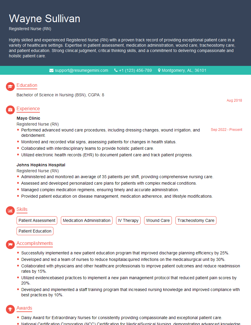
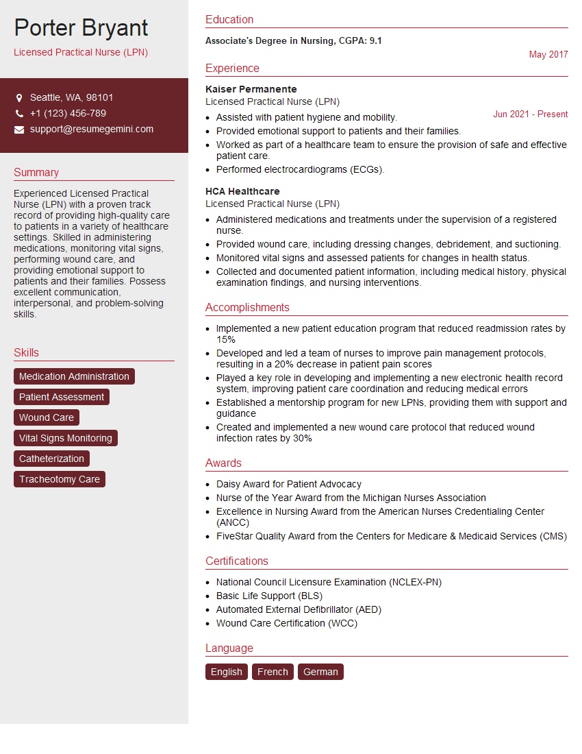
Mastering debridement techniques significantly enhances your career prospects in wound care, opening doors to specialized roles and advanced opportunities. A strong, ATS-friendly resume is crucial for showcasing your skills and experience to potential employers. To maximize your job search success, we strongly recommend using ResumeGemini to build a professional and impactful resume. ResumeGemini provides tools and resources to help you craft a compelling narrative, and we offer examples of resumes tailored specifically to Debridement professionals to help guide you.
Explore more articles
Users Rating of Our Blogs
Share Your Experience
We value your feedback! Please rate our content and share your thoughts (optional).
What Readers Say About Our Blog
This was kind of a unique content I found around the specialized skills. Very helpful questions and good detailed answers.
Very Helpful blog, thank you Interviewgemini team.
