Every successful interview starts with knowing what to expect. In this blog, we’ll take you through the top Neonatal Intubation interview questions, breaking them down with expert tips to help you deliver impactful answers. Step into your next interview fully prepared and ready to succeed.
Questions Asked in Neonatal Intubation Interview
Q 1. Describe the indications for neonatal endotracheal intubation.
Neonatal endotracheal intubation, the insertion of a tube into a newborn’s trachea (windpipe), is indicated when the infant is unable to maintain adequate oxygenation and ventilation on their own. This can stem from various causes.
- Respiratory distress syndrome (RDS): A common condition affecting premature infants, where the lungs lack surfactant, leading to collapsed alveoli.
- Meconium aspiration syndrome (MAS): Where meconium (fetal stool) is aspirated into the lungs during delivery, causing airway obstruction and inflammation.
- Congenital diaphragmatic hernia (CDH): A birth defect where the diaphragm is incomplete, allowing abdominal organs to enter the chest cavity and compromise lung development.
- Apnea and bradycardia: Episodes of cessation of breathing and slowed heart rate requiring immediate intervention.
- Choking or aspiration of foreign bodies: These events can obstruct the airway and necessitate immediate removal and intubation.
- Severe prematurity or birth asphyxia: Infants born prematurely or experiencing oxygen deprivation during birth often require assistance with ventilation.
Essentially, intubation becomes necessary when the infant’s spontaneous breathing is insufficient to provide adequate oxygen levels and remove carbon dioxide.
Q 2. What are the different types of endotracheal tubes used in neonates?
Several types of endotracheal tubes (ETTs) are used, tailored to the neonate’s size and gestational age. They primarily differ in their internal diameter (ID) and length.
- Uncuffed ETTs: Primarily used in smaller neonates (less than 28 weeks gestational age or less than 1000 grams) to avoid tracheal damage. The lack of cuff means reliance on a proper fit to prevent air leaks.
- Cuffed ETTs: Employed in larger, more mature neonates (typically over 28 weeks gestation or greater than 1000 grams) to provide a better seal and prevent air leaks around the tube. Proper cuff inflation is critical to prevent tracheal injury.
- Murphy ETTs: These feature a built-in, reinforced end, reducing the risk of kinking or damage. This is especially helpful in managing delicate neonate airways.
- Endotracheal tubes with integrated fiberoptic stylet: These are used in particularly challenging intubation scenarios, allowing for direct visualization of the airway.
The choice of tube depends on the individual neonate’s characteristics and the clinical situation. Careful selection ensures optimal airway management and minimizes the risk of complications.
Q 3. Explain the steps involved in performing neonatal endotracheal intubation.
Neonatal intubation is a procedure requiring a skilled and experienced practitioner. The steps involved are:
- Preparation: Assemble all necessary equipment (ETT of appropriate size, laryngoscope with appropriate blade, suction equipment, monitoring devices, etc.). Ensure the infant is properly positioned and supported.
- Preoxygenation: Before initiating the procedure, provide supplemental oxygen to the infant to maximize oxygen stores.
- Laryngoscopy: Gently insert the laryngoscope blade into the infant’s mouth, visualizing the vocal cords.
- ETT Placement: Advance the ETT into the trachea, visualizing its passage through the vocal cords. Avoid pushing too hard and causing trauma.
- Confirmation of placement: Confirm proper placement using auscultation (listening for bilateral breath sounds), chest rise and fall, and capnography (measuring end-tidal CO2). Consider chest x-ray for definitive placement confirmation.
- Securing the ETT: Secure the tube in place using appropriate tape or fixation device to prevent accidental dislodgement.
- Connection to ventilator or bag-mask: Connect the tube to a ventilator or bag-mask for assisted ventilation.
The entire process demands meticulous attention to detail, ensuring minimal trauma and effective airway management. Proper training and practice are essential.
Q 4. What are the potential complications of neonatal endotracheal intubation?
Despite careful execution, neonatal endotracheal intubation carries potential complications:
- Tracheal trauma: Bleeding, perforation, or other injuries to the trachea from the tube itself.
- Esophageal intubation: Accidental placement of the tube into the esophagus instead of the trachea, resulting in inadequate ventilation and potential aspiration.
- Bradycardia: Slowed heart rate often triggered by vagal stimulation during intubation.
- Hypoxemia: Low blood oxygen levels due to inadequate ventilation or prolonged apneic periods.
- Hyperinflation of the lungs: Excessive ventilation leading to lung injury (barotrauma) or pneumothorax.
- Infection: Increased risk of respiratory infections due to an invasive procedure.
- Subglottic stenosis: Narrowing of the trachea below the vocal cords, potentially leading to long-term breathing difficulties.
Minimizing these risks hinges on precise technique, appropriate equipment selection, and vigilant monitoring of the infant’s vital signs during and after the procedure.
Q 5. How do you confirm proper endotracheal tube placement?
Confirming proper ETT placement is critical. Multiple methods should be employed:
- Auscultation: Listening for equal bilateral breath sounds over the lungs, indicating that air is entering both lungs equally. Absent breath sounds could suggest esophageal intubation or unilateral lung collapse.
- Chest rise and fall: Observing symmetrical chest expansion with each breath, suggesting proper ventilation.
- Capnography: Measuring the end-tidal carbon dioxide (EtCO2) level. A waveform and a value above 35 mmHg usually confirms that the ETT is in the trachea and carbon dioxide is being eliminated from the lungs.
- Chest X-ray: A chest radiograph provides definitive confirmation of ETT position, confirming its placement within the trachea and ensuring proper distance from the carina (the bifurcation of the trachea into the main bronchi).
Combining these methods offers a reliable assessment of ETT placement, minimizing the chance of misplacement.
Q 6. What are the signs of inadequate ventilation in a neonate?
Signs of inadequate ventilation in a neonate can be subtle yet critical to recognize:
- Respiratory distress: Increased respiratory rate, grunting, nasal flaring, retractions (sucking in of the skin between the ribs or above the sternum), and cyanosis (bluish discoloration of the skin).
- Bradycardia: Slow heart rate indicative of poor oxygenation or cardiac compromise.
- Acidosis: A build-up of acid in the blood, reflecting inadequate removal of carbon dioxide.
- Decreased oxygen saturation (SpO2): Lower than normal levels of oxygen in the blood, readily monitored via pulse oximetry.
- Absent or diminished breath sounds: Suggesting airway obstruction or pneumothorax.
- Apnea: Temporary cessation of breathing.
Prompt recognition of these signs is vital, requiring continuous monitoring and immediate intervention if necessary.
Q 7. Describe the management of a difficult airway in a neonate.
Managing a difficult airway in a neonate presents significant challenges, demanding rapid assessment and appropriate intervention. Strategies include:
- Alternative airway techniques: If direct laryngoscopy fails, consider using an alternative technique, such as fiberoptic bronchoscopy, which allows for direct visualization of the airway and precise ETT placement.
- LMA (Laryngeal Mask Airway): A supraglottic airway device that may be used as an alternative to ETT in some cases.
- Needle cricothyrotomy: A procedure involving inserting a needle into the cricothyroid membrane to create an airway, providing emergency oxygenation in life-threatening situations.
- Surgical airway: As a last resort, a surgical airway (tracheostomy) may be necessary if other methods fail. This is a major procedure and necessitates a surgical team.
- Team approach: Successful management hinges on teamwork. Assemble a skilled and experienced team with expertise in neonatal resuscitation and airway management.
Difficult airway scenarios necessitate a structured approach, often requiring escalation to more advanced techniques and a multidisciplinary approach. Prior planning and thorough training are essential to maximize the chances of a successful outcome.
Q 8. How do you manage accidental extubation?
Accidental extubation in a neonate is a critical event requiring immediate action. The first priority is to maintain airway patency. This involves quickly assessing the infant’s respiratory status – looking for signs of respiratory distress such as retractions, grunting, or cyanosis.
Immediate Steps:
- Assess the baby: Check heart rate, respiratory rate, oxygen saturation, and color.
- Provide positive pressure ventilation (PPV): Using a bag-valve mask (BVM) with supplemental oxygen, provide effective ventilation until re-intubation is possible.
- Attempt re-intubation: If possible, immediately attempt re-intubation using the same or a new endotracheal tube (ETT). Consider using a smaller size if initial attempts are unsuccessful.
- Prepare for advanced support: If re-intubation is unsuccessful or the infant remains unstable, prepare for advanced resuscitation efforts including chest compressions and medications.
- Document everything: Meticulously document the time of extubation, the interventions taken, the infant’s response, and the eventual outcome.
Preventing Future Extubations: After successful re-intubation, consider measures to reduce the risk of recurrent extubation such as securing the ETT appropriately, using a smaller size tube if appropriate, optimizing patient positioning, and careful handling of the tube.
I remember a case where a premature infant unexpectedly extubated. We immediately initiated PPV and successfully re-intubated within seconds. The quick response minimized the impact on the infant’s oxygen saturation and prevented further complications.
Q 9. What are the indications for using a laryngeal mask airway (LMA) in neonates?
Laryngeal Mask Airways (LMAs) are less commonly used in neonates compared to endotracheal tubes (ETTs) due to the unique anatomical challenges presented by their small airways. However, there are limited situations where an LMA might be considered.
- Brief procedures: In situations where short-term airway support is needed for a very brief procedure (e.g., short surgical procedure under sedation), an LMA might be a less invasive option than an ETT, particularly if the risk of intubation is high.
- Failed intubation: In cases where conventional intubation attempts have failed repeatedly, an LMA could provide temporary airway support while alternative strategies are planned.
- Specific situations: Certain specialized centers may utilize LMAs in specific neonates with severe anatomical anomalies affecting endotracheal intubation.
It’s crucial to remember that the use of LMAs in neonates is highly debated and situation-dependent. The risks and benefits must be carefully weighed against the risks and benefits of ETT intubation for each individual neonate.
Q 10. What are the advantages and disadvantages of using an LMA compared to ETT?
The choice between an LMA and an ETT in neonates is a critical one, determined by individual patient needs and the specific clinical circumstances.
Advantages of LMA over ETT:
- Less invasive: Does not require tracheal intubation, reducing the risk of trauma to the airway.
- Faster placement: Generally easier and faster to place than an ETT, which is crucial in emergency situations.
Disadvantages of LMA over ETT:
- Higher risk of airway obstruction: LMA provides less secure airway control compared to an ETT and may be associated with a greater risk of aspiration and airway obstruction.
- Limited airway access: Does not allow for direct suctioning of secretions below the larynx.
- Potential for aspiration: Increased risk of aspiration compared to an ETT, particularly during events like vomiting or regurgitation.
- Less reliable airway seal: May not maintain a secure airway seal as reliably as an ETT.
- Limited use in neonates: The anatomy of the neonate makes its application challenging.
In summary, while an LMA might have some advantages in specific, limited situations, the ETT remains the gold standard for securing the airway in the vast majority of neonatal resuscitation and management scenarios.
Q 11. Describe the appropriate size and type of ETT for different gestational ages.
ETT size selection for neonates is crucial for effective ventilation and minimizing airway trauma. The appropriate size depends heavily on the gestational age and weight of the infant.
Gestational Age and ETT Size:
- Extremely premature (<28 weeks): 2.5-3.0 mm ID
- Premature (28-34 weeks): 3.0-3.5 mm ID
- Near-term/Term (34-40 weeks): 3.5-4.0 mm ID
Type of ETT: Uncuffed ETTs are generally preferred for neonates to minimize the risk of tracheal damage and necrosis. However, cuffed tubes may be considered in certain situations where a more secure airway seal is deemed essential.
Important Note: These are guidelines; the optimal size should be determined on a case-by-case basis, considering the individual infant’s weight and physical examination findings. Always verify placement through auscultation and chest rise.
Q 12. How do you calculate the appropriate ETT size for a neonate?
There are several formulas for estimating ETT size, but none are universally perfect. Accurate assessment based on the infant’s physical characteristics and clinical condition is vital.
Common Formulas:
- Weight-based: (Weight in kg) / 4 + 4 = Internal Diameter (mm)
- Age-based (approximate): Premature infants generally require smaller tubes (2.5-3.5mm) while term infants may require 3.5-4.0mm.
Practical Application: The formulas provide an initial estimate. However, final tube selection should always be adjusted based on the infant’s response. If there’s poor chest rise or air leak, a smaller tube should be considered. Conversely, if ventilation is difficult, a slightly larger tube might be needed. The primary goal is to select a tube that provides adequate ventilation while minimizing the risk of tracheal trauma.
Q 13. What are the key differences in intubating a term versus a preterm infant?
Intubating a term versus a preterm infant presents distinct challenges due to anatomical differences.
Term Infants:
- Larger airway: Easier to visualize the vocal cords.
- More developed cartilage: Provides better airway support.
- Generally less fragile: Can tolerate instrumentation better.
Preterm Infants:
- Smaller, less developed airway: Makes visualization more difficult.
- Floppier epiglottis: Increased risk of trauma.
- Greater risk of airway injury: Requires gentler handling and smaller tubes.
- Increased risk of bradycardia: Intubation can trigger vagal responses.
Key Differences in Technique: In preterm infants, the use of specialized equipment like a smaller laryngoscope blade (e.g., Miller 0 or 1) and a smaller ETT is crucial. A gentler approach and careful monitoring of the heart rate are essential to minimize the risk of bradycardia. Additionally, the use of video laryngoscopy can enhance visualization in difficult cases.
Q 14. Describe your experience with neonatal resuscitation and intubation.
I have extensive experience in neonatal resuscitation and intubation, having performed countless procedures during my career. My experience encompasses a wide range of situations, from routine intubations in term infants to challenging cases involving extremely premature and critically ill infants with complex congenital anomalies.
Experience Highlights:
- Routine intubations: I’ve performed a large number of successful routine endotracheal intubations for various neonatal indications.
- Difficult intubations: I am proficient in managing difficult intubations, utilizing advanced techniques such as video laryngoscopy and alternative airway management strategies.
- Neonatal resuscitation: I have significant experience with neonatal resuscitation, including providing bag-valve mask ventilation and initiating advanced life support measures in conjunction with intubation.
- Teaching and supervision: I actively participate in teaching and supervising junior colleagues in neonatal resuscitation and airway management techniques.
Throughout my practice, I have always placed a strong emphasis on minimizing any potential trauma during the intubation process. This involves meticulous preparation, gentle handling, and the careful selection of appropriately sized equipment. Prioritizing patient safety and providing optimal airway management remain paramount in my practice.
Q 15. Explain the role of positive pressure ventilation in neonatal intubation.
Positive pressure ventilation (PPV) in neonatal intubation is crucial because newborns often lack the strength to breathe effectively on their own. It artificially delivers breaths, mimicking the natural process. We use PPV to maintain adequate oxygen levels and remove carbon dioxide from the lungs, preventing life-threatening complications.
Think of it like this: a newborn’s lungs are like deflated balloons. PPV acts like an external pump, inflating the balloons (lungs) with air, ensuring oxygen reaches the bloodstream and carbon dioxide is expelled. The pressure we apply is carefully monitored and adjusted based on the baby’s individual needs to avoid lung injury. The type of PPV – whether it’s bag-mask ventilation or mechanical ventilation – depends on the severity of the respiratory distress and the baby’s overall condition.
Career Expert Tips:
- Ace those interviews! Prepare effectively by reviewing the Top 50 Most Common Interview Questions on ResumeGemini.
- Navigate your job search with confidence! Explore a wide range of Career Tips on ResumeGemini. Learn about common challenges and recommendations to overcome them.
- Craft the perfect resume! Master the Art of Resume Writing with ResumeGemini’s guide. Showcase your unique qualifications and achievements effectively.
- Don’t miss out on holiday savings! Build your dream resume with ResumeGemini’s ATS optimized templates.
Q 16. What are the common causes of neonatal respiratory distress?
Neonatal respiratory distress has many potential causes, broadly categorized as:
- Lung Diseases: Respiratory Distress Syndrome (RDS) – a deficiency in surfactant (a substance that helps keep the alveoli open), meconium aspiration syndrome (MAS) – inhalation of meconium (fetal stool) into the lungs, and pneumonia – lung infection.
- Cardiac Issues: Congenital heart defects can lead to poor oxygenation and respiratory distress.
- Neurological Conditions: Conditions affecting the brain’s respiratory centers can cause breathing difficulties.
- Other Factors: Birth asphyxia (oxygen deprivation during birth), pulmonary hemorrhage, and congenital diaphragmatic hernia can also contribute.
Diagnosing the exact cause often requires a combination of clinical examination, chest x-ray, blood gas analysis, and potentially other specialized tests.
Q 17. How do you manage neonatal apnea?
Managing neonatal apnea involves immediate action to restore breathing. The approach is hierarchical, starting with the most basic interventions and escalating as needed.
- Tactile Stimulation: Gently rubbing or tapping the baby’s feet or back can stimulate breathing.
- Oxygen Supplementation: Providing supplemental oxygen via a mask or nasal cannula increases oxygen levels in the blood.
- Positive Pressure Ventilation (PPV): If stimulation and oxygen fail, PPV with a bag-mask or mechanical ventilator is necessary.
- Medications: In severe cases, medications like caffeine or theophylline might be used to stimulate breathing.
- Continuous Monitoring: Apnea monitors are frequently used to detect episodes and trigger interventions.
The specific management strategy is dictated by the underlying cause of apnea, which requires careful assessment and investigation.
Q 18. What is your approach to managing a neonate with meconium aspiration syndrome?
Meconium aspiration syndrome (MAS) requires a multifaceted approach. The key is early intervention to minimize lung damage.
- Immediate Assessment: Assess the newborn’s respiratory status immediately after birth. The presence of meconium-stained amniotic fluid is a key indicator.
- Suctioning: Suctioning the meconium from the airway before the baby takes its first breath is often performed in the delivery room.
- Respiratory Support: This might involve PPV with a bag-mask or mechanical ventilation. Surfactant replacement therapy may be necessary if RDS is present.
- Antibiotics: Broad-spectrum antibiotics are often administered to prevent or treat pneumonia.
- Monitoring: Close monitoring of respiratory status, oxygen saturation, and blood gas levels is crucial.
The severity of MAS dictates the intensity of the interventions. Some babies require minimal support, while others may need prolonged mechanical ventilation and intensive care.
Q 19. Describe your experience with different types of ventilators used in the NICU.
My experience encompasses a range of ventilators commonly used in the NICU. These include conventional volume-cycled ventilators, high-frequency oscillatory ventilators (HFOV), and pressure-controlled ventilators. Each has unique capabilities and is selected based on the infant’s condition. For example, HFOV is often preferred for severe lung disease as it provides gentler ventilation, minimizing lung injury. Volume-cycled ventilators are more common for less severe cases, providing a set tidal volume.
I am also proficient with newer technologies that support patient-centered ventilation and provide real-time feedback to optimize ventilator settings.
Q 20. What are the settings you would use for a neonate on mechanical ventilation?
Ventilator settings for neonates are highly individualized and depend on factors such as gestational age, birth weight, and underlying disease. There’s no one-size-fits-all approach. However, typical parameters might include:
- Rate (breaths/minute): 20-40 breaths/minute, depending on the baby’s age and condition.
- Tidal Volume (mL): Usually calculated based on weight (4-6 mL/kg).
- FiO2 (fraction of inspired oxygen): Adjusted to maintain appropriate oxygen saturation levels (usually targeting 90-95%).
- PEEP (positive end-expiratory pressure): Provides support at the end of exhalation, preventing alveolar collapse. Values range from 3-6 cmH2O.
- Inspiratory Time (I-time): The duration of inspiration; usually 0.3-0.6 seconds.
These parameters are continuously monitored and adjusted based on the baby’s response and blood gas analysis.
Q 21. How do you monitor a neonate after intubation?
Post-intubation monitoring is critical to ensure the baby’s well-being and effective ventilation. This involves:
- Respiratory Monitoring: Continuous monitoring of heart rate, respiratory rate, and oxygen saturation (SpO2) is essential.
- Blood Gas Analysis: Regular blood gas samples are drawn to assess oxygen and carbon dioxide levels, pH, and other parameters.
- Chest X-ray: A chest x-ray helps to evaluate lung expansion, the presence of pneumothorax, and other complications.
- Clinical Assessment: Regular assessment of the baby’s overall clinical condition, including skin color, temperature, and level of alertness.
The frequency of monitoring depends on the baby’s condition. Critically ill neonates require more frequent and intensive monitoring.
Q 22. What are the signs of respiratory distress syndrome (RDS)?
Respiratory Distress Syndrome (RDS), also known as hyaline membrane disease, is a condition affecting premature infants due to a lack of surfactant in their lungs. Surfactant is a crucial substance that reduces surface tension in the alveoli (tiny air sacs), preventing them from collapsing. Signs of RDS typically appear shortly after birth and can include:
- Tachypnea: Rapid breathing rate, often exceeding 60 breaths per minute.
- Grunting: A characteristic sound made during expiration, an attempt to keep the alveoli open.
- Nasal flaring: Widening of the nostrils during inspiration, indicating increased respiratory effort.
- Retractions: Indrawing of the skin between the ribs or above the clavicles during breathing, reflecting the infant’s struggle to inflate their lungs.
- Cyanosis: Bluish discoloration of the skin and mucous membranes due to low blood oxygen levels.
- Apnea: Periods of cessation of breathing.
The severity of these signs varies, and early diagnosis and intervention are crucial to prevent further complications.
Q 23. What are the different modes of ventilation used in neonates?
Neonatal ventilation utilizes various modes, each tailored to the infant’s specific needs and respiratory status. Common modes include:
- Conventional Mechanical Ventilation (CMV): This delivers a set tidal volume (amount of air per breath) and respiratory rate. It’s a foundational mode but can be harsh on the lungs if not carefully managed.
- Synchronized Intermittent Mandatory Ventilation (SIMV): Allows the infant to breathe spontaneously between ventilator-delivered breaths. This supports the infant’s own respiratory efforts and is gentler than CMV.
- High-Frequency Oscillatory Ventilation (HFOV): Delivers small tidal volumes at very high frequencies. This mode is often used for infants with severe lung disease, as it minimizes lung trauma.
- Pressure Support Ventilation (PSV): Provides pressure assistance to each spontaneous breath the infant takes, helping them maintain adequate lung inflation.
- Non-invasive ventilation (NIV): Techniques like CPAP (Continuous Positive Airway Pressure) and BiPAP (Bilevel Positive Airway Pressure) can be used to support breathing without the need for an endotracheal tube.
The choice of ventilation mode depends on factors such as gestational age, the severity of respiratory distress, and the overall clinical picture. Careful monitoring and adjustments are necessary to optimize ventilation and minimize complications.
Q 24. How do you manage a pneumothorax in a neonate?
Pneumothorax, or air in the pleural space, is a serious complication in neonates, often requiring immediate intervention. Management involves:
- Immediate recognition: Signs include sudden respiratory deterioration, decreased breath sounds on the affected side, and increased chest asymmetry.
- Needle aspiration: A small needle is inserted into the pleural space to remove the air. This is a rapid and life-saving procedure often performed at the bedside.
- Chest tube insertion: If needle aspiration is unsuccessful or the pneumothorax recurs, a chest tube is inserted to allow continuous drainage of air and re-expansion of the lung.
- Supplemental oxygen: To maintain adequate oxygen levels.
- Monitoring: Close monitoring of respiratory status, oxygen saturation, and chest tube drainage is essential post-procedure.
Early diagnosis and prompt intervention are crucial to prevent further lung collapse and potential complications like tension pneumothorax (a life-threatening condition).
Q 25. Explain your understanding of neonatal airway anatomy.
Understanding neonatal airway anatomy is critical for successful intubation. Key differences from adult anatomy include:
- Smaller airway diameter: The neonate’s airway is significantly smaller and more prone to obstruction.
- Relatively large tongue: The tongue can easily obstruct the airway, requiring careful positioning during intubation.
- Cephalocaudal disproportion: The head is proportionally larger than the body, influencing the position of the airway.
- Short, flexible trachea: The trachea is shorter and more flexible than in adults, increasing the risk of accidental intubation of the right main bronchus.
- Anteriorly positioned epiglottis: The epiglottis is more floppy and anteriorly positioned, making visualization difficult.
These anatomical differences dictate the need for specialized equipment (smaller endotracheal tubes, smaller laryngoscope blades) and techniques for neonatal intubation.
Q 26. What are the ethical considerations of neonatal intubation?
Ethical considerations in neonatal intubation are significant and require careful consideration. Key factors include:
- Balancing benefits and risks: Intubation carries potential risks such as airway trauma, infection, and pneumothorax. These risks must be weighed against the potential benefits of improved oxygenation and ventilation.
- Parental involvement and consent: Parents should be fully informed about the procedure, its risks and benefits, and alternative options. Their input is essential in decision-making, especially in cases of uncertainty about the prognosis.
- Futile treatment: In situations where intubation offers little or no chance of survival or improvement in quality of life, ethical considerations regarding the continuation of life support should be discussed openly with the parents and the healthcare team.
- Resource allocation: The availability of resources such as personnel and equipment influences decisions regarding neonatal intensive care, including intubation.
A multidisciplinary approach, involving neonatologists, nurses, respiratory therapists, and ethicists, is crucial to ensure ethical and compassionate care.
Q 27. What are the common complications associated with prolonged intubation?
Prolonged intubation, while sometimes necessary, carries several potential complications:
- Tracheal stenosis: Narrowing of the trachea due to prolonged pressure or irritation from the endotracheal tube.
- Tracheomalacia: Softening and collapse of the tracheal wall.
- Subglottic stenosis: Narrowing of the airway below the vocal cords.
- Tracheal injury: Bleeding, ulceration, or perforation of the trachea.
- Infection: Increased risk of pneumonia and other respiratory infections.
- Laryngeal damage: Injury to the vocal cords, potentially leading to voice changes.
Minimizing the duration of intubation, careful tube placement, and diligent monitoring can help reduce the risk of these complications.
Q 28. How do you ensure proper suctioning techniques to avoid complications?
Proper suctioning techniques are crucial to avoid complications associated with intubation. Key considerations include:
- Sterile technique: Use sterile catheters and gloves to prevent infection.
- Appropriate catheter size: Use the smallest catheter possible to minimize trauma to the airway.
- Limited suction time: Suction for only a few seconds at a time to avoid hypoxia (low oxygen levels).
- Gentle insertion and withdrawal: Avoid forceful insertion or withdrawal to prevent airway damage.
- Pre-oxygenation: Administer 100% oxygen before and after suctioning to prevent hypoxemia.
- Monitoring: Closely monitor heart rate and oxygen saturation during and after suctioning.
The goal is to effectively remove secretions without causing further damage to the fragile neonatal airway. Regular assessment of the need for suctioning and careful technique are paramount.
Key Topics to Learn for Neonatal Intubation Interview
- Anatomy and Physiology of the Neonatal Airway: Understanding the unique anatomical differences in the neonatal airway compared to adults, including the size and shape of the larynx, trachea, and epiglottis. This is crucial for successful intubation.
- Equipment Selection and Setup: Mastering the proper selection and setup of laryngoscopes, endotracheal tubes, and other essential equipment. Practice efficient and sterile techniques.
- Intubation Techniques: Familiarize yourself with various intubation techniques, including direct and indirect laryngoscopy. Understand the advantages and disadvantages of each method and when to apply them.
- Confirmation of Tube Placement: Know the different methods for confirming proper endotracheal tube placement, including auscultation, chest rise and fall, and capnography. Understand the implications of incorrect placement.
- Management of Difficult Airways: Prepare to discuss strategies for managing difficult airways in neonates, including the use of alternative techniques and emergency airway management procedures. This demonstrates problem-solving skills.
- Post-Intubation Care: Understand the essential post-intubation care, including securing the endotracheal tube, monitoring respiratory status, and managing potential complications.
- Complications and Troubleshooting: Be prepared to discuss potential complications of neonatal intubation (e.g., esophageal intubation, tracheal trauma, hypoxemia) and how to troubleshoot these situations effectively.
- Ethical and Legal Considerations: Familiarize yourself with the ethical and legal implications surrounding neonatal intubation, particularly informed consent and documentation.
Next Steps
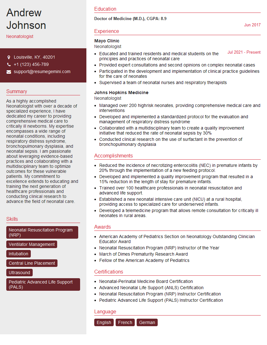
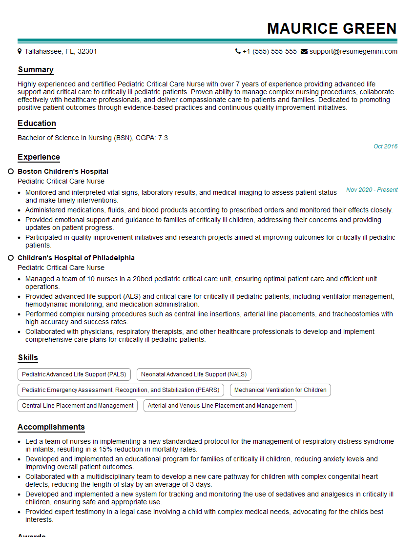
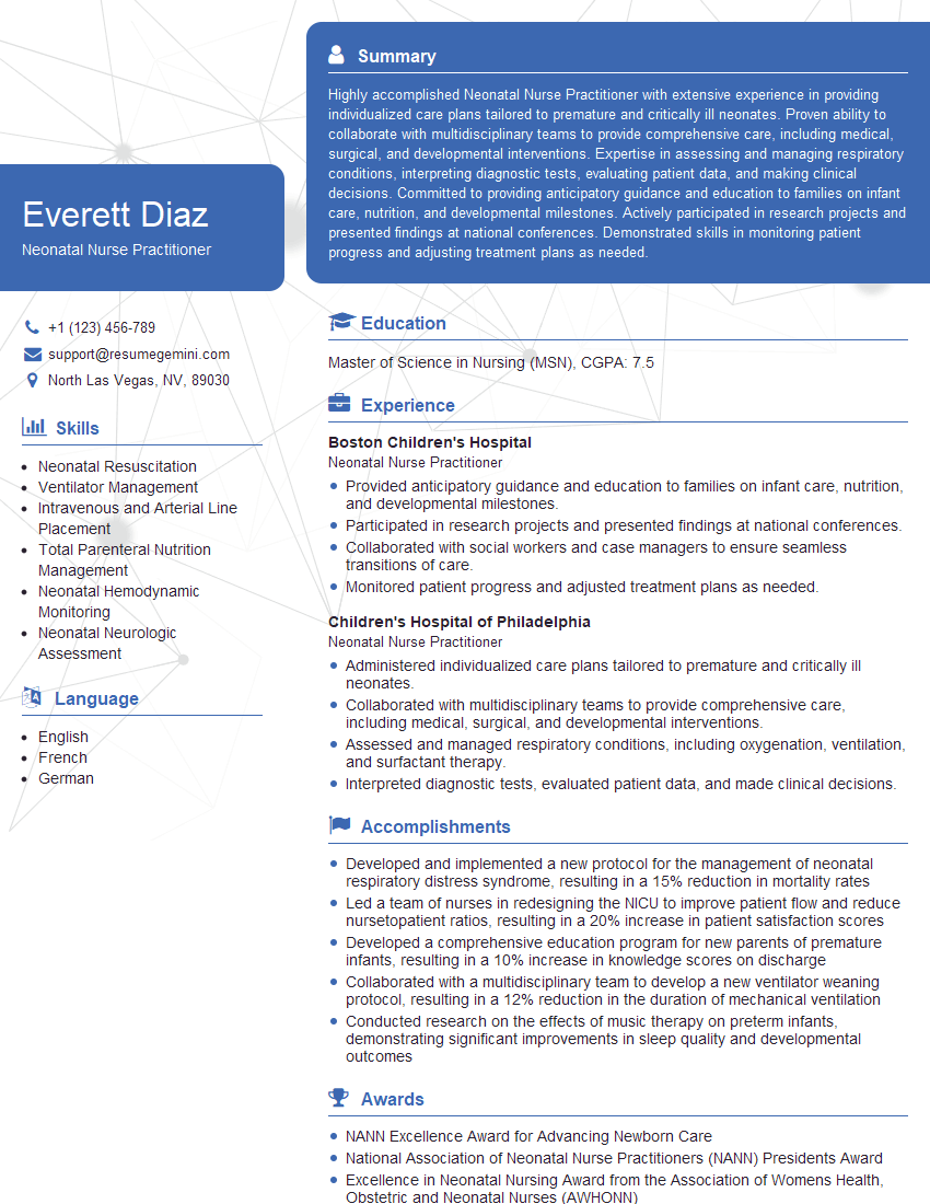
Mastering neonatal intubation significantly enhances your skills and opens doors to advanced roles and greater responsibility within neonatal care. To maximize your job prospects, it’s crucial to present your expertise effectively. An ATS-friendly resume is key to getting your application noticed by recruiters and hiring managers. We highly recommend using ResumeGemini to build a professional and impactful resume that highlights your skills and experience in neonatal intubation. ResumeGemini provides examples of resumes tailored to this specialization to guide you in creating a compelling application. This investment in your resume will significantly increase your chances of securing your dream position.
Explore more articles
Users Rating of Our Blogs
Share Your Experience
We value your feedback! Please rate our content and share your thoughts (optional).
What Readers Say About Our Blog
This was kind of a unique content I found around the specialized skills. Very helpful questions and good detailed answers.
Very Helpful blog, thank you Interviewgemini team.


