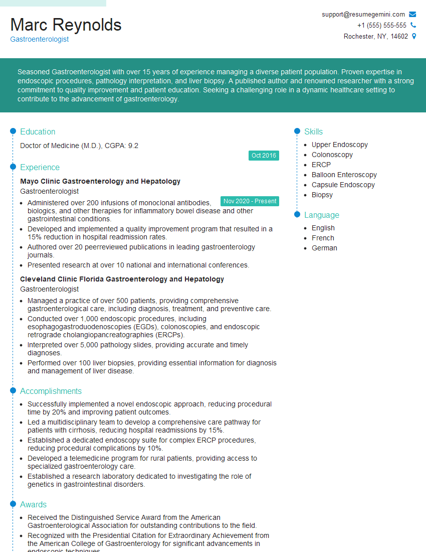
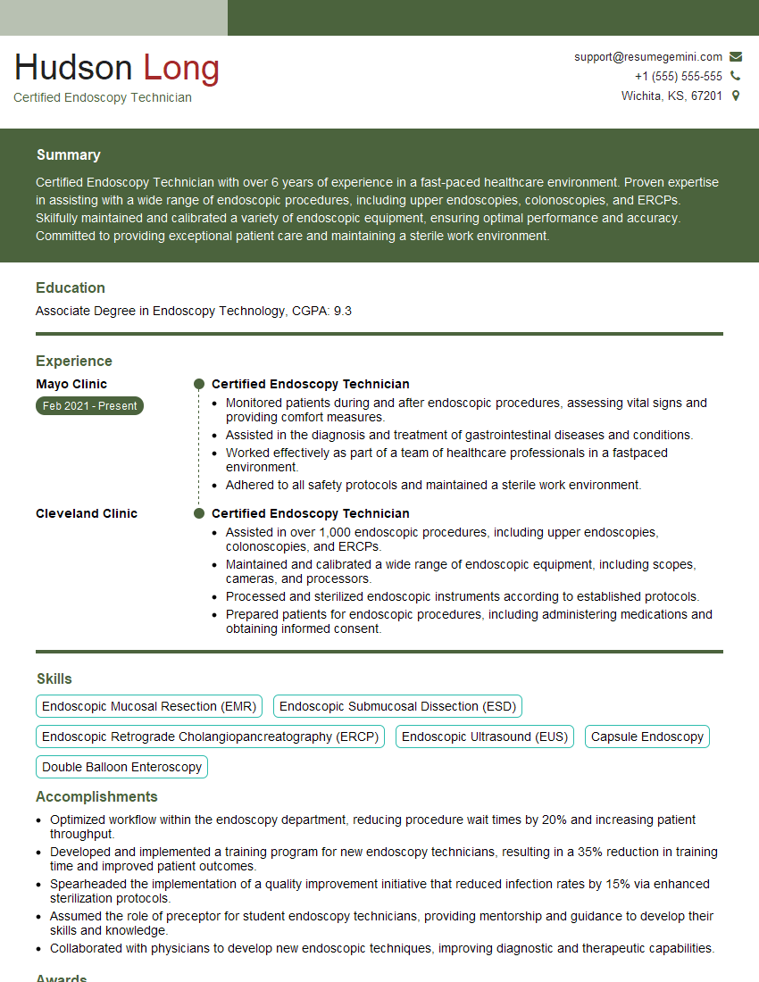
The right preparation can turn an interview into an opportunity to showcase your expertise. This guide to Upper Gastrointestinal Endoscopy interview questions is your ultimate resource, providing key insights and tips to help you ace your responses and stand out as a top candidate.
Questions Asked in Upper Gastrointestinal Endoscopy Interview
Q 1. Describe the procedure for performing an upper endoscopy.
Upper endoscopy, or esophagogastroduodenoscopy (EGD), is a minimally invasive procedure allowing visualization of the esophagus, stomach, and duodenum. The patient is typically sedated for comfort. A thin, flexible endoscope, equipped with a camera and light source, is gently passed through the mouth and into the upper GI tract. The physician carefully navigates the endoscope, inflating the lumen with air to provide optimal visualization. During the procedure, biopsies can be taken, polyps removed, or other therapeutic interventions performed as needed. After the procedure, the patient is monitored until the sedation wears off and is advised to avoid eating until the gag reflex returns.
Think of it like a tiny, flexible camera snake carefully exploring the inner lining of your digestive system. The procedure is generally well-tolerated, and patients often report minimal discomfort.
Q 2. What are the common indications for an upper endoscopy?
Upper endoscopy is indicated for a wide range of conditions. Common indications include:
- Investigating dyspepsia (indigestion): To rule out causes like ulcers, gastritis, or esophageal disorders.
- Abdominal pain of unknown origin: Endoscopy can identify sources of pain such as ulcers, tumors, or inflammation.
- Gastroesophageal reflux disease (GERD): To assess the severity of reflux and identify complications like Barrett’s esophagus.
- Suspected upper gastrointestinal bleeding: To locate the source of bleeding and initiate appropriate treatment.
- Evaluation of dysphagia (difficulty swallowing): To identify anatomical abnormalities or esophageal strictures.
- Screening for and surveillance of esophageal or gastric cancer: Regular endoscopies are crucial in high-risk individuals and those with Barrett’s esophagus.
- Diagnosis of H. pylori infection: Biopsies can be taken to detect the presence of this bacterium.
Essentially, any time there’s an unexplained upper GI symptom or a suspicion of a serious condition, endoscopy is a powerful diagnostic tool.
Q 3. Explain the different types of biopsy forceps used in upper endoscopy and their applications.
Various biopsy forceps are used, each designed for specific applications:
- Standard biopsy forceps: These are the most common type, used to obtain small tissue samples from flat areas. They have serrated jaws to securely grasp the tissue.
- Rat-tooth forceps: These forceps have multiple small teeth, ideal for grasping and removing larger or fragile lesions.
- Hemostatic forceps: These are specifically designed to grasp and control bleeding vessels. They often have a larger jaw and may incorporate a mechanism to cauterize the bleeding site.
- Small-bowel biopsy forceps: These forceps have a longer shaft, allowing access to deeper areas of the gastrointestinal tract.
- Suction biopsy forceps: These combine suction and biopsy capabilities, useful for obtaining tissue samples from lesions that are difficult to grasp.
The choice of forceps depends on the size, location, and nature of the lesion being biopsied. For example, a small, flat lesion might only need standard biopsy forceps, while a large, fragile polyp might require rat-tooth forceps.
Q 4. How do you manage a perforation during an upper endoscopy?
A perforation during upper endoscopy is a serious complication requiring immediate management. The key is early recognition and prompt intervention. Signs include sudden severe abdominal pain, hemodynamic instability (low blood pressure, rapid heart rate), and air in the peritoneal cavity (detected on X-ray).
Management steps typically involve:
- Immediate cessation of the procedure: The endoscope should be removed carefully.
- Intravenous fluids and hemodynamic support: To stabilize the patient’s condition.
- Broad-spectrum antibiotics: To prevent peritonitis (infection of the abdominal cavity).
- Surgical consultation: Most perforations require surgical repair to close the hole. The approach depends on the size and location of the perforation.
- Close monitoring in the intensive care unit: Continuous monitoring is critical.
The goal is to minimize the risk of peritonitis and mortality. Early recognition and prompt surgical intervention are crucial for a successful outcome.
Q 5. What are the signs and symptoms of esophageal varices, and how are they managed endoscopically?
Esophageal varices are abnormally dilated veins in the lower esophagus, often a complication of portal hypertension (increased pressure in the portal vein). Patients may be asymptomatic initially, but they can present with:
- Acute upper gastrointestinal bleeding: This is the most serious and life-threatening manifestation, presenting with hematemesis (vomiting blood) and melena (black, tarry stools).
- Dysphagia: Difficulty swallowing due to the large varices obstructing the esophageal lumen.
Endoscopic management involves:
- Banding: Placing small rubber bands around the varices, causing them to shrink and reduce the risk of bleeding.
- Sclerotherapy: Injecting a sclerosing agent into the varices, causing them to scar and obliterate.
- Transjugular intrahepatic portosystemic shunt (TIPS): A more complex procedure where a shunt is created to reduce portal vein pressure, typically performed by interventional radiology.
The choice of management depends on several factors, including the size and location of the varices and the severity of the bleeding. Preventing re-bleeding is a crucial aspect of long-term management.
Q 6. Describe the endoscopic appearance of Barrett’s esophagus.
Barrett’s esophagus is a precancerous condition where the normal squamous epithelium of the esophagus is replaced by columnar epithelium similar to that found in the stomach or intestine. Endoscopically, Barrett’s esophagus is characterized by:
- Reddish or salmon-pink mucosa: This is the hallmark appearance, contrasting with the normal pale pink of the esophagus.
- Irregular mucosa: The surface may appear granular or nodular.
- Longitudinal mucosal folds: These may be prominent.
It’s important to note that not all patients with Barrett’s esophagus will develop cancer, but regular surveillance endoscopy is crucial for early detection of dysplasia (precancerous changes) and potential malignancy.
Q 7. How do you differentiate between a gastric ulcer and a gastric cancer endoscopically?
Differentiating between a gastric ulcer and gastric cancer endoscopically can be challenging, requiring careful observation. Key features to consider include:
- Ulcers: Typically demonstrate a clean base, often with regular margins. They are often associated with Helicobacter pylori infection. Biopsies will show inflamed tissue with evidence of ulceration.
- Gastric Cancer: May present as an ulcerated lesion, but the margins are often irregular and raised. The base may be nodular or irregular, and surrounding mucosal changes may be present. Biopsies will reveal malignant cells.
Additional points to consider:
- Biopsies are crucial: Endoscopic appearance alone is insufficient for definitive diagnosis. Multiple biopsies are taken from the margins and base of the lesion.
- Imaging studies: Endoscopic ultrasound (EUS) or CT scans can be helpful in determining the depth of invasion and extent of the tumor.
In ambiguous cases, additional tests and specialist consultations are necessary for a definitive diagnosis.
Q 8. What are the potential complications of an upper endoscopy?
Upper endoscopy, while generally safe, carries a small risk of complications. These can range from minor discomfort to serious, life-threatening events. The frequency of these complications varies depending on patient factors, the experience of the endoscopist, and the complexity of the procedure.
- Bleeding: Minor bleeding is common and usually self-limiting. However, significant bleeding from ulcers or esophageal varices can necessitate immediate intervention.
- Perforation: This is a rare but serious complication, involving a hole in the esophagus, stomach, or duodenum. It requires immediate surgical repair.
- Pancreatitis: More common after ERCP (a procedure often done in conjunction with endoscopy), pancreatitis is inflammation of the pancreas, and can be mild or severe.
- Infection: Infections at the endoscopy insertion site are possible, although usually minor.
- Aspiration: Inhaling stomach contents into the lungs can cause pneumonia. This is particularly a risk in patients with reduced gag reflexes or altered mental status.
- Adverse reactions to sedation: Sedatives used to make the procedure comfortable can cause side effects like nausea, vomiting, headache, or, rarely, more serious respiratory or cardiovascular problems.
- Cardiac arrhythmias: Rare, but possible, particularly in patients with pre-existing heart conditions.
- Post-procedure discomfort: This is common and includes bloating, abdominal cramps, and throat soreness.
It’s crucial to remember that many of these complications are rare and that careful patient selection and meticulous technique significantly minimize the risk.
Q 9. How do you manage a bleeding ulcer during an upper endoscopy?
Managing a bleeding ulcer during an upper endoscopy depends on several factors, including the location, size, and rate of bleeding. The goal is to achieve hemostasis (stop the bleeding) as safely and effectively as possible.
- Hemoclipping: Small clips are applied to the bleeding vessel to occlude it. This is a highly effective method for many types of ulcers.
- Injection Therapy: Adrenaline (epinephrine) or other hemostatic agents are injected directly into the bleeding site. This constricts blood vessels and promotes clot formation.
- Thermal Coagulation: Using heat (e.g., argon plasma coagulation or bipolar electrocautery) to seal the bleeding vessel. This method cauterizes the tissue to stop the bleeding.
- Endoscopic Band Ligation: A rubber band is placed around the bleeding vessel, cutting off its blood supply. This is particularly useful for esophageal varices.
In cases of severe or uncontrollable bleeding, the patient may require immediate transfusion, surgery, or interventional radiology procedures, such as angioembolization.
Imagine a faucet dripping. Hemoclipping is like turning the faucet off completely. Injection therapy is like partially closing the faucet. Thermal coagulation is like melting the faucet head shut. The best approach depends on the specific situation.
Q 10. What is the role of chromoendoscopy in upper GI endoscopy?
Chromoendoscopy enhances the visualization of mucosal lesions during upper GI endoscopy. It involves applying dyes that change the appearance of abnormal tissues, making them easier to detect and characterize. This improves the diagnostic yield, particularly for early cancerous or precancerous lesions that might be otherwise missed.
- Lugol’s solution: This iodine-based dye stains normal tissue brown and leaves abnormal tissue unstained, highlighting areas of dysplasia or early cancer.
- Methylene blue: This dye highlights certain types of tumors and may be helpful in identifying small lesions.
- Indocyanine green (ICG): A fluorescent dye used to assess vascularity and perfusion of the tissues. This helps to identify areas at risk of bleeding.
For example, imagine a painting. Chromoendoscopy is like adding specific colored highlights to the areas of interest, making subtle details much more visible to the physician. It is essential for improved diagnostic accuracy and effective treatment planning.
Q 11. Explain the technique for performing endoscopic mucosal resection (EMR).
Endoscopic mucosal resection (EMR) is a technique used to remove superficial lesions from the gastrointestinal tract. It involves injecting submucosal saline to elevate the lesion, creating a cushion between the lesion and underlying layers. This prevents perforation during the resection.
The steps generally include:
- Injection: Saline is injected beneath the lesion using a needle or specialized device to lift it.
- Resection: A snare (a thin wire loop) is used to encircle the elevated lesion. The snare is then activated, either using diathermy (heat) or a cutting current, to dissect and remove the lesion.
- Hemostasis: Any bleeding is controlled using techniques mentioned earlier (e.g., hemostatic clips, injection therapy).
- Pathological evaluation: The resected tissue is sent for histological examination to confirm the diagnosis and rule out malignancy.
Think of it as carefully cutting out a small cookie from a larger cookie sheet—the saline injection is like lifting up the cookie to make it easier to cut, preventing you from accidentally cutting into the sheet itself.
Q 12. How do you manage aspiration during an upper endoscopy?
Aspiration, the inhalation of stomach contents into the lungs, is a serious complication of upper endoscopy. Preventing it is crucial. Strategies include:
- Proper patient positioning: The patient should be positioned appropriately to prevent regurgitation.
- Careful sedation: Using appropriate sedation minimizes the risk of depressed gag reflexes.
- Suctioning: Using suction during the procedure to remove any secretions or vomitus.
- Pre-procedural fasting: Ensuring the patient has fasted for the appropriate period before the procedure is critical to minimize the volume of gastric contents.
- Post-procedure monitoring: Close observation of the patient after the procedure helps to quickly identify and treat any aspiration that may occur.
If aspiration does occur, immediate management includes airway management, supplemental oxygen, and potential treatment for aspiration pneumonia with antibiotics if indicated. Prevention is key, as the treatment of aspiration can be challenging.
Q 13. What are the indications for endoscopic retrograde cholangiopancreatography (ERCP)?
Endoscopic retrograde cholangiopancreatography (ERCP) is a procedure to visualize and treat conditions of the bile and pancreatic ducts. Indications include:
- Choledocholithiasis (gallstones in the bile duct): ERCP is the gold standard for removing gallstones from the common bile duct.
- Cholangitis (bile duct infection): ERCP helps drain infected bile and relieve obstruction.
- Pancreatitis: In some cases, ERCP can help identify and treat the cause of pancreatitis.
- Bile duct strictures: Narrowing of the bile duct can be treated with stenting or dilation during ERCP.
- Ampullary tumors: Tumors at the junction of the bile and pancreatic ducts can be diagnosed and treated with ERCP.
- Pancreatic duct stones: Stones in the pancreatic duct can be removed using ERCP.
ERCP is a technically demanding procedure best performed by experienced endoscopists. The decision to perform ERCP depends on several factors including the clinical presentation, imaging findings, and the patient’s overall health.
Q 14. Describe the procedure for placing a nasogastric tube.
Placing a nasogastric (NG) tube involves inserting a flexible tube through the nose, down the esophagus, and into the stomach. This is commonly done for purposes of feeding, decompression, or medication administration.
- Preparation: Measure the length of the tube needed (from the nose to the xiphoid process), lubricate the tube, and explain the procedure to the patient.
- Insertion: Gently insert the lubricated tube through the nostril, advancing it along the posterior nasopharynx until it reaches the esophagus. Ask the patient to swallow sips of water to aid passage.
- Confirmation: Confirm proper placement through x-ray, measuring the aspirated gastric contents (pH check), or measuring the length of the tube from the nostril to the point where it exits the nose and verifying that this matches your pre-measured length. Never rely on solely clinical signs.
- Securing the tube: Once the correct placement is verified, secure the tube to the patient’s nose or face with tape to prevent dislodgement.
It’s vital to monitor patients with NG tubes for complications such as discomfort, tube displacement, and nasal irritation. Improper placement can lead to lung aspiration, which is a very serious complication.
Q 15. How do you assess the adequacy of bowel preparation before a colonoscopy?
Assessing bowel preparation adequacy before a colonoscopy is crucial for a successful procedure and accurate diagnosis. We primarily assess the clarity of the bowel lumen visualized during the procedure. Ideally, the colon should be completely clear, allowing for excellent visualization of the colonic mucosa. I use a validated scoring system, like the Boston Bowel Preparation Scale (BBPS), which assesses the presence of stool, liquid, and debris. A score of 0-5 indicates excellent preparation with 0 representing complete clarity. A score of > 5 usually indicates insufficient preparation and may necessitate procedure postponement. We also consider the patient’s history of bowel preparation compliance and any potential factors influencing bowel prep effectiveness, such as recent abdominal surgery, medication use (opioids can cause constipation), or underlying bowel disease. For example, a patient with a history of chronic constipation might require a more aggressive bowel preparation regimen, possibly involving a split-dose approach or stronger osmotic laxatives. Visual inspection during the procedure is the gold standard, but stool sampling can be used to supplement the assessment if there is significant doubt.
Career Expert Tips:
- Ace those interviews! Prepare effectively by reviewing the Top 50 Most Common Interview Questions on ResumeGemini.
- Navigate your job search with confidence! Explore a wide range of Career Tips on ResumeGemini. Learn about common challenges and recommendations to overcome them.
- Craft the perfect resume! Master the Art of Resume Writing with ResumeGemini’s guide. Showcase your unique qualifications and achievements effectively.
- Don’t miss out on holiday savings! Build your dream resume with ResumeGemini’s ATS optimized templates.
Q 16. What are the contraindications for upper endoscopy?
Contraindications for upper endoscopy are categorized into absolute and relative contraindications. Absolute contraindications are situations where the procedure should not be performed due to significant risk. These include: uncontrolled bleeding disorders (risk of perforation or hemorrhage), severe esophageal varices with high risk of bleeding, recent myocardial infarction (risk of cardiac stress), and inability to cooperate with the procedure, especially unsedated procedures. Relative contraindications are situations where the benefits of the procedure must be carefully weighed against the risks. Examples include severe cardiac or respiratory disease, severe esophagitis or esophageal stricture (increased risk of perforation), recent surgery or trauma to the upper gastrointestinal tract, and pregnancy (though the procedure can be performed in some instances). The decision on whether to proceed often involves consultation with other specialists (cardiologist, anesthesiologist, etc.) to assess the patient’s overall risk profile and ensure appropriate management during and after the procedure. Each patient’s clinical presentation is unique and requires careful assessment of the potential risks and benefits of proceeding with the endoscopy.
Q 17. Explain the principles of sedation for upper endoscopy.
Sedation for upper endoscopy aims to provide patient comfort and cooperation during the procedure while maintaining airway safety and hemodynamic stability. The principles involve titrating the sedative medication to achieve conscious sedation, where the patient can respond to verbal commands but is relaxed and comfortable. A common approach uses a combination of a benzodiazepine (like midazolam) for anxiolysis and amnesia, and an opioid (like fentanyl or meperidine) for analgesia. We carefully monitor the patient’s vital signs (heart rate, blood pressure, oxygen saturation, and respiratory rate) throughout the procedure. Pulse oximetry is essential to detect any signs of hypoxemia. Capnography is used in many centers to monitor the patient’s ventilation and avoid hypoventilation. An anesthesiologist is consulted when the patient requires more extensive monitoring or has significant comorbidities. Before administering sedation, a thorough evaluation of patient history (including allergies, medical conditions, and medications) is crucial. The sedation level needs to be tailored based on the patient’s individual needs and response to medication, making the procedure as safe and comfortable as possible. Post-procedure monitoring in a recovery area is critical until the effects of sedation have worn off sufficiently.
Q 18. Describe your experience with different types of endoscopes.
My experience encompasses a wide range of endoscopes, including standard video endoscopes (both gastroscopes and duodenoscopes), narrow-band imaging (NBI) endoscopes, and chromoendoscopy systems. Standard video endoscopes provide excellent visualization of the upper GI tract, allowing for the detection of various abnormalities. I regularly utilize NBI endoscopes which enhance the visualization of mucosal capillaries and surface structures, improving the detection of subtle lesions, particularly in the early detection of cancers. Chromoendoscopy, which involves using dyes to highlight lesions, further aids in the diagnosis and targeting of lesions. I’ve also worked extensively with therapeutic endoscopes, equipped with channels for instrument passage, allowing for procedures like polypectomy, band ligation, and stent placement. My familiarity with these different types of endoscopes ensures the selection of the appropriate instrument for each case, maximizing diagnostic accuracy and treatment effectiveness. The technological advancements in endoscopy constantly present exciting new tools. I always stay updated on the latest developments and incorporate them into my practice to improve patient care.
Q 19. How do you interpret endoscopic findings and correlate them with clinical symptoms?
Interpreting endoscopic findings requires a thorough understanding of normal anatomy, pathology, and the correlation with the patient’s clinical presentation. I use a systematic approach, beginning with an assessment of the mucosa for any abnormalities like erythema, edema, ulcerations, or masses. The location, size, and characteristics of any lesion are meticulously documented. For example, the presence of an ulcer in the duodenum might be suggestive of a peptic ulcer disease, whereas a mass in the stomach might indicate a possible gastric cancer. The endoscopic findings are then correlated with the patient’s symptoms. A patient with epigastric pain and a duodenal ulcer is a strong clinical correlation. The endoscopic findings may also guide the choice of biopsies or other procedures. In cases of uncertainty, I utilize additional imaging modalities like endoscopic ultrasound (EUS) or computed tomography (CT) scans to further clarify the diagnosis. Furthermore, I consider the patient’s medical history, age, and risk factors. A young, healthy patient with gastritis may have a different prognosis than an older patient with a similar finding, especially if other risk factors for cancer are present. It’s a holistic approach of combining the visual findings with the patient’s medical profile and performing necessary additional tests to arrive at an accurate diagnosis and formulate a management plan.
Q 20. What is your experience with endoscopic ultrasound (EUS)?
I have extensive experience with endoscopic ultrasound (EUS), a powerful technique that combines endoscopy with ultrasound imaging. EUS allows for precise assessment of the layers of the gastrointestinal wall, providing invaluable information for staging tumors, evaluating the depth of invasion, and detecting lymph node involvement. This is particularly useful for esophageal, gastric, and pancreatic cancers, where accurate staging is crucial for treatment planning. I use EUS for various purposes, including the diagnosis and staging of pancreatic masses, evaluation of mediastinal lymph nodes, and the assessment of inflammatory bowel disease. In some cases, we perform EUS-guided fine-needle aspiration (EUS-FNA) to obtain tissue samples for cytological or histological analysis. The combination of real-time imaging and tissue sampling makes EUS an indispensable tool in the management of many upper GI conditions. EUS requires specialized training and expertise, but the ability to provide accurate staging and facilitate targeted biopsies significantly improves patient outcomes. This detailed evaluation often helps the medical team determine the most appropriate treatment plan, which can include surgical or nonsurgical options.
Q 21. How do you manage a patient with a severe reaction to sedation during endoscopy?
Managing a patient experiencing a severe reaction to sedation during endoscopy is a critical situation requiring immediate attention. The reaction’s severity dictates the response. Mild reactions, such as hypotension or bradycardia, might be managed with supportive measures such as intravenous fluids and monitoring. However, severe reactions, like respiratory depression or cardiac arrest, require immediate resuscitation efforts. This involves securing the airway (intubation if necessary), initiating cardiopulmonary resuscitation (CPR) if needed, administering oxygen, and supporting blood pressure. If appropriate, medications like naloxone (for opioid overdose) or flumazenil (for benzodiazepine overdose) might be administered. Close monitoring of vital signs is essential throughout the resuscitation and recovery. We also ensure appropriate personnel are present to support the resuscitation efforts, including an anesthesiologist or other intensivist specialists. The patient should be transferred to an intensive care unit (ICU) for close monitoring and further management of complications. Following the incident, a thorough review of the case is mandatory to determine the cause of the reaction, assess the effectiveness of the resuscitation, and prevent similar occurrences in the future. This involves reviewing the patient’s medical history, the medications administered, and the monitoring data to help prevent similar incidents. Proper documentation is essential for medical-legal reasons.
Q 22. Describe your experience with advanced endoscopic techniques such as argon plasma coagulation.
Argon plasma coagulation (APC) is an advanced endoscopic technique I use frequently to treat various upper gastrointestinal lesions. It involves using an argon gas plasma beam to ablate tissue. Think of it like a very precise, controlled burn. This differs from traditional methods like electrocautery, offering a more precise and less-destructive approach.
My experience encompasses its use in treating various conditions, including bleeding ulcers, angiodysplasia (abnormal blood vessels), and early esophageal cancers. For instance, I’ve successfully used APC to stop bleeding in a patient with a large peptic ulcer, avoiding the need for more invasive surgery. The procedure’s precision minimizes damage to surrounding healthy tissue, leading to faster healing and reduced complications. I’m proficient in selecting appropriate settings for different tissue types and lesion characteristics, ensuring optimal treatment efficacy and patient safety.
I also regularly utilize APC in conjunction with other endoscopic techniques such as endoscopic mucosal resection (EMR) to enhance the removal of larger lesions. The controlled ablation provided by APC helps to improve the resection process by hemostasis and making the lesion easier to remove. It is a vital tool in my endoscopic arsenal.
Q 23. How do you maintain the cleanliness and sterilization of endoscopes?
Maintaining the cleanliness and sterilization of endoscopes is paramount to prevent infections and ensure patient safety. Our unit strictly adheres to internationally recognized guidelines and utilizes a multi-step process. This begins with thorough pre-cleaning immediately after each procedure, involving manual removal of visible debris and rinsing. This is followed by high-level disinfection using an automated endoscope reprocessor which utilizes enzymatic detergents and high-temperature disinfection.
We meticulously monitor and record all aspects of the reprocessing cycle – including temperature, pressure, and duration – ensuring every endoscope meets strict sterility standards. Regular quality control measures, including microbiological testing, are undertaken to verify the efficacy of our sterilization procedures. Every step is documented, creating a comprehensive audit trail that’s crucial for infection control and regulatory compliance. We also regularly service and maintain our equipment according to the manufacturer’s recommendations, ensuring optimal functioning and longevity of our endoscopes.
Q 24. Explain your understanding of infection control protocols in the endoscopy unit.
Infection control is a top priority in our endoscopy unit. We follow stringent protocols based on guidelines established by relevant organizations such as the CDC and WHO. These include hand hygiene, the use of personal protective equipment (PPE) including gloves, gowns, and masks, and the proper handling and disposal of sharps. Our staff undergoes regular training on these protocols, encompassing every aspect of the procedure, from preparation to post-procedure cleanup.
We have a robust system for monitoring and tracking potential infections. Any suspected cases are immediately investigated, and appropriate measures are taken to prevent further spread. We meticulously manage the disinfection and sterilization of endoscopes and all other equipment. Our unit is also equipped with specialized ventilation systems to minimize airborne contamination. All staff members are well-versed in infection control principles and understand the importance of their roles in preventing the transmission of infections.
For example, we carefully manage patients with known or suspected infections, taking extra precautions to isolate them during procedures and prevent cross-contamination. Regular audits and reviews of our infection control policies help us identify areas for improvement and ensure continued adherence to the highest standards. This multi-faceted approach helps to minimize risks of infection and deliver high-quality and safe care.
Q 25. Describe your experience documenting findings and procedures during endoscopy.
Accurate and comprehensive documentation is essential in endoscopy. Following each procedure, I meticulously record all relevant information in the patient’s electronic health record (EHR). This includes a detailed description of the procedure performed, the findings obtained, any biopsies taken, and the disposition of the patient. The report includes details such as the location, size, and appearance of any lesions, including photographs or videos obtained during the procedure.
I also meticulously document any complications, interventions, and the patient’s response to the procedure. For example, if a polyp was removed, I would detail its size, location, and histological type after pathology results are available. This documentation ensures continuity of care and facilitates communication among healthcare providers. We utilize standardized reporting templates to maintain consistency and accuracy across all procedures. This system ensures that all critical information is readily accessible to the patient’s physician and other members of the care team.
Q 26. How do you communicate findings to the patient and physician?
Communicating findings clearly and effectively to both the patient and the physician is crucial. For patients, I use plain language, avoiding medical jargon, to explain the findings in a way they can understand. I ensure they are comfortable asking questions and address any concerns they may have. I use visual aids like photographs or diagrams where appropriate to help them visualize the findings and understand the implications of the results.
With the physician, I provide a comprehensive report, including all relevant details about the procedure and the findings. This may involve a formal written report, but often includes verbal communication detailing the procedure and any immediate concerns or action items. I use professional terminology and ensure accuracy in my reports. For example, if I discovered a concerning lesion, I would immediately discuss the findings with the physician, outlining my recommendations for further investigation or management. Clear and timely communication ensures coordinated and effective patient care.
Q 27. How do you handle difficult or unexpected situations during an endoscopy?
Handling difficult or unexpected situations during endoscopy requires a calm, methodical approach. My training emphasizes preparedness for complications, and I’m proficient in managing various emergencies that may occur. These include perforation, bleeding, and aspiration. For example, if unexpected bleeding occurs during a polypectomy, I’m trained in various techniques to control the bleeding – from injection therapy to APC or even snare polypectomy.
A structured approach is crucial. I prioritize immediate patient stabilization, summoning assistance as needed. If a perforation is suspected, I would immediately stop the procedure, notify the surgical team, and prepare for potential surgical intervention. My experience allows me to quickly assess the situation, select appropriate intervention, and ensure patient safety. Post-procedure, I would meticulously document all events in the patient’s record.
Q 28. What are your professional development goals related to upper GI endoscopy?
My professional development goals center around enhancing my expertise in advanced endoscopic techniques and improving patient outcomes. I aim to become proficient in newer technologies like endoscopic submucosal dissection (ESD) and further develop my skills in managing complex cases. I am interested in pursuing further training and certification in advanced therapeutic endoscopy, particularly in the management of early-stage esophageal and colorectal cancers.
Furthermore, I’m keen on contributing to research in the field, potentially focusing on improving the safety and efficacy of endoscopic procedures. Staying updated with the latest advancements through continuing medical education and participation in professional societies is a continuous goal. This commitment ensures that I remain at the forefront of the field and am able to provide my patients with the best possible care.
Key Topics to Learn for Upper Gastrointestinal Endoscopy Interview
- Endoscopic Anatomy and Physiology: Understanding the normal anatomy of the esophagus, stomach, and duodenum, including variations and common abnormalities. This includes knowledge of mucosal patterns and vascular supply.
- Instrumentation and Technique: Mastering the practical aspects of endoscopy, including insertion, navigation, suctioning, biopsy techniques, and polypectomy. Consider the differences between various endoscopes and their applications.
- Indications and Contraindications: Knowing the appropriate scenarios for performing an upper GI endoscopy and recognizing situations where it is contraindicated or requires special precautions.
- Common Pathologies and Their Endoscopic Features: Developing the ability to recognize the endoscopic appearances of esophageal varices, ulcers, gastritis, tumors, and other common GI pathologies. Practice differentiating between benign and malignant lesions.
- Endoscopic Procedures: Familiarize yourself with common endoscopic procedures like polypectomy, hemostasis, dilation, and stent placement. Understand the principles and complications associated with each.
- Complications and Management: Thoroughly understand potential complications of upper GI endoscopy, such as perforation, bleeding, and aspiration, and how to manage them effectively. This includes pre-procedure patient assessment and post-procedure care.
- Image Interpretation and Reporting: Develop skills in accurately interpreting endoscopic images and writing clear and concise reports that effectively communicate findings to referring physicians.
- Patient Communication and Counseling: Learn how to effectively communicate with patients before, during, and after the procedure, addressing their concerns and providing appropriate instructions.
- Advanced Endoscopic Techniques (optional): Explore advanced techniques like endoscopic ultrasound (EUS), endoscopic mucosal resection (EMR), and argon plasma coagulation (APC) depending on the level of the position you are applying for.
Next Steps
Mastering Upper Gastrointestinal Endoscopy is crucial for career advancement in gastroenterology and related fields. A strong understanding of these procedures and their complexities will significantly enhance your marketability and open doors to exciting opportunities. To maximize your chances of success, crafting an ATS-friendly resume is essential. ResumeGemini offers a valuable resource to help you build a professional and effective resume that highlights your skills and experience. ResumeGemini provides examples of resumes tailored to Upper Gastrointestinal Endoscopy, ensuring your application stands out from the competition. Take the next step toward your dream career today!
Explore more articles
Users Rating of Our Blogs
Share Your Experience
We value your feedback! Please rate our content and share your thoughts (optional).
What Readers Say About Our Blog
To the interviewgemini.com Webmaster.
Very helpful and content specific questions to help prepare me for my interview!
Thank you
To the interviewgemini.com Webmaster.
This was kind of a unique content I found around the specialized skills. Very helpful questions and good detailed answers.
Very Helpful blog, thank you Interviewgemini team.

