Feeling uncertain about what to expect in your upcoming interview? We’ve got you covered! This blog highlights the most important Necropsy examination interview questions and provides actionable advice to help you stand out as the ideal candidate. Let’s pave the way for your success.
Questions Asked in Necropsy examination Interview
Q 1. Describe the procedure for performing a complete necropsy on a canine.
A complete canine necropsy is a systematic examination of the deceased animal to determine the cause of death. It’s a meticulous process, much like solving a complex puzzle. We start with a thorough external examination, noting any injuries, unusual markings, or body condition. Then, we move to the internal examination.
- External Examination: This involves careful observation of the animal’s overall appearance, including body weight, coat condition, any external wounds or abnormalities.
- Internal Examination: This begins with opening the body cavities (thoracic, abdominal, and pelvic). Each organ is examined in situ (in its natural place) for size, shape, color, and texture. We look for any abnormalities. Then, we carefully remove each organ and examine it closely. This involves weighing certain organs (e.g., liver, kidneys) for comparative analysis.
- Sampling: Throughout the process, we collect tissue samples for further histopathological (microscopic) examination.
- Documentation: Every step is meticulously documented, including photographs and detailed descriptions of findings.
Imagine it as a detective investigating a crime scene. Each observation, however small, can be a vital clue.
Q 2. Explain the significance of collecting tissue samples during a necropsy.
Collecting tissue samples is crucial for confirming our gross necropsy findings and for identifying microscopic lesions which may not be visible to the naked eye. These samples allow for further diagnostic testing such as histopathology (microscopic examination of tissues), microbiology (identifying bacteria, viruses, or fungi), and toxicology (detecting toxins or poisons).
For instance, a liver that appears normal on gross examination might show evidence of chronic inflammation or subtle cellular damage only detectable through microscopic analysis. Similarly, microbiology can identify the presence of infectious agents, providing a definitive diagnosis in suspected infectious diseases. Toxicology can reveal the presence of poisons, drugs, or other toxins that might have contributed to the death.
In essence, tissue sampling provides a more complete picture of the animal’s health status before death and allows for a more accurate diagnosis.
Q 3. How do you differentiate between post-mortem changes and ante-mortem lesions?
Differentiating between post-mortem changes and ante-mortem lesions is essential for accurate diagnosis. Post-mortem changes are alterations that occur after death, while ante-mortem lesions are present before death.
- Post-mortem changes include things like rigor mortis (stiffening of muscles), livor mortis (blood settling), and putrefaction (decomposition). They are typically diffuse and predictable, following a characteristic timeline.
- Ante-mortem lesions, on the other hand, are usually localized, show evidence of a healing response (inflammation, scarring), and might be related to a specific disease process. For example, a localized area of inflammation would suggest an ante-mortem event such as an infection or injury, whereas generalized autolysis is a post-mortem change.
Understanding these differences requires experience and careful observation. For example, a heart with a recent rupture will show signs of blood clotting and inflammation in the area of the rupture. A simple blood clot after death may only have a few changes compared to one prior to death. A skilled pathologist will be able to tell the difference based on the appearance of the surrounding tissue.
Q 4. What are the common causes of sudden death in cattle?
Sudden death in cattle can have various causes. Some of the most common include:
- Bloat (Rumen Tympany): A condition where gas builds up in the rumen (the largest stomach compartment), leading to respiratory distress and death.
- Heart Failure: Various heart conditions can cause sudden death, often without any prior obvious clinical signs.
- Toxicosis: Poisoning from plants, pesticides, or other toxins can cause rapid death.
- Infectious Diseases: Certain infections, such as anthrax or bovine leukemia virus, can cause sudden death.
- Trauma: Accidents or injuries can cause immediate death.
- Lightning strike: Although infrequent, this can be a cause of sudden death in herds.
Diagnosing the cause requires a careful necropsy examination, often including laboratory tests, to identify the underlying problem. For example, we might find evidence of significant rumen distension with a bloat, or cardiac lesions with heart failure.
Q 5. Describe the techniques used for preserving necropsy samples for histopathology.
Preserving necropsy samples for histopathology is essential for obtaining high-quality microscopic sections. The most common method involves fixation in 10% neutral buffered formalin. This solution stops autolysis (self-digestion of tissues) and preserves tissue architecture for microscopic evaluation.
- Fixation: Tissue samples should be small (no more than 1cm thick) to ensure proper penetration of formalin. The ratio of formalin to tissue should be at least 10:1 (10 parts formalin to 1 part tissue). Fixation usually takes 24-72 hours.
- Processing: After fixation, the tissue undergoes processing, which involves dehydration with alcohol, clearing with xylene, and embedding in paraffin wax. This prepares the tissue for sectioning on a microtome.
- Sectioning: Thin sections (typically 3-5 micrometers thick) are cut and mounted onto glass slides.
- Staining: Finally, the sections are stained (hematoxylin and eosin are commonly used), to enhance contrast and make the cellular structures clearly visible under a microscope.
Proper preservation is paramount because it directly affects the quality of the microscopic examination and the reliability of the diagnosis.
Q 6. How do you approach a necropsy when dealing with a highly contagious disease?
Necropsies involving highly contagious diseases require strict biosecurity protocols to prevent the spread of the disease. This involves the use of personal protective equipment (PPE), including disposable coveralls, gloves, masks, eye protection, and possibly respirators, depending on the specific disease. The necropsy should be performed in a designated area with proper ventilation and decontamination procedures in place.
All instruments and surfaces must be thoroughly disinfected after use, and all waste materials, including tissue samples, must be disposed of properly according to relevant guidelines. The area should be meticulously cleaned and disinfected once the procedure is complete.
In some cases, it might be necessary to send samples to a specialized laboratory under strict biosecurity conditions, to avoid any risk of spreading a contagious agent.
It’s essential to follow all relevant protocols to prevent the risk of infecting other animals or personnel.
Q 7. Explain the role of gross necropsy in diagnostic pathology.
Gross necropsy, the visual examination of tissues and organs without the aid of microscopy, plays a critical role in diagnostic pathology. It provides the initial assessment and forms the foundation for subsequent investigations. It’s the first step in solving the “puzzle” of the animal’s death.
The observations made during the gross examination dictate which tissues should be sampled for further testing (histopathology, microbiology, toxicology, etc.) and direct the pathologist toward potential causes of death. For example, if a gross necropsy reveals a large, firm mass in the liver, this might prompt the pathologist to collect samples for histopathological examination, looking for evidence of a tumor.
In short, gross necropsy is crucial for generating hypotheses about the cause of death, directing further testing, and allowing us to form a comprehensive diagnosis.
Q 8. What are the limitations of necropsy examination?
Necropsy, while a powerful tool, has inherent limitations. One major limitation is the post-mortem changes that occur after death. These changes, like autolysis (self-digestion) and putrefaction (decomposition), can obscure subtle findings and make it difficult to determine the exact cause of death. For example, autolysis can mimic certain diseases, leading to misdiagnosis. Another limitation is the potential for artifacts introduced during the necropsy process itself. Improper handling of tissues or instruments can damage organs or create misleading appearances. Finally, some diseases leave few visible macroscopic lesions, requiring advanced techniques like histopathology (microscopic examination of tissues) or toxicology (analysis of toxins) for diagnosis. Think of it like trying to solve a crime scene where the evidence has been partially destroyed or contaminated – you get a partial picture at best.
Q 9. How do you interpret organ weights during a necropsy?
Organ weights are crucial in necropsy. We compare the weights to established reference ranges for the species, age, sex, and body size of the animal. Significant deviations from these ranges can indicate disease. For instance, an enlarged heart (cardiomegaly) might suggest heart failure, while a shrunken liver (cirrhosis) points to chronic liver disease. However, interpreting organ weight requires caution. Factors like dehydration or edema (fluid accumulation) can artificially alter weights. We also need to consider the overall context of the necropsy findings; a slightly elevated liver weight alone doesn’t automatically indicate disease, but in conjunction with other findings, it becomes more significant. It’s a piece of the puzzle, not the whole picture.
Q 10. Describe the different types of tissue fixation methods and their applications.
Tissue fixation is essential for preserving tissue morphology for later microscopic examination. The most common method is fixation in formalin (formaldehyde solution). Formalin cross-links proteins, preventing autolysis and preserving the cellular architecture. This is ideal for routine histopathology. However, formalin can generate artifacts, particularly with certain specialized stains, and it is also a carcinogen, requiring careful handling. Other fixation methods include alcohol-based fixatives (like ethanol), which are useful for preserving certain enzymes or antigens, and Bouin’s solution, which is excellent for preserving tissue morphology but may hinder some staining techniques. The choice of fixation method depends on the specific diagnostic goals. For example, if immunohistochemistry is needed, special fixation methods are employed to preserve the target antigen optimally.
Q 11. Explain the importance of proper sample labeling and chain of custody.
Proper sample labeling and chain of custody are paramount to maintaining the integrity of the necropsy findings and ensuring the admissibility of the results in legal contexts. Each sample must be clearly labeled with a unique identifier (e.g., case number, animal ID, tissue type, date) and the individual who collected it. A chain of custody document tracks the movement and handling of each sample from collection to analysis, ensuring no unauthorized access or tampering has occurred. Think of it as a detailed record of where every sample was at all times – critical for maintaining the credibility and legal validity of the necropsy results, which could have immense implications if the findings are used in litigation.
Q 12. How do you determine the cause of death in a case of suspected poisoning?
Suspected poisoning cases require a multi-faceted approach. The necropsy itself may reveal gross lesions suggestive of certain toxins (e.g., burns in the esophagus from caustic substances). However, definitive diagnosis relies on toxicological analysis of tissues (liver, kidney, stomach contents) using techniques like chromatography and mass spectrometry. These analyses can identify specific toxins and their concentrations. In addition, a detailed history from the owner or keeper can be immensely helpful, noting any exposure to potential toxins or unusual behaviors of the animal. The combination of necropsy findings, clinical history, and toxicological results is crucial for establishing the cause of death in a poisoning case.
Q 13. Describe your experience with performing necropsies on various species (e.g., birds, fish, reptiles).
My experience spans a wide range of species. While the fundamental principles of necropsy remain consistent, the techniques and focus differ based on the species. Performing a necropsy on a bird requires a delicate approach due to their fragile bones and small organs. Fish necropsies often involve assessing the gills for parasites or signs of water quality issues. Reptiles present unique challenges due to their specialized anatomy and slower rates of autolysis. For instance, determining liver disease in a reptile often requires more in-depth histopathology than it would in a mammal due to the reptile’s unique hepatic structure. In each case, careful observation and knowledge of the species’ normal anatomy and physiology are crucial for accurate interpretation.
Q 14. What are the key differences between necropsy techniques in human and veterinary pathology?
Human and veterinary necropsy share fundamental principles but differ significantly in their scope, regulatory framework, and procedures. Human necropsies are highly regulated, often requiring legal authorization, and prioritize meticulous documentation for legal and ethical reasons. The focus is often on identifying disease processes for public health implications. Veterinary necropsies, while still demanding accuracy, are often less strictly regulated. The focus frequently involves disease diagnosis for herd or population health, identification of zoonotic diseases (diseases transmissible from animals to humans) and support for clinical case management. For instance, a human necropsy might involve a detailed neurological examination, while a veterinary necropsy might emphasize the assessment of the gastrointestinal tract in a suspected poisoning case. The differences are not so much in the techniques themselves but in the reason for doing the procedure and what information needs to be focused on.
Q 15. How do you handle unexpected findings during a necropsy?
Unexpected findings during a necropsy are common and often crucial for accurate diagnosis. My approach involves a methodical process. First, I meticulously document the unexpected finding, including its location, size, color, and any associated lesions. Then, I carefully consider the animal’s history, clinical signs, and any other diagnostic information available. This helps determine if the finding is related to the suspected cause of death or represents a separate, concurrent condition. I might take additional samples for further analysis, including histology, microbiology, or toxicology, depending on the nature of the finding. For example, if I unexpectedly find a large mass in an organ, I would collect samples for histopathology to determine its nature (e.g., benign or malignant tumor). The unexpected finding becomes part of a more comprehensive picture, and its inclusion is vital to provide the most accurate and complete report.
Let’s say I’m performing a necropsy on a bird and find significant air sacculitis – an inflammation of the air sacs – which wasn’t initially suspected. I’d document it, collect samples for microbial culture to determine the causative agent, and re-evaluate my initial assessment considering the infection’s potential role in the bird’s demise. This careful reevaluation ensures a comprehensive explanation of the cause of death.
Career Expert Tips:
- Ace those interviews! Prepare effectively by reviewing the Top 50 Most Common Interview Questions on ResumeGemini.
- Navigate your job search with confidence! Explore a wide range of Career Tips on ResumeGemini. Learn about common challenges and recommendations to overcome them.
- Craft the perfect resume! Master the Art of Resume Writing with ResumeGemini’s guide. Showcase your unique qualifications and achievements effectively.
- Don’t miss out on holiday savings! Build your dream resume with ResumeGemini’s ATS optimized templates.
Q 16. Explain your knowledge of common necropsy instruments and their uses.
A necropsy requires a range of specialized instruments. These can be broadly categorized:
- Sharp Instruments: Scalpels, scissors (e.g., Mayo scissors for tissue dissection, Metzenbaum scissors for delicate work), bone cutters (for larger animals), and bone rongeurs (for smaller bones). Scalpels allow precise incisions; scissors are used for cutting through tissue, and bone cutters and rongeurs facilitate bone removal and examination.
- Blunt Instruments: Tissue forceps (various sizes for handling delicate or tougher tissues), retractors (to hold open body cavities), and probes (to explore body cavities and structures). Forceps aid in manipulating organs and tissues, while retractors improve visibility during dissection.
- Measuring and Sampling Instruments: Rulers for measuring lesions, syringes and needles for collecting samples, and specimen containers (appropriate for different samples). Accurate measurements are essential, while syringes preserve samples effectively.
- Other essential tools: A necropsy table, suitable lighting, a camera for documentation, and personal protective equipment (PPE) like gloves, gowns, and masks are absolutely crucial for safety and effective necropsy.
Proper handling and sterilization of these instruments are critical to maintain the integrity of the necropsy and prevent contamination.
Q 17. Describe your experience with performing microscopic examination of necropsy samples.
Microscopic examination of necropsy samples is an essential part of my work, often providing crucial details unavailable through gross examination. I have extensive experience preparing and examining tissue samples using standard histological techniques. This involves fixing the tissues in formalin (a chemical fixative), embedding them in paraffin wax, sectioning them using a microtome (producing very thin slices), staining them with various dyes (e.g., hematoxylin and eosin, special stains for specific tissues or microorganisms), and then examining the slides under a light microscope. I am proficient in identifying various microscopic lesions, such as inflammation, necrosis (cell death), neoplasia (tumors), and parasitic infections. I’ve used this extensively in diagnosing various conditions, such as infectious diseases, autoimmune disorders, and various types of cancers.
For example, in a case of suspected feline infectious peritonitis (FIP), histopathological examination of affected organs might reveal characteristic histiocytic aggregates, which are diagnostic. Gross lesions alone might be insufficient to reach a definitive diagnosis.
Q 18. How do you interpret histopathological findings in the context of gross necropsy findings?
Interpreting histopathological findings in conjunction with gross necropsy findings is vital for a complete diagnosis. Gross findings provide an overview of the macroscopic changes in the body, while histopathology provides the microscopic detail. The two complement each other. For instance, if gross examination reveals an enlarged liver, histopathology could reveal the cause — fatty change, inflammation, or a tumor. The combination provides a much more complete understanding of the disease process. It’s like having a broad aerial view (gross necropsy) and a detailed street-level view (histopathology). Both views are essential for a comprehensive picture.
Consider a case with gross evidence of pneumonia (consolidated lung tissue). Histopathology might then reveal the specific type of pneumonia (e.g., bacterial, viral, fungal) by identifying the inflammatory cells and the presence of microorganisms.
Q 19. What are the ethical considerations related to necropsy examination?
Ethical considerations are paramount in necropsy examinations. Respect for the animal and its owner are central. This involves obtaining proper consent (if possible) from the owner before proceeding. Handling the carcass with respect and dignity is essential throughout the procedure. Maintaining confidentiality of the findings and only sharing them with authorized individuals is also a primary concern. Furthermore, accurate and unbiased reporting is crucial, irrespective of external pressure. Finally, adhering to all relevant regulations and guidelines regarding animal welfare and biosafety is non-negotiable.
A situation where owner consent is difficult – perhaps an animal found dead – still requires responsible and ethically sound handling, respecting the deceased animal’s remains.
Q 20. How would you approach a difficult or complex necropsy case?
A difficult or complex necropsy case requires a systematic approach. First, I thoroughly review the animal’s history, clinical signs, and any diagnostic tests already performed. I then meticulously perform the necropsy, paying close attention to detail and documenting every finding. I collect numerous samples for various diagnostic tests, including microbiology, virology, toxicology, and histopathology, as needed. If necessary, I consult with specialists in relevant fields (pathologists, toxicologists) for assistance in interpretation. Sometimes, further investigation may be required even after the initial necropsy. This might involve additional testing, reviewing medical records, or contacting other professionals. It is about building a puzzle, one piece at a time, until a conclusive picture emerges.
For example, a case with multiple organ system failure might require extensive investigation including toxicology and sophisticated microbiology to determine if the organ damage was due to a toxin, an infection or a combination of both.
Q 21. Describe your proficiency in using relevant software for necropsy data management and reporting.
I’m proficient in using various software for necropsy data management and reporting. I use specialized veterinary pathology information systems (VPIS) for secure storage, retrieval and analysis of necropsy data. These systems allow for the creation of detailed digital reports with images, tables, and graphs. I’m also familiar with laboratory information management systems (LIMS) for tracking samples and their corresponding results. My proficiency in these systems includes data entry, report generation, and data analysis to identify trends and patterns. These digital tools significantly improve efficiency and facilitate collaboration with other veterinarians and specialists.
For example, a VPIS allows efficient storage of images from the necropsy, alongside detailed descriptions, which aids in later review and comparison with other cases. This also facilitates the creation of a standardized report format ensuring consistency and high quality of documentation.
Q 22. What are the best practices for maintaining a safe and sterile necropsy environment?
Maintaining a safe and sterile necropsy environment is paramount to prevent both the spread of infectious agents and the contamination of the specimen. This involves a multi-layered approach focusing on personal protective equipment (PPE), facility design, and diligent disinfection practices.
- PPE: This is the first line of defense. It typically includes: gloves (nitrile or latex, changing frequently), lab coat or gown (disposable is preferred), eye protection (goggles or face shield), and a mask (at least an N95 respirator, possibly higher depending on the suspected agent). We always wear appropriate footwear, and consider using shoe covers.
- Facility Design: The necropsy room should ideally be dedicated to this purpose, with features like negative air pressure to prevent the escape of aerosols, adequate lighting, and easily cleanable surfaces (stainless steel is ideal). Proper ventilation systems are crucial.
- Disinfection: Rigorous disinfection protocols are essential. We use a broad-spectrum disinfectant, such as diluted bleach solution, before, during, and after the procedure. All instruments and surfaces must be thoroughly disinfected, and disposable materials must be properly disposed of in designated biohazard containers. We follow a strict protocol of cleaning surfaces starting from the cleanest to the dirtiest areas to prevent cross-contamination.
- Waste Disposal: All waste generated during a necropsy, including tissues, organs, and fluids, is categorized and disposed of according to local and national regulations, usually in autoclaved biohazard bags and containers.
Think of it like operating a surgical suite; every step is carefully planned to minimize risks.
Q 23. How do you address potential biohazards during a necropsy?
Addressing biohazards is an integral part of every necropsy. It’s not just about protecting ourselves; it’s about protecting the environment and the community. We employ a multi-pronged strategy:
- Risk Assessment: Before commencing, we carefully assess the potential biohazards based on the animal’s history, clinical signs, and preliminary findings. This helps us select the appropriate PPE and safety measures.
- Containment Strategies: We use appropriate containment strategies such as biohazard bags, sharps containers, and proper disposal methods to contain the potential infectious agents. The use of personal protective equipment is mandatory. Specific procedures are modified if dealing with zoonotic diseases, such as rabies or avian influenza.
- Decontamination Procedures: Decontamination procedures are crucial. This involves the thorough disinfection of all equipment, surfaces, and even the air (if necessary) using appropriate disinfectants like sodium hypochlorite solutions or other validated agents.
- Post-Necropsy Cleaning: A meticulous post-necropsy cleaning is performed following a strict protocol, ensuring all potentially contaminated materials and areas are effectively decontaminated and waste is disposed of appropriately.
- Emergency Response Plan: A pre-established emergency response plan addresses accidental spills, injuries, or exposure incidents. Having a detailed plan in place makes handling any emergency situation less stressful and more efficient.
For example, if we suspect rabies, we utilize enhanced PPE, including full-body protection suits, and employ extra precautions during brain tissue sampling to avoid aerosolisation.
Q 24. What are the legal and regulatory requirements related to necropsy procedures?
Legal and regulatory requirements surrounding necropsy procedures vary by jurisdiction but generally center on animal welfare, biosafety, and the disposal of biological waste. Key aspects include:
- Animal Welfare Legislation: Procedures must comply with laws and guidelines related to humane animal handling and euthanasia. The focus is always on minimizing suffering.
- Biosafety Regulations: These regulations address the safe handling of potentially infectious materials, the use of PPE, and the appropriate disposal of biohazardous waste. Compliance with OSHA (Occupational Safety and Health Administration) guidelines is very important in the US, with similar equivalents in other countries.
- Waste Management Regulations: Stringent regulations govern the disposal of biological waste generated during necropsies, ensuring it doesn’t contaminate the environment or pose a health risk. Disposal methods typically involve incineration or autoclaving.
- Record Keeping: Accurate and detailed records must be kept, including the necropsy protocol, findings, and the disposal method of the waste. This documentation is essential for traceability and potential legal situations.
- Licensing and Accreditation: Depending on the location and the type of work, licenses or accreditations may be required to perform necropsies. This would typically involve fulfilling specific training and safety standards.
Ignorance of these regulations can lead to serious legal consequences and compromise public safety.
Q 25. Describe your experience with writing necropsy reports.
Necropsy reports are the cornerstone of communicating our findings. Over the years, I’ve developed a structured approach to ensure clarity, accuracy, and completeness. Each report includes:
- Case Identification: Clear identification of the animal (species, age, sex, identification number). The date of the necropsy and the submitting veterinarian should be included.
- History: A concise summary of the animal’s history, including clinical signs, treatment, and the reason for the necropsy.
- Gross Findings: A detailed description of the macroscopic findings during the examination. I use anatomical terminology and precise language to avoid ambiguity. Photographs and diagrams are frequently included to support the written description.
- Microscopic Findings (if applicable): If histopathology was performed, this section details the microscopic examination findings, referring to specific stains and microscopic changes.
- Diagnostic Tests (if applicable): Results of any supplementary diagnostic tests such as microbiology, serology, or toxicology are included.
- Diagnosis: A conclusive statement of the cause of death or the primary disease process. It needs to correlate directly with the findings.
- Additional Comments: Any other relevant observations or comments.
My reports are written in a clear and concise manner, understandable to both veterinarians and clients. I utilize standardized terminology to ensure consistency and facilitate the communication of our findings to others. A well-written report is a vital tool in diagnostic decision-making.
Q 26. How do you communicate your findings to clients or other stakeholders?
Communicating necropsy findings requires adapting our approach to the audience. With clients, I prioritize clarity and empathy. Technical jargon is minimized, and I use plain language to explain the cause of death and any significant findings. I usually start with a summary of the cause of death and gradually proceed to more specific details if necessary.
With other stakeholders, such as veterinarians or researchers, I can use more technical language and include more detailed information. I will frequently send additional documentation, or images to support the findings.
In both cases, transparency and open communication are crucial. I encourage questions and ensure the client or stakeholder completely understands the report. I often follow up to address any further queries or concerns.
Q 27. How do you stay updated with the latest advancements in necropsy techniques and diagnostic tools?
Staying current in this rapidly evolving field demands continuous learning. I actively engage in several strategies:
- Professional Organizations: Active membership in professional organizations like the American College of Veterinary Pathologists provides access to journals, conferences, and continuing education opportunities. These opportunities allow me to engage with leading professionals in the field.
- Scientific Literature: Regularly reviewing peer-reviewed journals and scientific publications keeps me abreast of the latest research and advancements in necropsy techniques, diagnostic tools, and interpretations.
- Conferences and Workshops: Attending national and international conferences and workshops allows for hands-on learning, networking, and the exchange of information with colleagues from different institutions.
- Online Resources: I utilize online platforms, such as veterinary medical websites and databases, to access continuing education materials and stay up to date on new developments in the field.
- Mentorship and Collaboration: Collaborating with experienced colleagues, engaging in mentorship programs, and actively participating in case discussions greatly enhances my understanding and skills.
Continuous learning isn’t just about keeping my skills sharp; it’s about providing the best possible service and diagnosis.
Q 28. Describe a time you had to overcome a technical challenge during a necropsy.
During a necropsy on a large marine mammal, we encountered significant challenges in accessing the internal organs due to the animal’s size and the thickness of its blubber layer. Traditional surgical instruments proved inadequate for this task.
To overcome this, we had to improvise. We utilized specialized tools, originally designed for other purposes, alongside adapted veterinary surgical instruments. We worked collaboratively, applying different levels of expertise to find the most effective approach. Our multidisciplinary team, including marine mammal specialists, veterinary pathologists, and surgeons, worked together to develop a novel approach to gain access to the critical organs. It was a long, challenging procedure that required careful planning and skillful execution. This experience enhanced our problem-solving abilities and underscored the importance of teamwork and resourcefulness in overcoming complex technical challenges.
Key Topics to Learn for Necropsy Examination Interview
- External Examination: Thorough understanding of techniques for assessing body condition, identifying external injuries and lesions, and documenting findings accurately.
- Internal Examination: Mastering the systematic approach to organ dissection, including proper incision techniques, organ weight measurement, and detailed description of macroscopic findings.
- Histopathology Correlation: Ability to integrate macroscopic findings with microscopic pathology results to form a comprehensive diagnosis.
- Toxicology and Chemistry: Knowledge of sample collection, handling, and interpretation of results from toxicology and chemistry analyses in relation to necropsy findings.
- Microbial Culture and Diagnostics: Understanding the process of collecting samples for bacterial, fungal, and viral cultures, and interpreting the results to aid in diagnosis.
- Parasitology: Familiarity with common parasites and their identification in necropsy specimens.
- Case Reporting and Documentation: Proficiency in preparing clear, concise, and comprehensive necropsy reports that accurately reflect findings and interpretations.
- Legal and Ethical Considerations: Awareness of relevant regulations, ethical guidelines, and best practices in necropsy procedures and reporting.
- Problem-Solving and Critical Thinking: Applying knowledge to diagnose complex cases, integrate multiple sources of data, and formulate well-reasoned conclusions.
- Instrumentation and Techniques: Demonstrating practical skills and familiarity with the tools and equipment used in a necropsy examination.
Next Steps
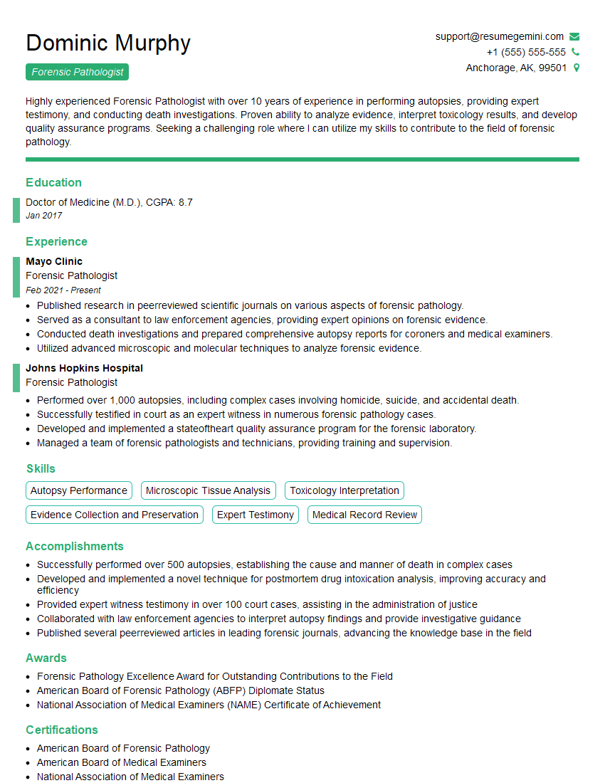
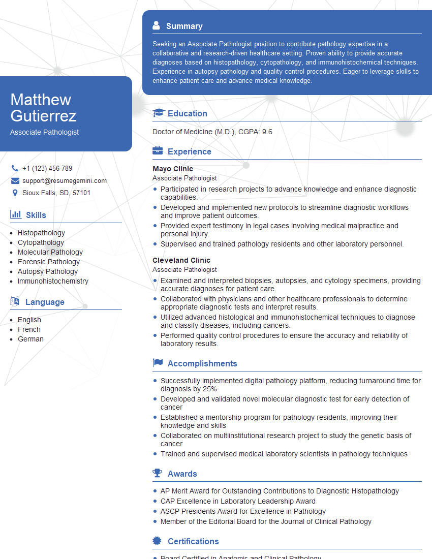
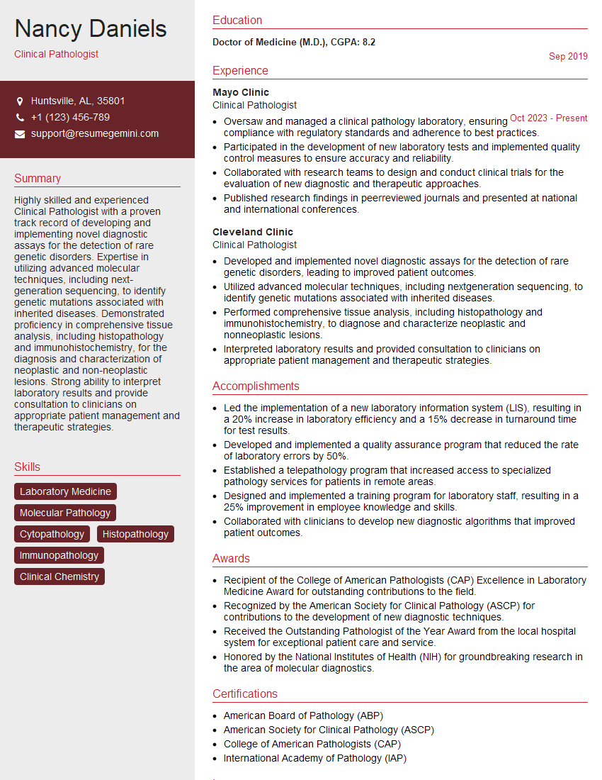
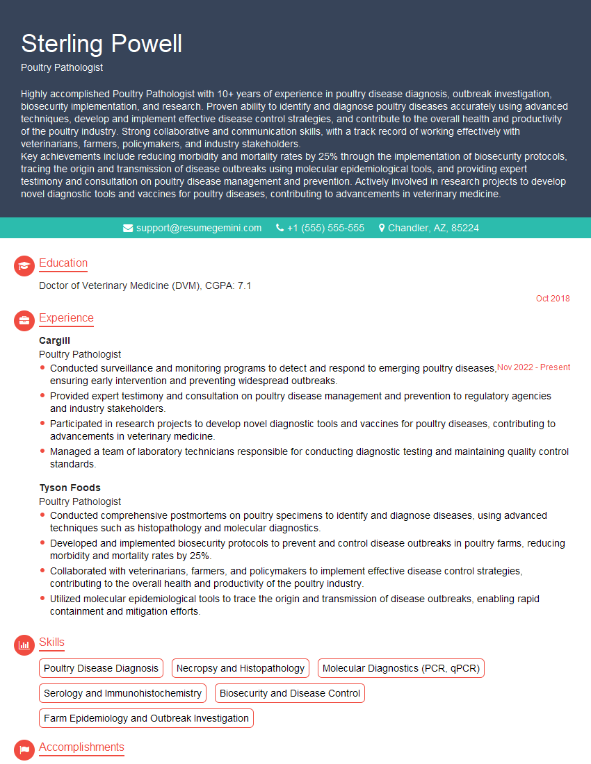
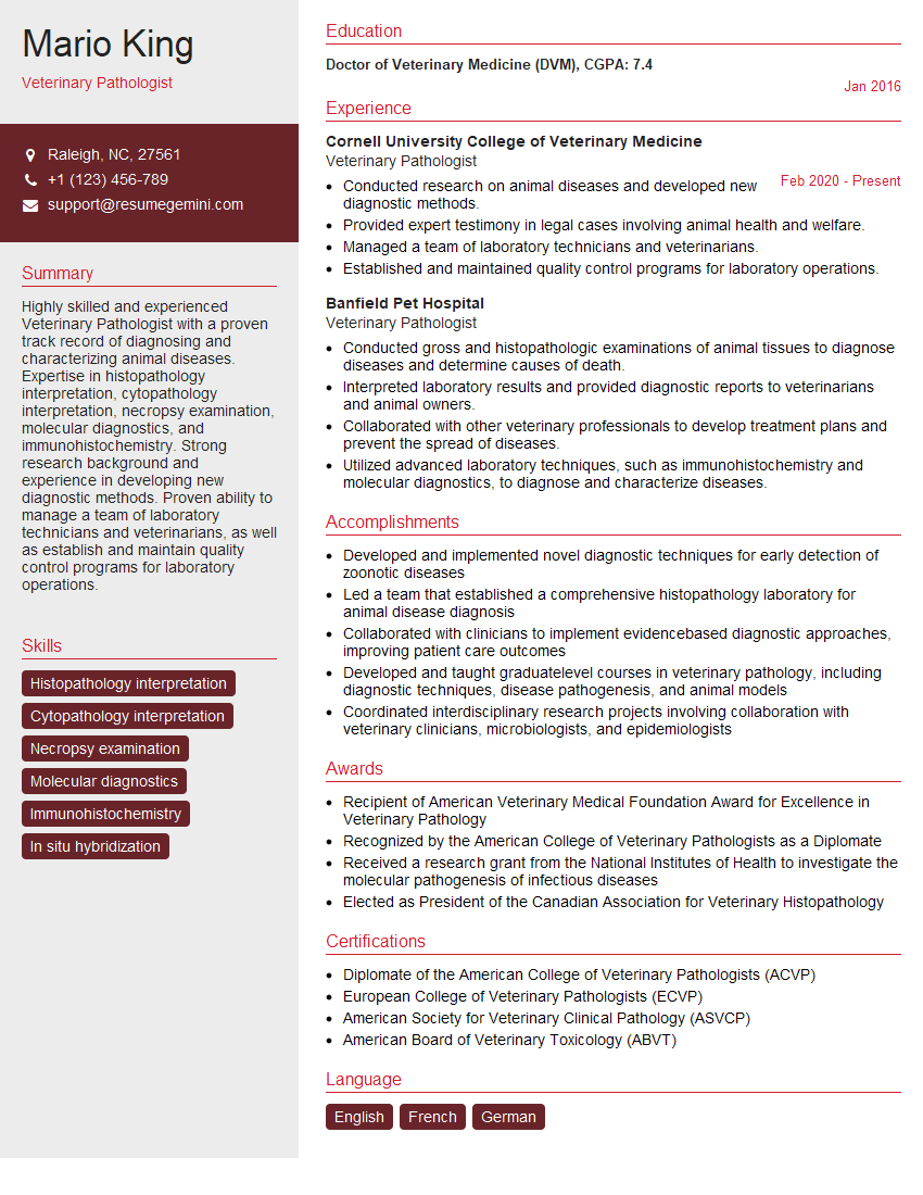
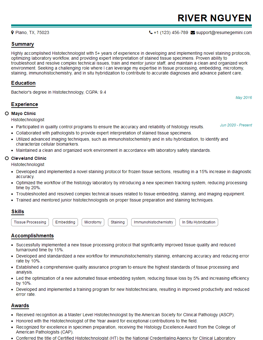
Mastering necropsy examination techniques is crucial for career advancement in veterinary pathology, forensic science, or related fields. A strong understanding of these principles demonstrates expertise and attention to detail, highly valued by employers. To maximize your job prospects, crafting an ATS-friendly resume is essential. ResumeGemini is a trusted resource that can help you build a professional and impactful resume, showcasing your skills and experience effectively. Examples of resumes tailored specifically to necropsy examination positions are available through ResumeGemini, providing valuable templates to guide your resume creation.
Explore more articles
Users Rating of Our Blogs
Share Your Experience
We value your feedback! Please rate our content and share your thoughts (optional).
What Readers Say About Our Blog
To the interviewgemini.com Webmaster.
Very helpful and content specific questions to help prepare me for my interview!
Thank you
To the interviewgemini.com Webmaster.
This was kind of a unique content I found around the specialized skills. Very helpful questions and good detailed answers.
Very Helpful blog, thank you Interviewgemini team.