Unlock your full potential by mastering the most common Management of Foot and Ankle Sports Injuries interview questions. This blog offers a deep dive into the critical topics, ensuring you’re not only prepared to answer but to excel. With these insights, you’ll approach your interview with clarity and confidence.
Questions Asked in Management of Foot and Ankle Sports Injuries Interview
Q 1. Describe your experience diagnosing common foot and ankle injuries in athletes.
Diagnosing foot and ankle injuries in athletes requires a systematic approach combining a thorough history, physical examination, and often, imaging studies. I begin by understanding the mechanism of injury – how did it happen? What was the athlete doing at the time? This helps pinpoint potential injury types. The physical exam is crucial, evaluating range of motion, tenderness to palpation (touch), stability, and neurological function. I look for swelling, bruising, deformity, and gait abnormalities. Common injuries I diagnose include ankle sprains (high and low), plantar fasciitis, stress fractures, tendon injuries (Achilles tendinitis, peroneal tendonitis), and turf toe. For example, a basketball player landing awkwardly might present with an ankle sprain, while a runner might complain of gradual onset heel pain indicative of plantar fasciitis. The history and physical examination findings guide my choice of imaging studies.
Q 2. Explain the difference between a high ankle sprain and a low ankle sprain.
The key difference between high and low ankle sprains lies in the affected ligaments. A low ankle sprain involves injury to the ligaments on the outside of the ankle (anterior talofibular ligament, calcaneofibular ligament, and posterior talofibular ligament). These are the most common ankle sprains and often occur during activities involving inversion (rolling the ankle inward). A high ankle sprain, on the other hand, involves the syndesmosis, a ligamentous complex connecting the tibia and fibula. This injury results from excessive external rotation or dorsiflexion (pointing the toes upward) of the foot, often seen in football or soccer.
Think of it like this: a low ankle sprain is like twisting a shoelace, while a high ankle sprain is like forcefully separating the two parts of a shoelace.
High ankle sprains generally involve more significant injury and longer recovery times than low ankle sprains, often requiring more extensive immobilization and rehabilitation.
Q 3. What are the common causes of plantar fasciitis and its treatment approaches?
Plantar fasciitis is an inflammation of the plantar fascia, a thick band of tissue on the bottom of the foot that runs from the heel to the toes. It’s a common cause of heel pain, especially in runners, athletes with flat feet, or individuals who spend prolonged periods standing or walking. Common causes include overuse, excessive pronation (rolling inward of the foot), inappropriate footwear, and tight calf muscles. The plantar fascia acts like a bowstring, and repetitive stress can cause micro-tears, leading to inflammation and pain.
Treatment involves a multi-faceted approach. Conservative management typically starts with rest, ice, compression, and elevation (RICE). Other interventions include orthotics (arch supports), stretching exercises (especially calf stretches and plantar fascia stretches), night splints to keep the foot dorsiflexed overnight, physical therapy, and anti-inflammatory medications (NSAIDs). In some cases, corticosteroid injections may provide temporary pain relief, but this is often avoided due to long-term risks. Surgery is rarely necessary but may be considered as a last resort for chronic, refractory cases.
Q 4. Discuss the role of imaging (X-ray, MRI, Ultrasound) in assessing foot and ankle injuries.
Imaging plays a vital role in assessing foot and ankle injuries, helping to differentiate between soft tissue and bony injuries and guide treatment decisions. X-rays are useful for detecting fractures, dislocations, and osteoarthritis. They don’t visualize soft tissues well. MRI provides detailed images of soft tissues, making it ideal for evaluating ligament sprains, tendon tears, and bone bruises. It’s excellent for assessing high ankle sprains and assessing the integrity of the ligaments. Ultrasound is a cost-effective, readily available modality useful for evaluating acute injuries, guiding injections, and assessing tendon pathology. It is particularly helpful for identifying tendon tears and assessing the extent of inflammation.
The choice of imaging modality depends on the clinical suspicion. For example, a suspected ankle sprain may start with X-rays to rule out fracture before proceeding to MRI if needed. A suspected stress fracture might initially be evaluated with X-rays and then possibly with a bone scan if X-rays are negative.
Q 5. How do you differentiate between a stress fracture and a tendon injury in the foot?
Differentiating between a stress fracture and a tendon injury in the foot can be challenging, often requiring a combination of clinical assessment and imaging. Stress fractures typically present with localized pain that worsens with activity and improves with rest. The pain may be subtle initially, and the athlete might attribute it to fatigue. Tendon injuries, on the other hand, often present with pain localized to the tendon itself, sometimes with palpable tenderness and crepitus (a crackling sensation). They are usually aggravated by specific movements that stress the tendon.
X-rays may not always show stress fractures early on. MRI or bone scans are often needed for confirmation. Ultrasound can be valuable in assessing tendon injuries directly. The clinical history, physical examination, and imaging findings are combined to reach an accurate diagnosis.
Q 6. Explain your approach to managing acute ankle sprains, including bracing and rehabilitation.
Management of acute ankle sprains follows the PRICE protocol: Protection, Rest, Ice, Compression, and Elevation. Early immobilization with a brace or splint is crucial to stabilize the joint and reduce pain and swelling. The type and duration of immobilization depend on the severity of the sprain. For minor sprains, a simple compression bandage and elevation might suffice. More severe sprains may require a rigid ankle brace or even a walking boot for several weeks. Early mobilization and weight-bearing as tolerated, guided by pain levels, are encouraged to prevent stiffness and promote healing.
Rehabilitation is key to regaining full function and preventing recurrence. This includes a structured program of range-of-motion exercises, strengthening exercises (especially for the calf muscles and peroneal muscles), proprioceptive training (balance exercises), and gradually increasing activity levels. Physical therapy plays a crucial role in guiding this process. The athlete should progressively return to sports activities only after complete pain resolution and restoration of full functional capacity.
Q 7. Describe your experience with surgical interventions for foot and ankle injuries.
My experience with surgical interventions for foot and ankle injuries involves a range of procedures. I typically recommend surgery only when conservative management fails to provide adequate pain relief and functional improvement. Common surgical procedures include ligament reconstruction (for chronic ankle instability), tendon repair (for significant tendon tears), fracture fixation (for displaced fractures or non-union), and osteotomy (bone realignment). I’ve also been involved in procedures to address conditions such as hallux valgus (bunion), plantar fasciitis refractory to conservative treatment and arthritis.
The decision to proceed with surgery is carefully considered, weighing the potential benefits against the risks and recovery time. Postoperative management includes pain control, wound care, early mobilization as appropriate, and a tailored rehabilitation program to optimize functional recovery. Close collaboration with the patient and the rehabilitation team is essential to ensure the best possible outcome.
Q 8. What are the common complications associated with ankle fractures?
Ankle fractures, while often treatable, can lead to several complications. These complications can significantly impact long-term function and quality of life. The severity of the complication depends on factors such as the type and severity of the fracture, the patient’s overall health, and the effectiveness of treatment.
- Malunion: This occurs when the fractured bone heals in an incorrect position, leading to deformity, pain, and reduced ankle function. Imagine trying to fit a puzzle piece that’s slightly bent – it just won’t fit right. This requires further surgical intervention to correct the alignment.
- Nonunion: This is when the fractured bone fails to heal completely. This can result in persistent pain, instability, and the need for bone grafting or other surgical procedures to stimulate healing. Think of it like a crack in a wall that never quite mends.
- Arthritis: Fractures, especially those involving the joint surface, can lead to early-onset osteoarthritis. The rough edges of the fractured bone can cause irritation and inflammation in the joint, ultimately leading to cartilage degeneration and pain.
- Chronic Pain: Even with successful healing, some individuals experience persistent pain, stiffness, and limited range of motion. This is often due to nerve damage or scar tissue formation.
- Compartment Syndrome: This is a serious condition where swelling within the muscle compartments of the lower leg compresses blood vessels and nerves, potentially leading to tissue damage and limb loss. Early recognition and treatment are crucial.
- Infection: Open fractures or surgical interventions increase the risk of infection, which can have serious consequences if left untreated. Prophylactic antibiotics are commonly used to reduce this risk.
Careful management and rehabilitation are crucial to minimize these risks and optimize patient outcomes. This includes appropriate fracture fixation, meticulous wound care, and a comprehensive physiotherapy program.
Q 9. How do you assess and manage compartment syndrome in the lower leg?
Compartment syndrome is a serious condition requiring immediate attention. It occurs when swelling within the muscle compartments of the lower leg increases pressure, compromising blood supply and nerve function. Early diagnosis and treatment are vital to prevent permanent damage.
Assessment involves a thorough clinical examination focusing on the ‘6 Ps’: Pain, Paresthesia (numbness or tingling), Pallor (pale skin), Paralysis (weakness or inability to move the affected muscles), Pulselessness (absence of palpable pulse), and Poikilothermia (coolness of the limb). Measurement of compartment pressures using a pressure monitor provides objective data. We always compare findings with the unaffected side.
Management is primarily surgical. A fasciotomy, which involves making incisions in the fascia (the tough connective tissue surrounding the muscle compartments), is performed to relieve the pressure. This procedure allows the tissues to expand and restore blood flow. Pre-operative assessments, like Doppler ultrasound can assess blood flow, and patient history is crucial. Post-operative care includes wound management and physiotherapy to restore function.
For example, an athlete sustaining a significant tibial fracture may experience rapid swelling leading to compartment syndrome. Immediate surgical intervention is necessary to avoid lasting nerve damage or muscle necrosis. Timely intervention will improve the likelihood of a good outcome.
Q 10. Discuss the role of physiotherapy and rehabilitation in recovering from foot and ankle injuries.
Physiotherapy and rehabilitation play an integral role in recovering from foot and ankle injuries. They are not merely an adjunct to treatment but are essential for restoring function, strength, and mobility. A well-structured program is tailored to the individual patient’s needs and injury type.
- Early stages focus on pain management, reducing swelling (edema), and improving range of motion. Techniques include gentle range-of-motion exercises, ice therapy, and elevation of the limb. Protecting the area and managing pain is critical to avoid discomfort and promote early engagement.
- Intermediate stages involve progressive strengthening exercises, proprioceptive training (enhancing balance and coordination), and neuromuscular re-education. This might involve using resistance bands for strengthening or balance boards for improving stability.
- Later stages focus on return to functional activities, such as walking, running, or sports-specific training. This is a gradual process, progressing as tolerated, with monitoring for any pain or discomfort, and modification as needed.
Imagine rebuilding a house after a fire – you wouldn’t rush the process. Rehabilitation is equally gradual, progressing through controlled stages to ensure optimal recovery and minimize the risk of re-injury.
For instance, following a lateral ankle sprain, early physiotherapy helps reduce swelling and pain, while later stages focus on strengthening the peroneal muscles, improving balance, and gradually resuming athletic activities. A personalized plan is crucial to ensure the patient’s safe return to full function.
Q 11. Describe your experience prescribing and managing orthotics for foot and ankle problems.
Orthotics are custom-made or prefabricated shoe inserts designed to correct biomechanical problems of the feet and ankles, often improving comfort and reducing pain. My experience encompasses both assessment and management of orthotic prescription for various foot and ankle conditions.
Assessment starts with a detailed history, including the patient’s symptoms, activity levels, and footwear habits. A thorough physical examination includes assessing gait, foot posture, range of motion, and muscle strength. I often use gait analysis (observation of walking patterns) and static postural assessment. Occasionally, I will order additional diagnostic imaging such as x-rays, ultrasound, or MRI scans.
Management involves selecting the appropriate type of orthotic based on the individual’s needs. Custom orthotics offer the most precise correction, but prefabricated orthotics provide a more affordable and readily available option. Factors such as arch support, heel cushioning, and metatarsal support are carefully considered. I also educate patients about proper orthotic care, ensuring they wear them appropriately and maintain them.
For example, a patient with plantar fasciitis might benefit from custom orthotics with increased arch support and heel cushioning. An athlete with recurrent ankle sprains may require orthotics to improve foot stability and proprioception. I often follow up to monitor effectiveness and adjust the prescription if necessary.
Q 12. What are the common causes of Achilles tendinopathy and its treatment strategies?
Achilles tendinopathy is a common condition characterized by pain and inflammation in the Achilles tendon, the strong tendon connecting the calf muscles to the heel bone. It’s often seen in athletes and active individuals.
Common causes include overuse, inadequate warm-up, improper footwear, and sudden increases in activity level. Underlying biomechanical factors, such as foot pronation (the inward rolling of the foot during walking), can also contribute.
Treatment strategies aim to reduce pain and inflammation and restore tendon function. They typically include:
- Conservative management: This forms the cornerstone of treatment and usually involves rest, ice, compression, and elevation (RICE). Non-steroidal anti-inflammatory drugs (NSAIDs) may help to alleviate pain and inflammation. Eccentric exercises (lengthening the muscle while contracting) are often prescribed to strengthen and rehabilitate the tendon.
- Extracorporeal shock wave therapy (ESWT): This non-invasive procedure uses sound waves to stimulate healing and reduce pain. It’s not a first-line approach, typically used after conservative methods have plateaued.
- Surgical intervention: This is usually reserved for cases that do not respond to conservative management. Surgical options may include debridement (removal of damaged tissue) or tenotomy (surgical lengthening of the tendon).
A patient presenting with Achilles tendinopathy would undergo a thorough assessment including palpation, range-of-motion testing, and possibly imaging studies. Treatment would be tailored to their specific condition and activity level. Early intervention and adherence to the treatment plan are crucial for optimal outcomes.
Q 13. How do you manage chronic ankle instability in athletes?
Chronic ankle instability (CAI) is a persistent condition characterized by recurrent ankle sprains and a feeling of ‘giving way’ of the ankle. Managing CAI in athletes requires a multi-faceted approach.
Management often involves a combination of:
- Prophylactic bracing: Ankle braces provide external support and reduce the risk of further sprains. This is particularly useful for athletes returning to sports.
- Proprioceptive training: Exercises designed to improve balance and coordination are essential. Examples include balance boards, wobble boards, and single-leg stance exercises. These work to improve neuromuscular control around the ankle joint.
- Strengthening exercises: Strengthening the muscles around the ankle joint, particularly the peroneal muscles, improves stability and reduces the risk of re-injury. I emphasize both eccentric and concentric muscle strengthening.
- Functional rehabilitation: A gradual return to sports-specific activities, guided by the athlete’s progress and tolerance, is critical. We start with low-impact activities and gradually increase intensity and complexity.
- Surgical intervention: In cases of significant ligament damage or persistent instability despite conservative management, surgical reconstruction of the ligaments may be considered. This is typically a last resort.
A tailored approach ensures the athlete safely returns to their sport while minimizing the risk of recurrent injuries. The plan should address the specific biomechanical deficits and the athlete’s sport-specific demands.
Q 14. What are your preferred methods for assessing range of motion and strength in the foot and ankle?
Assessing range of motion (ROM) and strength in the foot and ankle is crucial for diagnosis and monitoring progress. I use a combination of methods.
Range of motion is assessed using a goniometer to measure the angles of dorsiflexion, plantarflexion, inversion, and eversion. Visual inspection and palpation are also used to identify any limitations or joint abnormalities. I always compare the affected side to the unaffected side for reference.
Strength is assessed using manual muscle testing (MMT), where I apply resistance to specific muscle groups while the patient performs active movements. For more objective measurements, I may use handheld dynamometers to quantify muscle strength. I often utilize functional testing such as hop tests and timed tasks that can gauge the patient’s functional ability and identify deficits in specific muscle groups.
For instance, a patient with an ankle sprain might demonstrate reduced dorsiflexion and plantarflexion range of motion and weakness in the peroneal muscles. These findings inform the rehabilitation plan to restore optimal function and strength.
Q 15. Describe your experience with non-surgical management of plantar fasciitis.
Plantar fasciitis, a common cause of heel pain, is often successfully managed non-surgically. My approach focuses on a conservative, multi-pronged strategy aimed at reducing inflammation, improving flexibility, and correcting biomechanical issues.
Rest and Ice: Initial management involves rest, avoiding activities that aggravate the pain, and applying ice packs several times a day to reduce inflammation. Think of it like giving your foot a much-needed ‘time-out’ to heal.
Stretching and Strengthening: I prescribe specific stretches targeting the plantar fascia and calf muscles. These exercises are crucial for improving flexibility and range of motion. Imagine the plantar fascia as a tight rubber band – stretching it regularly helps restore its elasticity. Strengthening exercises, focusing on intrinsic foot muscles, are also vital to support the arch and prevent recurrence.
Orthotics and Footwear: Custom or over-the-counter orthotics provide arch support and cushioning, reducing strain on the plantar fascia. Proper footwear with adequate cushioning and support is essential to prevent further irritation. Think of orthotics as personalized shock absorbers for your feet.
Medication: Nonsteroidal anti-inflammatory drugs (NSAIDs) like ibuprofen or naproxen can help manage pain and inflammation in the short term. I carefully monitor medication use to minimize side effects.
Injections: In some cases, corticosteroid injections can provide temporary pain relief. However, this isn’t a long-term solution and overuse can weaken the plantar fascia.
Physical Therapy: A tailored physical therapy program often incorporates manual therapy, modalities like ultrasound, and a progressive exercise program. A physical therapist can guide you through the process, ensuring correct technique and gradual progression.
The success of non-surgical management depends on patient compliance with the prescribed treatment plan and addressing the underlying biomechanical factors contributing to the condition.
Career Expert Tips:
- Ace those interviews! Prepare effectively by reviewing the Top 50 Most Common Interview Questions on ResumeGemini.
- Navigate your job search with confidence! Explore a wide range of Career Tips on ResumeGemini. Learn about common challenges and recommendations to overcome them.
- Craft the perfect resume! Master the Art of Resume Writing with ResumeGemini’s guide. Showcase your unique qualifications and achievements effectively.
- Don’t miss out on holiday savings! Build your dream resume with ResumeGemini’s ATS optimized templates.
Q 16. Explain your approach to managing a Lisfranc fracture-dislocation.
A Lisfranc fracture-dislocation, an injury involving the midfoot, requires a prompt and precise approach. The severity dictates the treatment plan.
Diagnosis: Accurate diagnosis is paramount, utilizing X-rays, and sometimes CT scans, to assess the extent of the injury. Missed or improperly treated Lisfranc injuries can lead to long-term complications like arthritis.
Non-operative Management: In minimally displaced fractures without significant instability, non-operative management might involve immobilization with a cast or surgical boot, followed by gradual weight-bearing and physical therapy. However, this approach is less common due to the high risk of malunion and long-term instability.
Operative Management: Most Lisfranc injuries require surgical intervention to restore anatomical alignment and stability. The surgical technique is tailored to the specific injury pattern, often involving open reduction and internal fixation (ORIF) using screws, plates, or other implants to stabilize the affected bones. Post-operative management includes immobilization, followed by a gradual, supervised rehabilitation program.
Rehabilitation: Regardless of the treatment method, rigorous rehabilitation is crucial for restoring function and preventing long-term complications. This includes range-of-motion exercises, strengthening, and gradually increasing weight-bearing activities under the guidance of a physical therapist.
Return to sport is a gradual process, depending on the complexity of the injury and the patient’s response to treatment. I carefully monitor patients’ progress using clinical assessments and imaging studies.
Q 17. How do you determine the appropriate return-to-play criteria for athletes with foot and ankle injuries?
Determining return-to-play (RTP) criteria for athletes with foot and ankle injuries requires a holistic approach, considering several crucial factors.
Complete Healing: The injury must be fully healed, with no residual pain or inflammation. Imaging studies (X-rays, MRI) can help assess the healing process.
Full Range of Motion (ROM): The athlete must have regained full ROM in the affected joint, comparable to the uninjured side. Restricted ROM can significantly impair athletic performance and increase the risk of re-injury.
Strength and Balance: Muscle strength and balance must be restored to pre-injury levels. Functional strength testing and balance assessments are key to ensuring the athlete is ready to return to sport.
Functional Testing: Performing sport-specific drills and exercises is crucial to evaluate the athlete’s ability to perform without pain or limitations. This might involve jumping, cutting, and landing maneuvers.
Pain-Free Movement: The athlete should be pain-free during activity. Pain is a significant indicator of incomplete healing and should not be ignored.
Athlete Readiness: The athlete’s own perception of readiness and confidence are also critical factors. Returning prematurely due to pressure can compromise the healing process and increase the risk of re-injury. A psychological readiness component is extremely important.
The RTP decision is a collaborative process involving the athlete, the physician, and other healthcare professionals (physical therapists, athletic trainers).
Q 18. Describe your experience with different types of ankle bracing and their applications.
Ankle bracing plays a significant role in managing various ankle injuries and providing support. The type of brace used depends on the specific injury and the athlete’s needs.
Lace-up Ankle Braces: These provide moderate support and are suitable for mild sprains or for athletes seeking prophylactic protection. They are readily available and relatively inexpensive.
Air Stirrup Ankle Braces: These offer adjustable compression and support, useful for managing moderate to severe sprains and providing stability. The inflatable chambers allow for customized support and comfortable fit.
Rigid Ankle Braces: These provide significant support and immobilization, often used after surgery or for severe injuries. They limit ankle movement and promote healing.
Hinged Ankle Braces: These offer support and controlled range of motion, often used during the rehabilitation phase to help improve stability while allowing for controlled movement.
I carefully consider factors like the severity of the injury, the athlete’s activity level, and individual preferences when recommending a specific type of ankle brace. The goal is to optimize support, minimize the risk of re-injury, and facilitate rehabilitation.
Q 19. What are the common causes of hallux valgus (bunion) and its treatment options?
Hallux valgus, or bunion, is a deformity characterized by a prominent bump on the joint at the base of the big toe. Several factors contribute to its development.
Genetics: A family history of bunions significantly increases the risk. In essence, some individuals inherit a predisposition to this condition.
Foot Structure: Certain foot types, like flat feet or excessively flexible feet, are more prone to bunion formation. The biomechanics of the foot play a significant role.
Footwear: Wearing tight, narrow shoes, particularly high heels, puts excessive pressure on the big toe joint and contributes to the progressive development of bunions. Think of it as constant pressure on a vulnerable area.
Arthritis: Underlying joint conditions, such as osteoarthritis, can exacerbate bunion formation and increase pain.
Treatment options vary depending on the severity of the deformity and the patient’s symptoms.
Conservative Management: This includes wearing wider shoes, using bunion pads or splints to cushion the area, and using NSAIDs for pain relief. Conservative management is effective in managing the condition early in its development.
Surgical Intervention: If conservative measures fail to alleviate symptoms or the deformity is severe, surgical correction may be necessary. Several surgical techniques are available, depending on the specific anatomy and the severity of the condition. Surgery is indicated in advanced cases that significantly impair function or cause persistent pain.
Q 20. Discuss the role of medication (NSAIDs, analgesics) in managing foot and ankle injuries.
NSAIDs (non-steroidal anti-inflammatory drugs) and analgesics play an important, but often temporary, role in managing foot and ankle injuries. They help manage pain and inflammation.
NSAIDs (Ibuprofen, Naproxen): These reduce both pain and inflammation, making them effective in the initial stages of an injury. However, prolonged use can have side effects, including gastrointestinal problems and kidney issues. I always emphasize responsible use.
Analgesics (Acetaminophen): These primarily address pain without significantly reducing inflammation. They are often used in conjunction with NSAIDs or when inflammation is not a primary concern. They are generally safer for longer-term use compared to NSAIDs but aren’t as effective for inflammation.
Medication is often used as a temporary measure, alongside other treatment modalities like rest, ice, physical therapy, and possibly surgery. I emphasize the importance of a holistic approach, using medication judiciously and focusing on addressing the underlying cause of the injury.
Q 21. How do you manage patients with diabetes and foot ulcers?
Managing patients with diabetes and foot ulcers requires a highly specialized approach due to the increased risk of complications like infection and amputation. The management involves a multidisciplinary team including podiatrists, endocrinologists, and infectious disease specialists.
Wound Care: Proper wound care is paramount, involving debridement (removal of dead tissue), infection control (antibiotics), and appropriate dressings to promote healing. Regular monitoring of the ulcer is essential to track progress and identify any complications.
Offloading: Reducing pressure on the ulcer is crucial to promote healing. This may involve using custom-made orthotics, specialized footwear, or total contact casts to redistribute weight and prevent further damage.
Glycemic Control: Strict blood glucose control is crucial to improve circulation and facilitate healing. Regular monitoring of blood sugar levels and adjustments to diabetes management are essential.
Infection Control: Prompt detection and treatment of infection are vital to prevent serious complications. Regular assessment of the wound and cultures are essential to guide antibiotic treatment.
Vascular Assessment: Assessing vascular health is important as compromised circulation can hinder healing. Techniques like Doppler ultrasound are used to evaluate blood flow. In cases of severe vascular disease, interventions like angioplasty or bypass surgery might be necessary to improve circulation.
Patient Education: Educating patients on proper foot care, including daily inspection, hygiene, and footwear choices, is crucial in preventing future ulcers.
The goal of management is to promote wound healing, prevent infection, and minimize the risk of amputation. A proactive, multidisciplinary approach significantly improves outcomes.
Q 22. Explain your approach to managing ingrown toenails.
Managing ingrown toenails involves a multifaceted approach prioritizing conservative management whenever possible. Initially, we focus on improving nail hygiene and proper trimming techniques. This includes teaching patients to cut their nails straight across, avoiding overly short cuts, and keeping the area clean and dry. We often recommend soaking the affected toe in warm, soapy water several times a day to soften the skin and reduce inflammation.
If conservative measures fail, we may consider minor surgical interventions. A simple procedure involves partially removing the ingrown portion of the nail, using a specialized instrument to create a small space between the nail and the surrounding skin. In severe cases, or cases of recurrent ingrown toenails, a more involved procedure might be necessary, potentially involving chemical or surgical nail matrixectomy to prevent future ingrowth. Post-operative care is crucial, involving regular cleaning and dressing changes to prevent infection. We work closely with podiatrists to ensure proper treatment and follow-up care.
For example, I recently treated a young athlete who had developed a severely infected ingrown toenail. Initial conservative measures failed due to delayed presentation. We proceeded with partial nail avulsion and post-operative antibiotics. His recovery was rapid and uneventful. This highlights the importance of early intervention and appropriate treatment strategy choice based on individual presentation.
Q 23. Discuss your experience with the use of injections (cortisone, PRP) in the management of foot and ankle conditions.
Cortisone injections are frequently used to manage inflammatory conditions in the foot and ankle, such as plantar fasciitis, tendonitis, and bursitis. The cortisone reduces inflammation, providing pain relief and improved function. The effects, however, are often temporary. PRP (Platelet-Rich Plasma) therapy, on the other hand, is a regenerative treatment utilizing the patient’s own blood platelets to promote healing and tissue regeneration. It has shown promise in managing chronic tendon injuries and other recalcitrant conditions where cortisone has proven insufficient.
My experience highlights that the decision to use injections must be individualized and carefully considered. Cortisone is a powerful anti-inflammatory but carries the risk of tendon rupture and tissue weakening with repeated injections. PRP, while promising, requires a careful assessment of patient suitability and has shown variable success rates depending on the specific condition. We always consider the long-term implications of each treatment and thoroughly discuss the potential benefits and risks with the patient.
For instance, I’ve successfully used cortisone injections to provide short-term pain relief for an athlete with acute plantar fasciitis prior to a major competition, while also initiating a comprehensive physiotherapy program to address the underlying biomechanical issues. For a different athlete with a chronic Achilles tendon injury unresponsive to conservative management, PRP therapy proved to be more beneficial in fostering long-term tissue repair.
Q 24. What is your experience with conservative versus surgical management of different foot and ankle conditions?
The decision between conservative and surgical management of foot and ankle conditions hinges on several factors: the severity of the injury, the patient’s age and overall health, the specific condition, and the patient’s goals. Conservative treatment, encompassing rest, ice, compression, elevation (RICE), physiotherapy, bracing, orthotics, and medications, is generally the first-line approach for many conditions. Surgical intervention is reserved for cases where conservative management fails to provide adequate relief or when there is significant structural damage.
For example, a minor ankle sprain typically responds well to conservative management, whereas a severe ankle fracture requiring ligament repair necessitates surgical intervention. Similarly, plantar fasciitis often responds to conservative measures, but some cases may require surgical release of the plantar fascia if conservative treatments are unsuccessful. I prioritize conservative management whenever possible, as surgery often involves recovery time and the possibility of complications.
I frequently use a structured decision-making process which involves weighing the potential benefits and risks of both conservative and surgical options while carefully involving the athlete and considering their sporting goals, timeline and individual preferences. This shared decision-making ensures the athlete is actively involved in their care and understands the rationale behind the chosen treatment path.
Q 25. How do you counsel athletes regarding injury prevention strategies for the foot and ankle?
Counseling athletes on injury prevention involves a holistic approach emphasizing proper footwear, appropriate training regimens, and biomechanical analysis. We focus on education regarding the importance of gradual increases in training intensity, appropriate warm-up and cool-down routines, and attention to individual biomechanics. Proper footwear selection is crucial, matching the shoe to the activity and addressing any underlying foot deformities.
We also stress the importance of addressing any pre-existing foot conditions or risk factors like flat feet or hyperpronation, and recommend using appropriate orthotics or footwear modifications to mitigate these risks. We also encourage athletes to listen to their bodies and rest when necessary to prevent overuse injuries.
For instance, I regularly work with runners to analyze their gait and recommend appropriate modifications in their running technique, shoes, and training programs to minimize the risk of developing plantar fasciitis or stress fractures. Regular assessments and early intervention are essential for both preventing injuries and early treatment of niggles and injuries in competitive athletes.
Q 26. Describe your experience with different types of surgical techniques for foot and ankle injuries.
My experience encompasses a wide range of surgical techniques for foot and ankle injuries. This includes arthroscopic procedures for ankle injuries (like ligament repair or cartilage debridement), open reduction and internal fixation (ORIF) for fractures, tendon repairs and reconstructions, bunionectomies, and various procedures for treating deformities like hammertoes or flat feet. We use minimally invasive techniques whenever feasible to minimize scarring, reduce post-operative pain, and accelerate recovery.
Specific techniques such as plate and screw fixation for fractures, suture anchor techniques for ligament repair, and advanced techniques for tendon reconstruction demonstrate the variety of surgical tools used in this specialized field. The choice of surgical technique is always determined by the individual patient’s needs and the specific injury characteristics. We use a combination of imaging (X-rays, CT scans, MRIs) and a thorough physical exam to guide surgical decisions.
For example, I recently performed an arthroscopic ankle repair on a basketball player who sustained a significant lateral ankle sprain. The minimally invasive approach resulted in a quicker recovery and a faster return to sport compared to traditional open surgery. In contrast, for a patient with a severely displaced ankle fracture, ORIF with plate and screw fixation was necessary to restore the structural integrity of the joint.
Q 27. How do you work with other healthcare professionals (physicians, physical therapists, etc.) to provide comprehensive care for athletes with foot and ankle injuries?
Providing comprehensive care for athletes with foot and ankle injuries is a collaborative effort. I work closely with a multidisciplinary team including orthopedic surgeons, podiatrists, physical therapists, athletic trainers, and sports medicine physicians. Effective communication and shared decision-making are essential. We use a structured approach with regular communication to ensure continuity and coordinate care.
For example, after an athlete has undergone surgery, I collaborate closely with the physical therapist to develop a customized rehabilitation program. The physical therapist guides the rehabilitation, monitoring the athlete’s progress and making adjustments to the program as needed. I might also confer with sports medicine physicians to manage any related medical concerns, such as pain management or medication prescriptions. The athletic trainer plays a vital role in monitoring return to play, ensuring the athlete’s readiness for progressively more challenging activities.
This collaborative approach ensures that athletes receive the highest quality of care, maximizing their recovery and minimizing the risk of re-injury. Regular meetings and sharing of information across disciplines allows for a holistic approach.
Q 28. Explain your understanding of the biomechanics of the foot and ankle and how it relates to injury prevention and treatment.
Understanding the biomechanics of the foot and ankle is fundamental to both injury prevention and treatment. The foot and ankle are complex structures with numerous joints, ligaments, tendons, and muscles that work together to provide support, balance, and mobility. Abnormal biomechanics, such as overpronation, supination, or excessive foot motion, can increase the risk of injuries like plantar fasciitis, ankle sprains, and stress fractures.
Biomechanical assessments, including gait analysis and static postural assessments, help to identify these abnormalities. We use this information to guide the development of injury prevention strategies, such as orthotics, footwear recommendations, and therapeutic exercises to correct muscle imbalances and improve joint stability. In treating injuries, we tailor treatment plans to address the underlying biomechanical factors that may have contributed to the injury. This might involve the use of orthotics, gait retraining, and specialized exercises designed to restore normal biomechanics.
For example, an athlete with overpronation may benefit from custom-made orthotics to provide support and control the excessive foot motion, thereby reducing stress on the foot and ankle structures. This holistic understanding ensures optimal treatment and helps prevent recurrence. Early intervention and appropriate management are critical.
Key Topics to Learn for Management of Foot and Ankle Sports Injuries Interview
- Biomechanics of the Foot and Ankle: Understanding normal and abnormal movement patterns, common injury mechanisms, and the role of footwear and training.
- Diagnosis and Assessment: Mastering history taking, physical examination techniques, and the interpretation of imaging studies (X-ray, MRI, Ultrasound) to accurately identify injuries.
- Acute Management of Injuries: Practical application of the PRICE principle (Protection, Rest, Ice, Compression, Elevation), pain management strategies, and initial wound care.
- Conservative Treatment Modalities: Proficient knowledge of therapeutic exercises, manual therapy techniques, bracing, taping, and the use of assistive devices.
- Surgical Intervention: Understanding the indications and contraindications for surgical intervention, common surgical procedures, and postoperative management.
- Rehabilitation and Return to Sport: Developing individualized rehabilitation programs, incorporating progressive loading, and utilizing functional assessments to ensure safe and effective return to activity.
- Prevention Strategies: Designing and implementing injury prevention programs focusing on footwear, training techniques, and strengthening exercises.
- Common Foot and Ankle Injuries: In-depth knowledge of specific injuries such as ankle sprains, plantar fasciitis, stress fractures, Achilles tendinopathy, and turf toe.
- Differential Diagnosis: The ability to differentiate between various foot and ankle conditions and recognize when referral to a specialist is necessary.
- Evidence-Based Practice: Understanding and applying the latest research findings to clinical decision-making.
Next Steps
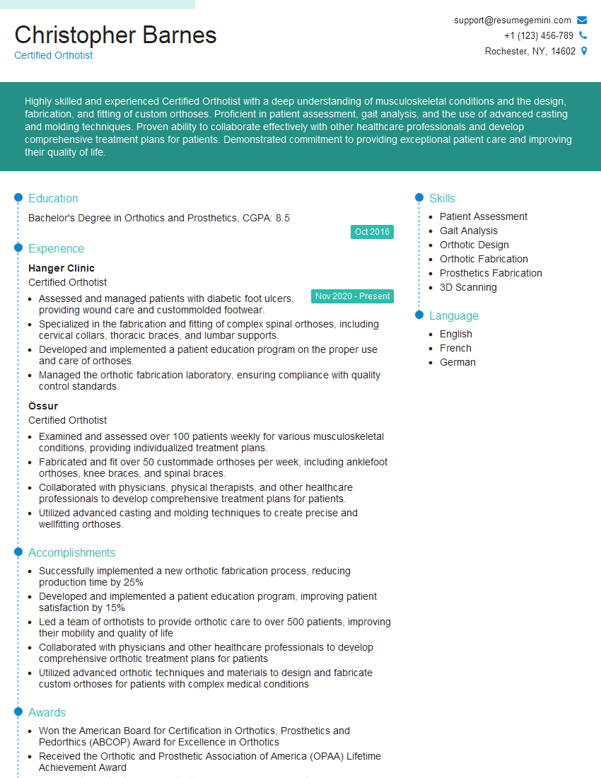
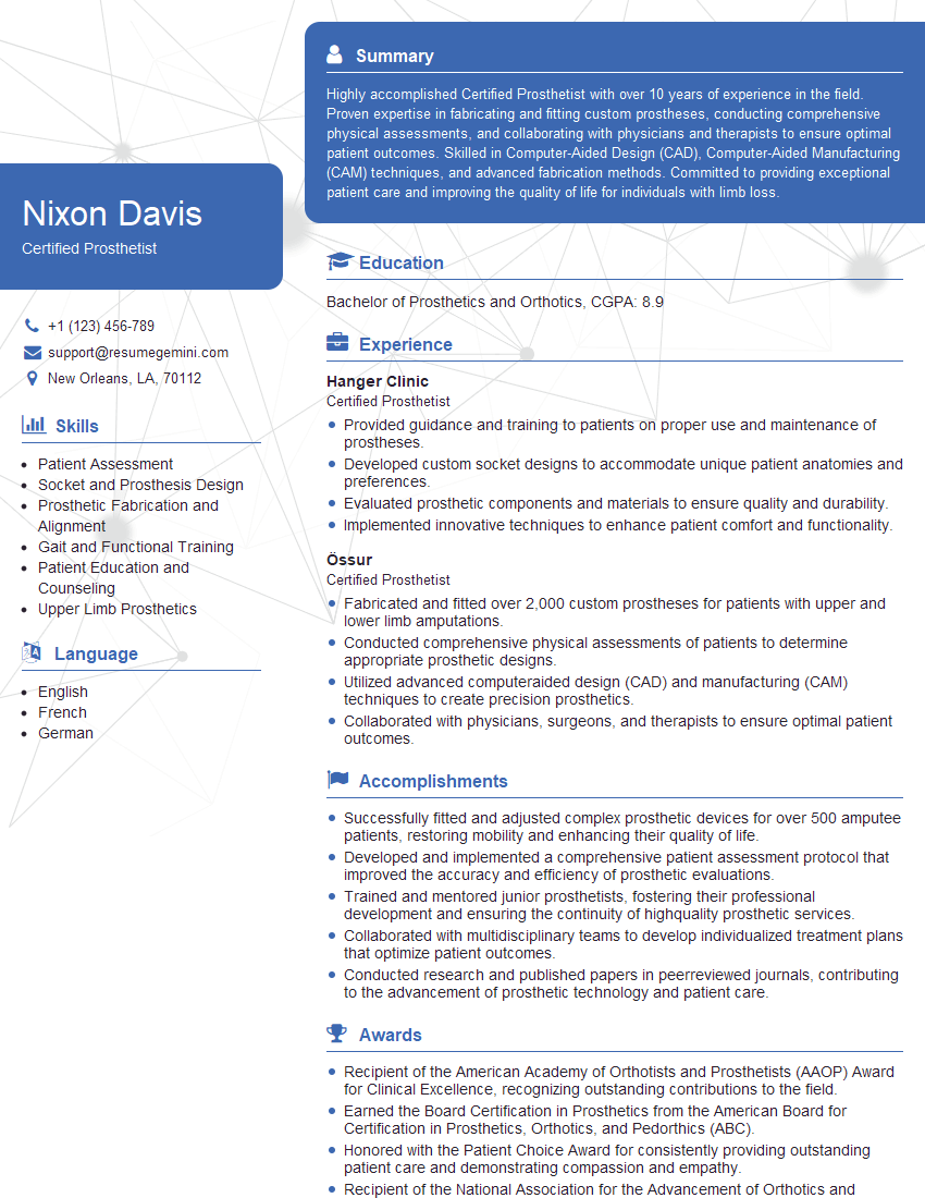
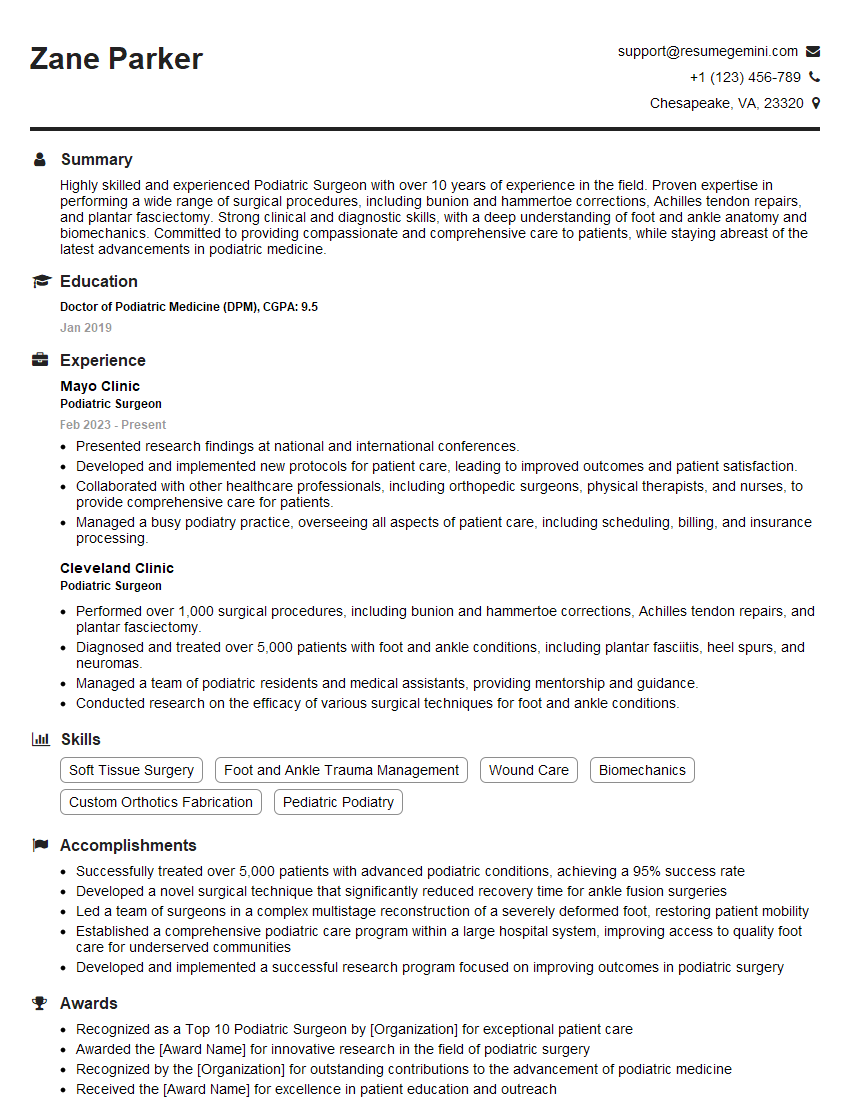
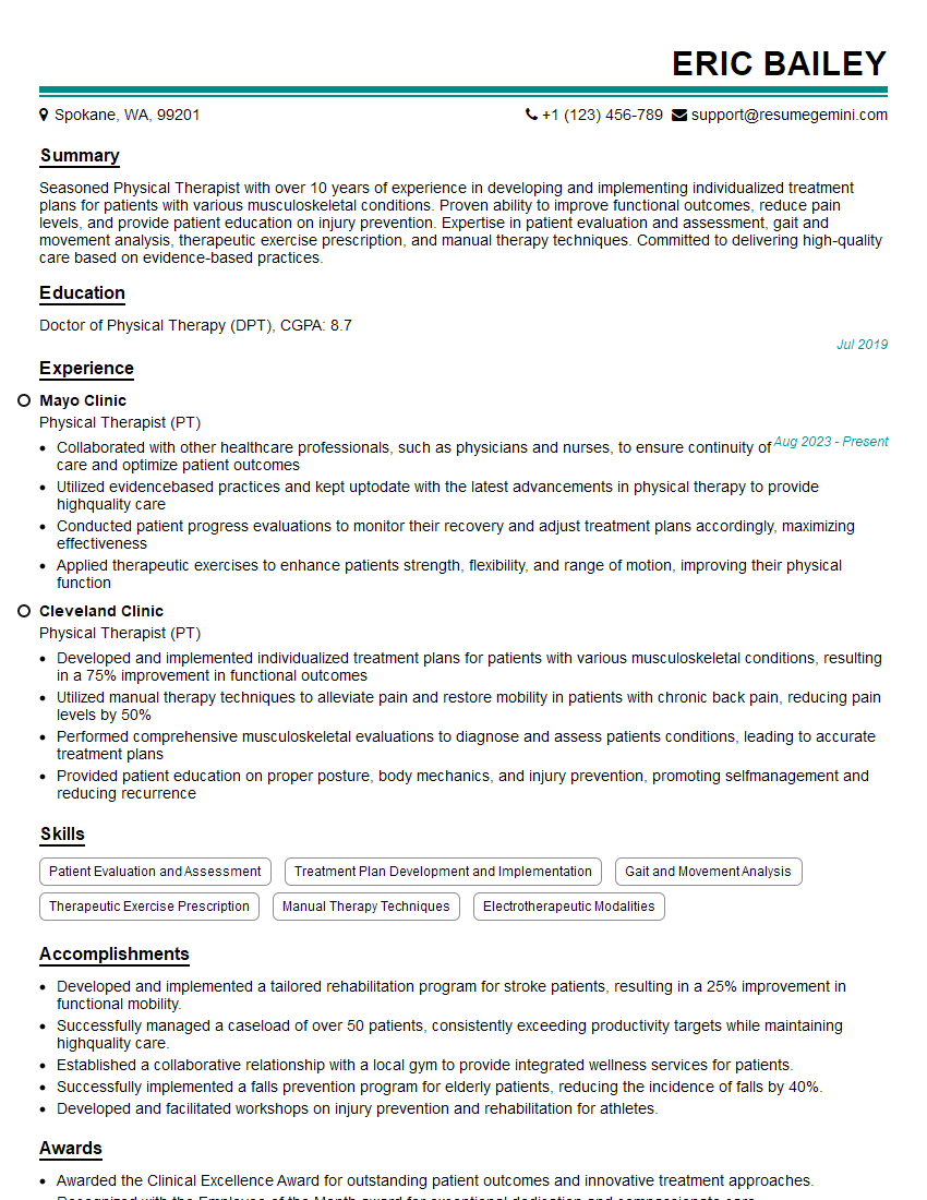
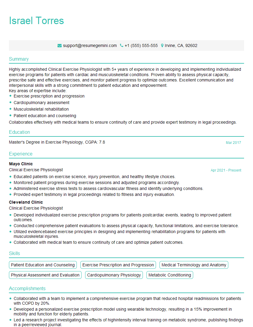
Mastering the management of foot and ankle sports injuries is crucial for career advancement in sports medicine and related fields. A strong understanding of these concepts will significantly enhance your interview performance and open doors to exciting opportunities. To maximize your job prospects, it’s essential to present your skills and experience effectively. Creating an ATS-friendly resume is key to getting your application noticed. ResumeGemini is a trusted resource to help you build a professional and impactful resume that highlights your expertise. Examples of resumes tailored to Management of Foot and Ankle Sports Injuries are available to guide you. Take the next step and build a resume that showcases your capabilities!
Explore more articles
Users Rating of Our Blogs
Share Your Experience
We value your feedback! Please rate our content and share your thoughts (optional).
What Readers Say About Our Blog
This was kind of a unique content I found around the specialized skills. Very helpful questions and good detailed answers.
Very Helpful blog, thank you Interviewgemini team.
