The right preparation can turn an interview into an opportunity to showcase your expertise. This guide to Tibialis Posterior Tendon Dysfunction interview questions is your ultimate resource, providing key insights and tips to help you ace your responses and stand out as a top candidate.
Questions Asked in Tibialis Posterior Tendon Dysfunction Interview
Q 1. Describe the anatomy and biomechanics of the Tibialis Posterior tendon.
The Tibialis Posterior (TP) tendon is a crucial structure in the foot and ankle, responsible for supporting the medial longitudinal arch. Anatomically, it originates from the posterior tibia and fibula, courses down the leg, and inserts onto several tarsal bones (navicular, cuneiforms, and cuboid). Its biomechanical function is complex, involving plantarflexion and inversion of the foot. Imagine it as the main supporting cable of a suspension bridge – the arch of your foot. It helps stabilize the foot during weight-bearing, especially during activities like walking and running, by resisting the forces that tend to flatten the arch. Weakness or dysfunction in this tendon leads to instability and collapse of the arch, resulting in a variety of problems.
Q 2. What are the common causes of Tibialis Posterior Tendon Dysfunction?
Tibialis Posterior Tendon Dysfunction (TPTD) has a multifactorial etiology. Common causes include overuse injuries, particularly in athletes who engage in activities placing high demands on the tendon. Obesity, which increases stress on the arch, is another significant risk factor. Rheumatologic conditions like rheumatoid arthritis can also contribute to tendon inflammation and weakening. Furthermore, anatomical variations like pes planus (flat feet) predispose individuals to TPTD. It’s often a cumulative process; repeated micro-tears over time, exacerbated by the above factors, ultimately lead to tendon damage. Think of it like a frayed rope; each strain weakens it further until it fails.
Q 3. Explain the different stages of Tibialis Posterior Tendon Dysfunction.
TPTD is often staged according to the degree of tendon degeneration and the resulting foot deformity. Stage 1 is characterized by tendon inflammation and mild tenosynovitis, often presenting with pain and mild swelling. Stage 2 shows tendon thickening and some structural degeneration, leading to more significant arch flattening. In Stage 3, the tendon is significantly degenerated or ruptured, resulting in marked arch collapse and a prominent medial bulge (often called a ‘too many toes’ sign) where the tendon used to be. Stage 4 represents the end-stage, with significant deformity and often accompanying arthritis of the midfoot. Early diagnosis and intervention are crucial, as progression through the stages often leads to more complex management and poorer outcomes. It’s like a building collapsing – the earlier you address the cracks, the better your chances of preventing a complete collapse.
Q 4. How do you clinically diagnose Tibialis Posterior Tendon Dysfunction?
Clinical diagnosis begins with a thorough history and physical examination. This includes assessing for pain along the course of the TP tendon, examining the arch height, evaluating for flexibility and strength deficits, and checking for any signs of midfoot instability. The ‘too many toes’ sign, where more than one toe is visible from the front view of the foot, is a hallmark sign of advanced disease. Palpation of the tendon can reveal thickening or tenderness. Special tests like the single-leg heel-raise assess the functional integrity of the tendon. The combination of these clinical findings paints a comprehensive picture, guiding further investigations.
Q 5. What are the key imaging findings in Tibialis Posterior Tendon Dysfunction?
Imaging plays a vital role in confirming the diagnosis and determining the stage of TPTD. Ultrasound is often the initial imaging modality, allowing for real-time visualization of tendon structure, thickness, and signal characteristics suggestive of inflammation or degeneration. MRI offers more detailed anatomical assessment, particularly for identifying tendon tears and associated bone changes. X-rays primarily demonstrate the degree of midfoot collapse and any secondary arthritic changes. Each modality provides complementary information. Think of it like assembling a puzzle; each imaging technique contributes a piece to the complete picture of the tendon’s condition.
Q 6. Describe the conservative management options for Tibialis Posterior Tendon Dysfunction.
Conservative management is the mainstay of treatment for early stages of TPTD. This involves rest, modification of activities that aggravate symptoms, and pain management, including NSAIDs and physical therapy. Physical therapy plays a pivotal role, focusing on strengthening the intrinsic and extrinsic foot muscles, improving flexibility, and providing customized orthotic support. Orthotics are crucial to redistribute weight-bearing stresses and provide arch support, preventing further deterioration of the tendon and the arch. In some cases, bracing or casting may be necessary to immobilize the foot and promote healing.
Q 7. What are the surgical indications for Tibialis Posterior Tendon Dysfunction?
Surgical intervention is considered when conservative measures fail to provide adequate pain relief or when there is significant progression of the deformity. Surgical options include tendon repair, reconstruction, or tenodesis, depending on the stage of disease. Surgical intervention is aimed at restoring the structural integrity of the tendon and addressing the associated deformity. The goal is to improve function, reduce pain, and prevent further deterioration of the foot and ankle. Surgical decisions are carefully individualized, considering the patient’s age, activity level, and the extent of the disease.
Q 8. Explain different surgical techniques used to treat Tibialis Posterior Tendon Dysfunction.
Surgical intervention for Tibialis Posterior Tendon Dysfunction (TPTD) is considered when conservative management fails. The choice of surgical technique depends on the severity of the tendon damage and the stage of the disease.
Tendon Repair: This involves directly repairing a torn or partially torn tendon. It’s suitable for early-stage TPTD with minimal tendon damage. The surgeon will carefully suture the tendon ends together, often using specialized sutures and techniques to maximize healing.
Tendon Transfer: In cases of significant tendon damage or retraction, a tendon transfer might be necessary. This involves transferring a healthy tendon (like the flexor digitorum longus or the peroneus brevis) to replace the function of the tibialis posterior tendon. This procedure redirects force to stabilize the midfoot and rearfoot.
Arthrodesis (Fusion): For severe, end-stage TPTD with significant joint deformity, arthrodesis might be considered. This involves surgically fusing one or more joints in the foot (often the subtalar joint) to provide stability and pain relief. It’s a more drastic procedure, typically reserved for patients who haven’t responded to other treatments and who have significant pain and functional limitations.
Osteotomy: In some cases, correcting bone alignment (osteotomy) might be necessary in conjunction with other procedures to improve the biomechanics of the foot and ankle and reduce stress on the affected tendon.
The specific surgical approach is personalized based on a thorough assessment of the patient’s individual condition, including the extent of tendon damage, the presence of any associated deformities, and the patient’s overall health and activity level. Post-operative management plays a crucial role in achieving a successful outcome.
Q 9. What are the potential complications of Tibialis Posterior Tendon Dysfunction?
Potential complications of TPTD, both with and without surgical intervention, can include:
Persistent Pain: Despite treatment, some patients may experience ongoing pain, though this is less frequent with appropriate treatment and rehabilitation.
Non-union or Re-rupture: In cases of tendon repair, there’s a risk the tendon may not heal properly or may re-rupture. This is more likely with significant tendon damage.
Infection: As with any surgery, there’s a risk of infection at the surgical site, which requires prompt medical attention.
Nerve Damage: There’s a small chance of nerve damage during surgery, potentially leading to numbness or altered sensation in the foot.
Stiffness or Limited Range of Motion: Post-operative stiffness and limited ankle or foot mobility are potential complications requiring focused physiotherapy.
Malunion or Non-union: In cases involving osteotomy, the bones may not heal correctly or may heal in a misaligned position.
Delayed Wound Healing: Factors such as diabetes or smoking can increase the risk of delayed wound healing.
Recurrence of Deformity: Although rare with proper treatment, the foot deformity may recur.
Careful surgical technique, pre-operative patient selection, and diligent post-operative rehabilitation significantly minimize the risk of these complications.
Q 10. How do you differentiate Tibialis Posterior Tendon Dysfunction from other foot and ankle conditions?
Differentiating TPTD from other foot and ankle conditions requires a comprehensive clinical evaluation that combines physical examination, imaging studies, and a detailed patient history.
Conditions that can mimic TPTD include:
Posterior Tibial Tendonitis: This is an inflammatory condition of the tendon that doesn’t involve complete rupture. It often presents with pain and tenderness along the tendon, but typically lacks the significant deformity associated with advanced TPTD.
Flatfoot: While TPTD often leads to a flatfoot deformity, other conditions, like plantar fasciitis or tarsal coalition, can also cause flat feet. Distinguishing TPTD involves assessing the specifics of the deformity (e.g., the presence of a midtarsal break), considering the patient’s history and physical findings, and using imaging studies such as X-rays and MRI.
Ankle Sprains: Ankle sprains are different as they predominantly involve the ligaments of the ankle joint, whereas TPTD focuses on the tendon itself.
Arthritis: Conditions like rheumatoid arthritis or osteoarthritis can impact the ankle and foot, causing pain and stiffness. However, the specific location and pattern of pain, along with the presence of deformities, are crucial in differentiating these conditions from TPTD.
Clinical tests (such as the single-leg heel-raise test and assessing the flexibility and stability of the foot) coupled with imaging, allow for a precise diagnosis. MRI is particularly useful in assessing the extent of tendon damage and ruling out other causes of pain.
Q 11. Describe the post-operative rehabilitation protocol for Tibialis Posterior Tendon Dysfunction surgery.
Post-operative rehabilitation for TPTD surgery is crucial for a successful outcome. The protocol varies depending on the surgical procedure performed and the individual patient’s needs, but generally includes the following phases:
Immediate Post-operative Phase (0-6 weeks): This involves pain management, wound care, elevation of the foot, and the use of a protective cast or boot. Early range-of-motion exercises are introduced gently to prevent stiffness.
Early Rehabilitation Phase (6-12 weeks): The protective cast or boot is often removed, and a gradual transition to weight-bearing is initiated. This phase emphasizes range-of-motion exercises, strengthening exercises for the foot and ankle, and proprioceptive training (exercises to improve balance and coordination).
Advanced Rehabilitation Phase (12-24 weeks or longer): This focuses on improving strength, endurance, and agility. Functional exercises that mimic activities of daily living are introduced. The patient may progress to more advanced exercises, such as plyometrics (jump training) and running, depending on their progress and goals.
Throughout the rehabilitation process, close monitoring by a physical therapist is essential to ensure proper progression, address any complications, and tailor the program to the patient’s individual needs. Regular follow-up visits with the surgeon are also important.
Q 12. What are the common functional limitations experienced by patients with Tibialis Posterior Tendon Dysfunction?
Patients with TPTD experience a range of functional limitations that significantly impact their daily lives. The severity of these limitations varies depending on the stage of the disease.
Pain: Pain is a common symptom, often aggravated by prolonged standing, walking, or weight-bearing activities. Pain can range from mild discomfort to severe, debilitating pain.
Difficulty Walking: Walking becomes difficult, particularly long distances or uneven surfaces. Patients may experience instability and a tendency to trip or stumble.
Limited Range of Motion: Restricted ankle and foot movement restricts the ability to perform activities such as bending, squatting, and climbing stairs.
Foot Deformity: The progressive nature of TPTD leads to noticeable foot deformities, like flatfoot, which can cause additional pain and discomfort, as well as impacting footwear choices.
Impaired Balance: TPTD affects balance and coordination, making patients more prone to falls, particularly on unstable surfaces.
Difficulties with Activities of Daily Living: Everyday activities such as putting on shoes, socks, or tying shoelaces become more challenging and even painful.
The impact of these functional limitations can extend beyond the physical, affecting a person’s social, emotional, and psychological well-being.
Q 13. How do you assess the functional outcome after treatment for Tibialis Posterior Tendon Dysfunction?
Assessing functional outcome after TPTD treatment involves a multi-faceted approach, combining objective measures and subjective patient-reported outcomes.
Clinical Examination: This includes evaluating range of motion, strength, balance, gait, and the presence of any residual deformity.
Foot and Ankle Outcome Scores: Standardized questionnaires like the Foot and Ankle Outcome Score (FAOS) or the American Orthopaedic Foot and Ankle Society (AOFAS) Ankle-Hindfoot Scale provide quantitative data on pain, function, and overall satisfaction.
Gait Analysis: This involves objective assessment of walking patterns to identify any gait deviations or compensatory movements.
Patient-Reported Outcome Measures (PROMs): These questionnaires directly capture the patient’s perspective on their pain levels, functional limitations, and overall quality of life following treatment. They offer valuable insight beyond objective clinical findings.
Imaging Studies: Post-operative X-rays can assess the alignment of bones and the healing of any osteotomies. MRI scans can be used to assess tendon healing in cases of repair.
Combining these assessment methods provides a comprehensive picture of the functional outcome, guiding ongoing management and identifying areas for further improvement.
Q 14. Discuss the role of bracing and orthotics in the management of Tibialis Posterior Tendon Dysfunction.
Bracing and orthotics play a significant role in both non-surgical and post-surgical management of TPTD. They are often used conservatively and as an adjunctive therapy post operatively.
Custom Orthotics: Custom-made orthotics provide support to the arch of the foot, helping to reduce stress on the tibialis posterior tendon and improve foot mechanics. They are often recommended for patients with early-stage TPTD or as part of post-operative rehabilitation.
Ankle-Foot Orthoses (AFOs): AFOs provide more rigid support to the ankle and foot, often used in more severe cases of TPTD or during the early stages of post-operative recovery. They help to control motion and prevent further deformity.
Functional Braces: These offer some support and control while allowing for more mobility compared to AFOs. These braces can help support the arch and improve comfort during weight-bearing activities.
The type of brace or orthotic used depends on several factors, including the severity of the condition, the patient’s activity level, and the stage of treatment. Proper fitting and adjustment are crucial to ensure effectiveness and comfort.
It’s important to note that bracing and orthotics are not a cure for TPTD but rather tools used to alleviate symptoms, improve function, and help prevent further progression of the deformity.
Q 15. Explain the use of extracorporeal shock wave therapy (ESWT) in Tibialis Posterior Tendon Dysfunction.
Extracorporeal shock wave therapy (ESWT) is a non-invasive treatment option for Tibialis Posterior Tendon Dysfunction (TPTD). It involves delivering focused acoustic waves to the affected tendon, aiming to stimulate healing and reduce pain. The mechanism isn’t fully understood, but it’s thought to promote angiogenesis (new blood vessel formation), reduce inflammation, and stimulate the production of growth factors. In TPTD, ESWT can be helpful in the early stages, particularly when there’s tendinopathy (tendon degeneration) but no significant tendon rupture. It’s often used in conjunction with other conservative treatments like physical therapy and bracing.
Think of it like giving the tendon a tiny, controlled ‘massage’ that encourages repair. However, it’s crucial to understand that ESWT isn’t a magic bullet. It’s most effective when used early in the disease process and in conjunction with other therapies. For patients with advanced TPTD, where the tendon is significantly damaged or ruptured, surgical intervention is usually necessary.
Career Expert Tips:
- Ace those interviews! Prepare effectively by reviewing the Top 50 Most Common Interview Questions on ResumeGemini.
- Navigate your job search with confidence! Explore a wide range of Career Tips on ResumeGemini. Learn about common challenges and recommendations to overcome them.
- Craft the perfect resume! Master the Art of Resume Writing with ResumeGemini’s guide. Showcase your unique qualifications and achievements effectively.
- Don’t miss out on holiday savings! Build your dream resume with ResumeGemini’s ATS optimized templates.
Q 16. What are the different types of tendon transfers used in Tibialis Posterior Tendon Dysfunction?
Tendon transfers are surgical procedures used in more advanced cases of TPTD to restore the function of the weakened or ruptured tibialis posterior tendon. The goal is to replace the action of the tibialis posterior with another tendon that can provide similar support to the medial longitudinal arch. Several tendons can be used, depending on the specifics of the case and surgeon preference. Commonly used transfers include:
- Flexor digitorum longus (FDL) transfer: This is a frequently used option, offering reliable strength and relatively predictable outcomes.
- Flexor hallucis longus (FHL) transfer: This is another popular choice, but it can impact the ability to flex the big toe, requiring careful patient selection.
- Tibialis anterior transfer: This is less common, mainly used in situations where the FDL and FHL are not suitable options.
The choice of tendon transfer depends on several factors including the extent of the TPTD, the patient’s age, activity level, and the presence of any other foot and ankle pathologies. A detailed pre-operative assessment is crucial to choose the optimal tendon transfer.
Q 17. How do you manage recalcitrant cases of Tibialis Posterior Tendon Dysfunction?
Managing recalcitrant cases of TPTD—those that haven’t responded to conservative management—requires a multi-faceted approach. If conservative measures such as physical therapy, bracing, and ESWT have failed, surgical intervention often becomes necessary. This could involve a tendon transfer, as discussed earlier, or other procedures such as tendon repair or reconstruction. Sometimes, even after surgery, patients might experience persistent pain or functional limitations.
In such cases, we explore further options such as:
- Pain management strategies: This might involve medications, injections, or even referral to a pain specialist.
- Further surgical interventions: In rare cases, additional surgery may be considered to address complications or residual deformities.
- Adaptive footwear and orthotics: Custom-made orthotics and supportive footwear can significantly reduce stress on the foot and ankle, improving comfort and function.
- Functional rehabilitation: Intensive physiotherapy focusing on strength, balance, and proprioception is essential post-surgery, and in recalcitrant cases, may need to be more prolonged and targeted.
It’s critical to maintain open communication with the patient, emphasizing realistic expectations and acknowledging that complete resolution isn’t always attainable in severe, long-standing cases.
Q 18. What are the prognostic factors influencing the outcome of Tibialis Posterior Tendon Dysfunction?
Several prognostic factors influence the outcome of TPTD treatment. These factors can significantly impact the success of both conservative and surgical management. Key prognostic factors include:
- Stage of the disease: Early-stage TPTD, characterized by tendinopathy, generally has a better prognosis than advanced stages involving tendon rupture and significant deformity.
- Age and activity level: Younger, more active individuals tend to have better outcomes after surgical intervention, whereas older patients with low activity levels may experience slower recovery and less improvement.
- Presence of other foot and ankle pathology: Co-existing conditions such as arthritis or other tendon injuries can complicate treatment and affect the overall outcome.
- Patient compliance with treatment: Diligent adherence to the prescribed rehabilitation protocol is crucial for successful recovery. Poor compliance can negatively impact outcomes.
- Surgical technique and surgeon experience: The skill and experience of the surgeon performing the tendon transfer or other surgical procedures are important factors in determining success.
Considering these factors allows for a more accurate prediction of treatment success and helps to tailor treatment plans to individual needs.
Q 19. Describe the patient education strategies you utilize for Tibialis Posterior Tendon Dysfunction.
Patient education is paramount in the management of TPTD. My approach involves a multi-pronged strategy designed to empower patients to actively participate in their recovery. This includes:
- Detailed explanation of the condition: Using clear, simple language, I explain the anatomy of the tibialis posterior tendon, its function, and how it’s affected in TPTD. I use visual aids like anatomical models or diagrams to enhance understanding.
- Discussion of treatment options: I outline the various treatment options, highlighting the benefits and risks of each, and collaboratively determine the best course of action based on the individual’s needs and preferences.
- Emphasis on adherence to the treatment plan: I stress the importance of regular physical therapy, adherence to bracing recommendations (if any), and the need for gradual weight-bearing progression.
- Realistic expectation setting: Openly discussing potential limitations and recovery timelines helps manage patient expectations and prevent frustration.
- Provision of written instructions and resources: I provide patients with written summaries of the treatment plan, home exercise programs, and links to reputable online resources for additional information.
Ultimately, a well-informed and motivated patient is more likely to achieve a successful outcome.
Q 20. What are the current research trends in Tibialis Posterior Tendon Dysfunction?
Current research trends in TPTD focus on several key areas:
- Improved diagnostic tools: Researchers are investigating better imaging techniques (beyond standard X-rays and MRIs) to detect and stage TPTD earlier and more accurately.
- Biologic therapies: Studies are exploring the use of biologics, such as platelet-rich plasma (PRP) and stem cells, to promote tendon healing and reduce inflammation.
- Minimally invasive surgical techniques: The aim is to develop less invasive surgical approaches that minimize recovery time and complications associated with traditional tendon transfer procedures.
- Personalized treatment strategies: Research is focusing on identifying specific biomarkers or genetic factors that could help predict the response to different treatments and allow for more tailored interventions.
- Longitudinal studies: There’s a growing need for larger-scale, long-term studies to assess the effectiveness of different treatments and identify factors that contribute to long-term outcomes.
These research avenues hold promise for improving the diagnosis, treatment, and overall management of TPTD.
Q 21. How would you approach a patient with a history of Tibialis Posterior Tendon Dysfunction and recent ankle sprain?
A patient presenting with a history of TPTD and a recent ankle sprain requires a thorough evaluation to determine the extent of the injuries and their interaction. The ankle sprain may be an independent event, or it may have exacerbated the pre-existing TPTD. The approach would involve:
- Comprehensive history taking: Detailed assessment of the ankle sprain, including mechanism of injury, symptoms, and any previous ankle injuries.
- Physical examination: Careful examination of the ankle and foot to assess the severity of the sprain and to evaluate the status of the tibialis posterior tendon.
- Imaging studies: X-rays are essential to rule out fractures. An MRI may be necessary to assess the integrity of the tibialis posterior tendon and evaluate the extent of both the sprain and any TPTD involvement.
- Treatment plan development: Based on the findings, a treatment plan would be created. This might involve conservative management for both injuries (e.g., immobilization, physiotherapy, pain management), or it could necessitate surgical intervention, especially if there’s significant damage to the tendon or a severe ankle sprain.
- Close monitoring: Regular follow-up appointments are critical to monitor progress and adjust the treatment plan as needed.
The goal is to manage both the acute ankle sprain and the chronic TPTD effectively, taking into account the potential interaction between the two conditions. The treatment approach would be individualized based on the patient’s specific situation.
Q 22. Discuss the role of physical therapy in managing Tibialis Posterior Tendon Dysfunction.
Physical therapy plays a crucial role in managing Tibialis Posterior Tendon Dysfunction (TPTD), aiming to reduce pain, improve function, and prevent further damage. The treatment plan is highly individualized, depending on the severity of the condition and the patient’s overall health.
- Early Stages: Focuses on conservative measures like rest, ice, compression, and elevation (RICE). Exercises emphasize strengthening the tibialis posterior muscle and improving ankle stability. This often involves eccentric exercises (lengthening the muscle under tension) which are particularly effective in strengthening the tendon. We also educate patients on proper footwear and activity modification to minimize stress on the tendon.
- Later Stages: As the condition progresses, more advanced techniques may be implemented. These can include manual therapy to address joint restrictions in the foot and ankle, therapeutic ultrasound to promote tissue healing, and custom orthotics to support the arch and correct foot posture. In some cases, we might use functional electrical stimulation to improve muscle activation and strength.
- Advanced Stages: If conservative treatment fails, surgical intervention might be considered. Even after surgery, extensive physical therapy is essential for rehabilitation, focusing on regaining range of motion, strength, and proprioception (awareness of body position).
For instance, a patient with mild TPTD might start with simple calf stretches and ankle strengthening exercises, while a patient with a more advanced stage might require a comprehensive program including manual therapy, orthotics, and a gradual return to activity under the therapist’s guidance. The goal is always to restore normal foot and ankle mechanics and prevent recurrence.
Q 23. Explain the importance of patient compliance in the treatment of Tibialis Posterior Tendon Dysfunction.
Patient compliance is paramount in the successful treatment of TPTD. This condition often requires long-term commitment to a home exercise program, consistent use of orthotics (if prescribed), and adherence to activity modifications. Without compliance, the chances of successful outcome diminish significantly.
Think of it like this: TPTD is a bit like a slow leak in a tire. If you ignore it, the tire eventually goes flat. Similarly, if a patient doesn’t consistently follow the prescribed exercises and lifestyle changes, the tendon will continue to deteriorate. We often see patients who initially adhere well to their programs but then gradually reduce their effort. This can lead to setbacks and a longer recovery period.
To encourage compliance, we work closely with patients, educating them thoroughly about the condition and its implications, setting realistic goals, and providing regular feedback. We tailor exercises to their individual needs and preferences, making the program as manageable and engaging as possible. This might involve visual aids, written instructions, and regular check-in appointments to assess progress and answer any questions. We also encourage patients to track their progress and to actively participate in their care.
Q 24. What are the common risk factors associated with Tibialis Posterior Tendon Dysfunction?
Several risk factors increase the likelihood of developing TPTD. These factors often interact and synergistically increase the risk.
- High impact activities: Activities placing repetitive strain on the foot and ankle, such as running, jumping, and certain sports.
- Flat feet (pes planus): This alters foot mechanics, overloading the tibialis posterior tendon.
- Overpronation: Excessive inward rolling of the foot during gait.
- Obesity: Increased weight places additional stress on the tendon.
- Rheumatoid arthritis: This inflammatory condition can weaken tendons.
- Age: Tendons tend to become less resilient with age, increasing vulnerability.
- Previous ankle injury: Prior injury can disrupt the normal biomechanics of the foot and ankle.
- Genetic predisposition: A family history of TPTD can increase the risk.
For example, a middle-aged overweight individual with flat feet who enjoys long-distance running is at a significantly higher risk compared to a young, healthy person with normal foot arches.
Q 25. How do you assess the severity of Tibialis Posterior Tendon Dysfunction using clinical examination?
Assessing the severity of TPTD involves a thorough clinical examination, looking for a combination of findings.
- Pain: Pain along the course of the tibialis posterior tendon, often aggravated by activity.
- Swelling: Palpable swelling or tenderness over the tendon.
- Tenderness to palpation: Identifying specific points of tenderness along the tendon.
- Foot posture: Assessing for pes planus (flat feet), excessive pronation, or valgus deformity (outward turning of the heel).
- Range of motion: Evaluating ankle and subtalar joint flexibility.
- Strength: Testing the strength of the tibialis posterior muscle.
- Flexibility: Assessing for tightness in the gastrocnemius and soleus muscles.
- Gait analysis: Observing the patient’s walking pattern to identify any abnormalities.
- Positive Thompson test: Squeezing the calf muscle to assess the integrity of the Achilles tendon, which may be involved in later stages of TPTD.
The combination of these findings helps determine the stage of TPTD. For instance, a patient with mild pain, subtle flattening of the arch, and normal strength might be classified as having early-stage TPTD, while a patient with severe pain, significant arch collapse, and weakness might be classified as having advanced-stage disease.
Q 26. Describe the role of imaging modalities (X-ray, MRI, Ultrasound) in diagnosing Tibialis Posterior Tendon Dysfunction.
Imaging modalities play a vital role in confirming the diagnosis and assessing the severity of TPTD. While a clinical exam is crucial, imaging helps visualize the tendon and surrounding structures.
- X-ray: X-rays primarily rule out other conditions and assess for bony changes associated with advanced TPTD, such as talonavicular joint collapse or osteoarthritis. They are less useful in evaluating the tendon itself.
- Ultrasound: Ultrasound is a highly valuable tool for visualizing the tendon, assessing its thickness, and identifying tears or inflammation. It’s non-invasive, relatively inexpensive, and readily available.
- MRI: MRI provides the most detailed images of the tendon and surrounding soft tissues. It can accurately depict tendon tears, inflammation, and assess the degree of associated bone changes. While more expensive and less readily available than ultrasound, MRI is often the gold standard for visualizing soft tissue structures.
We might use a combination of these modalities. For example, we might start with an X-ray to rule out fractures and then follow up with an ultrasound to assess the tendon itself. In more complex cases, or when a definitive diagnosis is needed, an MRI might be necessary.
Q 27. What are the long-term complications of untreated Tibialis Posterior Tendon Dysfunction?
Untreated TPTD can lead to several long-term complications, significantly impacting foot and ankle function and quality of life.
- Progressive flatfoot deformity: This leads to pain, instability, and altered gait mechanics.
- Pain and disability: Persistent pain limiting daily activities and reducing mobility.
- Arthritis: Degenerative changes in the joints of the foot and ankle, leading to further pain and stiffness.
- Limited mobility: Difficulty with walking, running, and other activities.
- Increased risk of falls: Foot instability increases the risk of falls, particularly in older adults.
- Painful hindfoot varus or valgus: Deformities affecting the heel bone.
- Subtalar and midfoot joint osteoarthritis: This may require additional treatment, including surgery.
Imagine a patient with untreated TPTD who eventually develops severe arthritis, making even simple activities like walking excruciating. This underscores the importance of early diagnosis and prompt management to prevent long-term complications.
Q 28. Explain your approach to managing a patient with a complex case of Tibialis Posterior Tendon Dysfunction involving multiple tendon involvement.
Managing a complex case of TPTD involving multiple tendon involvement requires a multi-faceted approach, combining expertise from different healthcare professionals.
My approach involves:
- Comprehensive assessment: A thorough clinical evaluation, along with appropriate imaging studies (ultrasound and/or MRI) to identify the extent of tendon involvement, associated joint pathology, and severity of the deformity. This might involve consultations with a podiatrist or orthopaedic surgeon specializing in foot and ankle surgery.
- Tailored treatment plan: A customized plan that addresses each involved tendon and associated problems. This might involve a combination of conservative and surgical interventions.
- Conservative management: This might include a tailored program of exercises, orthotics, and physical therapy. We might involve modalities such as extracorporeal shock wave therapy to stimulate tendon healing. Pain management will be a key component.
- Surgical intervention (if necessary): Surgical intervention might be considered if conservative treatment fails. This could involve tendon repair, reconstruction, or fusion procedures, depending on the specific needs of the patient. This would require close collaboration with the surgical team.
- Post-surgical rehabilitation: Intensive physical therapy is crucial post-surgery to regain strength, improve range of motion, and optimize function. This is a prolonged process, requiring patience and persistence from both the patient and the rehabilitation team.
For example, a patient with severe TPTD involving the tibialis posterior, flexor hallucis longus, and flexor digitorum longus tendons might require surgical reconstruction of the tendons along with a prolonged rehabilitation program, including custom orthotics to prevent recurrence.
Key Topics to Learn for Tibialis Posterior Tendon Dysfunction Interview
- Anatomy and Biomechanics: Understand the anatomy of the tibialis posterior tendon, its role in foot and ankle stability, and the biomechanical forces acting upon it.
- Pathophysiology: Master the mechanisms leading to tibialis posterior tendon dysfunction, including stages of the condition and associated risk factors.
- Clinical Presentation: Be prepared to discuss the typical symptoms, physical examination findings, and diagnostic imaging techniques used to identify TPTD.
- Differential Diagnosis: Know how to differentiate TPTD from other conditions with similar symptoms, demonstrating a comprehensive understanding of foot and ankle pathology.
- Conservative Management: Detail the various conservative treatment approaches, including bracing, orthotics, physical therapy modalities, and activity modification strategies.
- Surgical Management: Discuss the indications, techniques, and potential complications of surgical interventions for TPTD, demonstrating a nuanced understanding of surgical options.
- Rehabilitation and Recovery: Explain the principles of rehabilitation following both conservative and surgical management, highlighting the importance of progressive loading and functional restoration.
- Patient Education and Communication: Demonstrate your ability to effectively communicate complex medical information to patients, tailoring your approach based on their understanding and needs.
- Research and Current Trends: Stay updated on the latest research in TPTD treatment and management, showcasing your commitment to professional development.
- Problem-Solving: Practice applying your knowledge to case studies, demonstrating your ability to diagnose, treat, and manage patients with TPTD effectively.
Next Steps
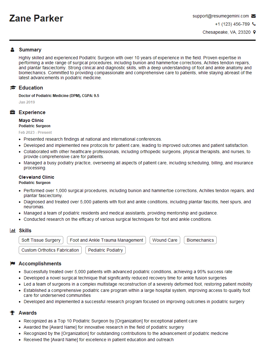
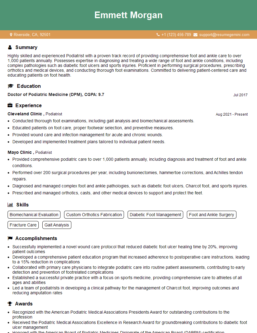
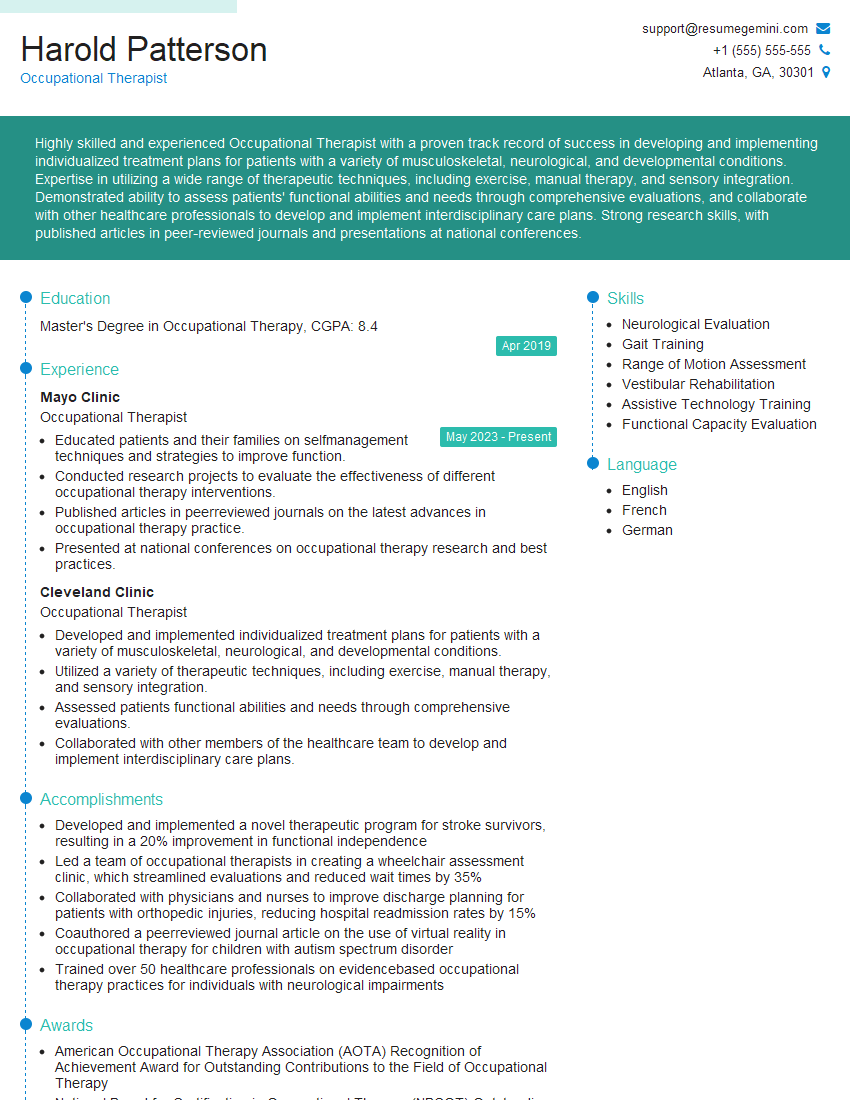
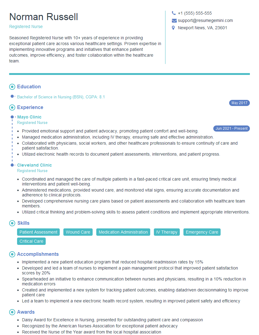
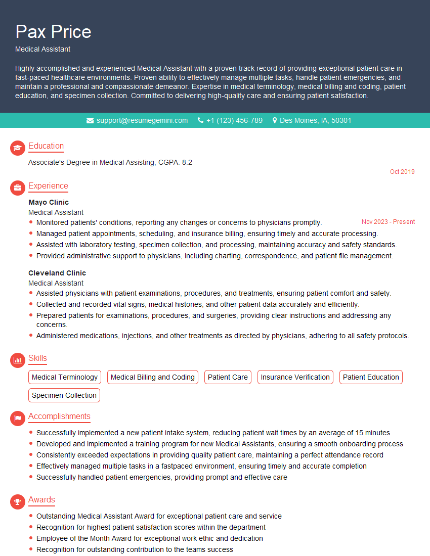
Mastering Tibialis Posterior Tendon Dysfunction is crucial for career advancement in podiatry, physical therapy, and related fields. A strong understanding of this condition demonstrates a high level of expertise and clinical competence, making you a highly sought-after candidate. To maximize your job prospects, invest time in creating an ATS-friendly resume that showcases your skills and experience effectively. ResumeGemini is a trusted resource for building professional resumes, and we provide examples tailored to Tibialis Posterior Tendon Dysfunction to help you get started. Use this opportunity to present yourself as a well-prepared and highly capable professional.
Explore more articles
Users Rating of Our Blogs
Share Your Experience
We value your feedback! Please rate our content and share your thoughts (optional).
What Readers Say About Our Blog
This was kind of a unique content I found around the specialized skills. Very helpful questions and good detailed answers.
Very Helpful blog, thank you Interviewgemini team.
