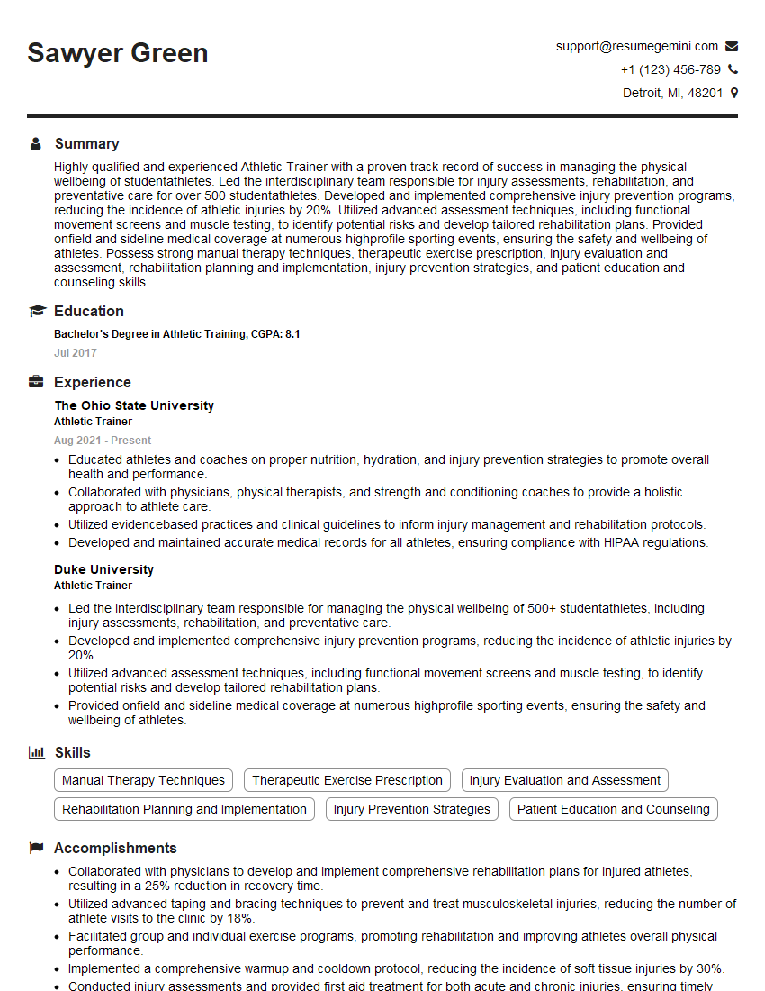
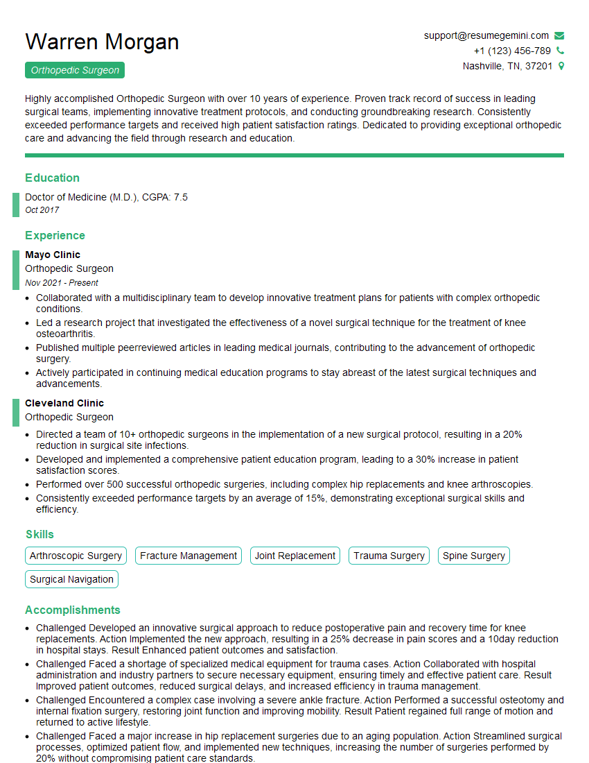
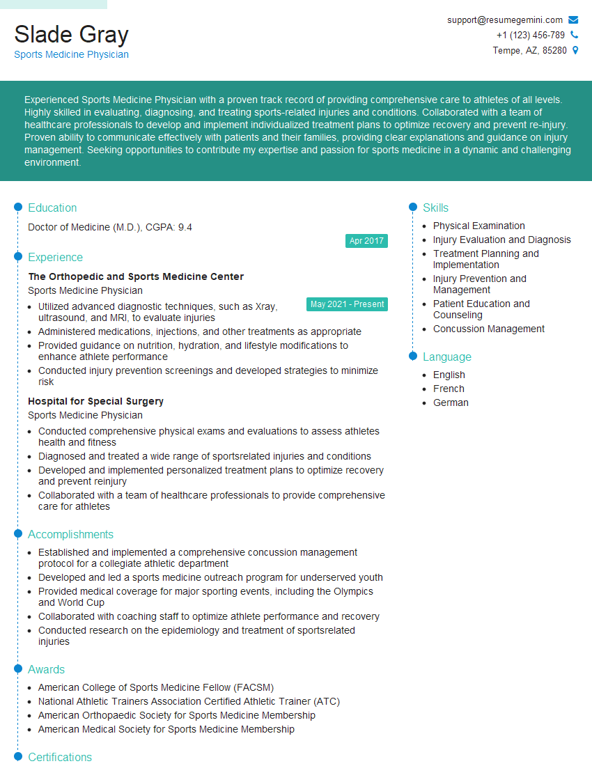
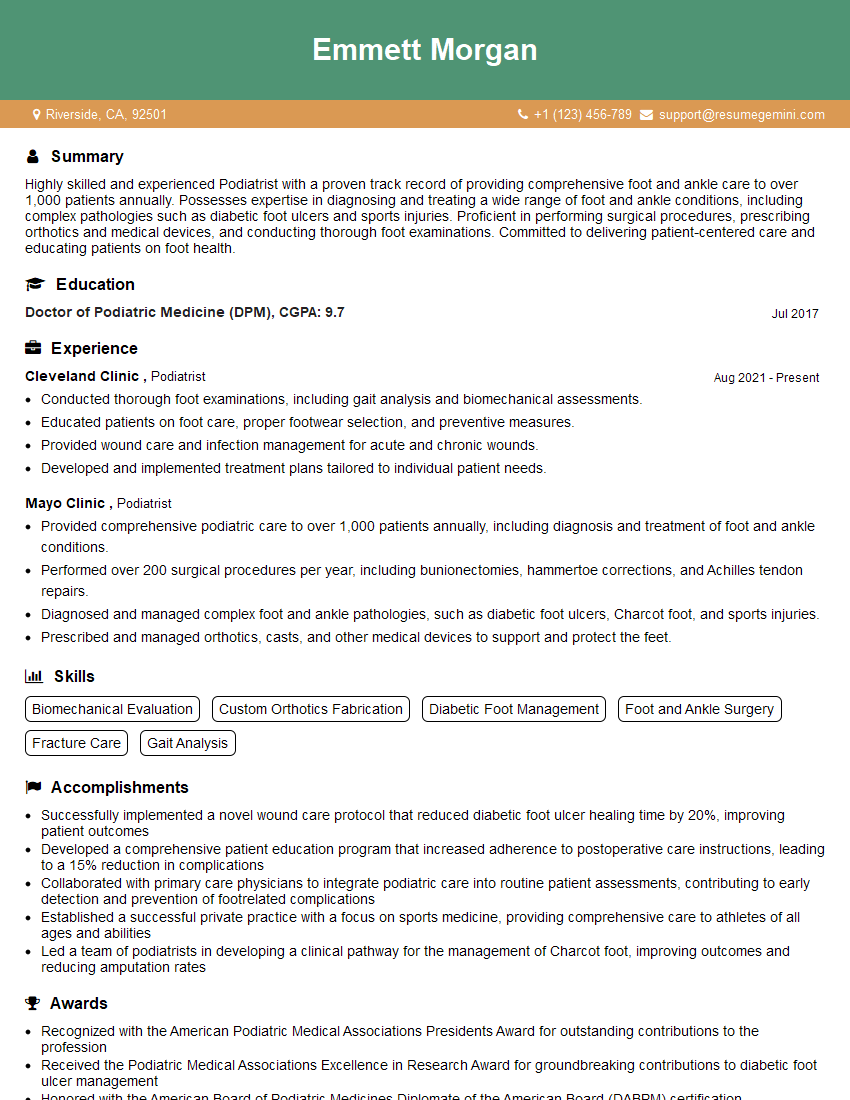
The right preparation can turn an interview into an opportunity to showcase your expertise. This guide to Foot and Ankle Fractures Management interview questions is your ultimate resource, providing key insights and tips to help you ace your responses and stand out as a top candidate.
Questions Asked in Foot and Ankle Fractures Management Interview
Q 1. Describe the different classifications of ankle fractures.
Ankle fractures are classified in several ways, depending on the bone involved and the nature of the fracture. A common system considers the location and the involvement of the three malleoli (bony prominences): the medial malleolus (part of the tibia), the lateral malleolus (part of the fibula), and the posterior malleolus (also part of the tibia).
- Unimalleolar fractures: Involve only one malleolus (e.g., a fracture of the lateral malleolus only).
- Bimalleolar fractures: Involve two malleoli (e.g., fractures of both the medial and lateral malleoli). These often result from a rotational injury.
- Trimalleolar fractures: Involve all three malleoli. These are usually more severe and complex, often requiring surgical intervention.
- Pilon fractures: These are severe fractures of the distal tibia, involving the articular surface of the ankle joint. They can be very complex and challenging to treat.
Another classification system considers the stability of the ankle joint after the fracture. A stable fracture typically maintains the alignment of the ankle joint, while an unstable fracture disrupts this alignment, often requiring surgical intervention to restore.
Q 2. Explain the Ottawa Ankle Rules and their significance.
The Ottawa Ankle Rules are a clinical decision rule used to determine whether an ankle X-ray is necessary after an ankle injury. They help to reduce unnecessary imaging and radiation exposure. The rules dictate that an X-ray is indicated if any of the following are present:
- Bone tenderness at the posterior edge or tip of the medial malleolus
- Bone tenderness at the posterior edge or tip of the lateral malleolus
- Inability to bear weight both immediately after the injury and in the examination room (four steps).
- Inability to weight bear for four steps in the ER.
Their significance lies in their ability to effectively identify patients who are unlikely to have an ankle fracture, thus reducing unnecessary healthcare costs and radiation exposure. While highly sensitive (meaning they rarely miss a fracture), they are not perfectly specific (meaning some patients may have a positive finding but not actually have a fracture).
Example: A patient presents after a twisting injury to their ankle. They have pain along the lateral malleolus but can bear weight immediately after the fall. According to the Ottawa Ankle Rules, an X-ray may not be necessary as the other criteria are not met.
Q 3. What imaging modalities are used to diagnose foot and ankle fractures?
Several imaging modalities are used for diagnosing foot and ankle fractures. The most common is:
- Plain radiography (X-rays): These are the initial imaging modality of choice. They provide excellent visualization of bones and can detect most fractures. A minimum of two views (anteroposterior and lateral) are required and often a mortise view is included for better assessment of the ankle joint.
- Computed tomography (CT): CT scans provide detailed three-dimensional images of the bones, allowing for a better assessment of complex fractures, articular involvement, and the presence of subtle fractures that may be missed on plain X-rays. It’s particularly useful for pilon fractures and complex fractures involving multiple bones.
- Magnetic resonance imaging (MRI): MRI provides excellent soft tissue detail and is useful for assessing ligament injuries, tendon tears, and cartilage damage associated with a fracture. It is less commonly used for initial fracture diagnosis unless there is a suspicion of occult (hidden) fractures or significant soft-tissue injury.
Q 4. How do you differentiate between a fracture and a sprain?
Differentiating between a fracture and a sprain involves a careful clinical examination and often imaging studies. A sprain is an injury to a ligament, while a fracture is a break in the bone.
- Pain: Both fractures and sprains cause pain, but fracture pain is often more intense and localized over the fracture site.
- Swelling: Swelling is common in both, but it tends to be more significant and rapid in fractures.
- Deformity: Visible deformity (a change in the normal shape of the limb) is a hallmark sign of a fracture. This is less common in sprains.
- Tenderness to palpation: Precise location of tenderness is important. Tenderness directly over a bone is suggestive of a fracture.
- Crepitus: A grating sensation felt when moving a fractured bone is a strong indicator of a fracture.
- Inability to bear weight: Difficulty or inability to bear weight strongly suggests a fracture but can also occur in severe sprains.
Ultimately, plain X-rays are the gold standard for confirming the presence of a fracture. A physical exam alone cannot reliably distinguish between the two, particularly in subtle cases.
Q 5. What are the common complications associated with Lisfranc injuries?
Lisfranc injuries are fractures and/or dislocations of the tarsometatarsal joints of the foot. These injuries can be devastating and often require surgical intervention. Common complications include:
- Malunion: The bones heal in a misaligned position, resulting in chronic pain, instability, and deformity.
- Nonunion: The fractured bones fail to heal, leading to persistent pain and instability.
- Arthritis: Damage to the articular surfaces of the tarsometatarsal joints can lead to early-onset arthritis, causing significant pain and stiffness.
- Avascular necrosis (AVN): Disruption of the blood supply to one or more of the tarsal bones can lead to bone death. This typically affects the second metatarsal.
- Chronic pain and instability: Even with successful treatment, some patients experience chronic pain and instability in their foot.
- Post-traumatic osteoarthritis: Long-term joint damage is common leading to significant joint pain.
These complications underscore the need for prompt diagnosis and appropriate treatment of Lisfranc injuries.
Q 6. Describe the surgical approach for a displaced talar neck fracture.
The surgical approach for a displaced talar neck fracture depends on several factors, including the degree of displacement, the presence of comminution (fragmentation of the bone), and the overall condition of the patient. A common approach involves an open reduction and internal fixation (ORIF).
Steps involved in ORIF for a displaced talar neck fracture:
- An incision is made: An incision is made over the talar neck to expose the fracture site.
- Reduction of the fracture: The fractured bone fragments are carefully manipulated and repositioned back into their anatomically correct alignment (reduction).
- Fixation: Once reduced, the fracture is stabilized using internal fixation devices such as screws, plates, or a combination of both. The choice of implants depends on the fracture pattern.
- Wound closure: Once the fracture is fixed, the wound is carefully closed in layers, and a sterile dressing is applied.
- Post-operative care: Post-operative care usually involves immobilization in a cast or splint, pain management, and physical therapy to regain ankle function.
The specific surgical technique and implants used may vary based on individual patient needs and the surgeon’s preference. The goal is to achieve anatomical reduction and stable fixation to allow for proper bone healing and restoration of ankle function.
Q 7. Explain the principles of fracture fixation.
The principles of fracture fixation guide the surgical management of fractures, particularly those requiring surgical intervention. The main goals are to restore anatomy, achieve stability, and promote healing.
- Anatomical reduction: The fractured bone fragments must be precisely aligned and restored to their original anatomical position. Imperfect reduction can lead to malunion and long-term complications.
- Stable fixation: Once reduced, the fracture must be held securely in place to prevent movement. This is typically achieved using internal fixation devices like plates, screws, or intramedullary nails. The stability required depends on the fracture pattern and bone healing potential.
- Minimally invasive techniques: Whenever feasible, minimally invasive techniques should be used to reduce soft tissue trauma and minimize the risk of infection and complications.
- Appropriate implant selection: The choice of implants should be guided by the type and location of the fracture, bone quality, and patient factors. Biocompatible materials are essential to minimize the risk of adverse reactions.
- Early mobilization: After achieving stable fixation, early mobilization and rehabilitation are encouraged to promote bone healing and restore function. The timing and intensity of mobilization will depend on the specifics of the injury and fracture healing.
These principles, while seemingly simple, require careful planning, precision, and expertise to achieve optimal outcomes. The ultimate aim is to allow for early weight-bearing and return to normal activity.
Q 8. What are the different types of fixation devices used in foot and ankle surgery?
Foot and ankle surgery utilizes a variety of fixation devices to stabilize fractures and promote healing. The choice of device depends on several factors, including the specific fracture pattern, bone quality, patient factors (e.g., age, comorbidities), and surgeon preference.
- Plates and Screws: These are commonly used for more complex fractures requiring rigid fixation. Plates provide structural support while screws provide compression across the fracture site. Different plate designs exist (e.g., locking plates, dynamic compression plates) to address various fracture patterns and bone qualities.
- Intramedullary Nails: These are long rods inserted into the medullary canal of the bone, providing axial stability. They are particularly useful in long bone fractures but can also be applied to certain foot and ankle fractures.
- Screws Alone: For simpler, less displaced fractures, screws alone might suffice to achieve adequate reduction and fixation. These can be cannulated screws (inserted over a guidewire) for minimally invasive surgery.
- K-wires (Kirschner wires): These are thin, smooth wires used for temporary fixation, often as part of a multi-pronged approach or in situations where a more definitive fixation is not immediately feasible. They are frequently used in pediatric patients or for minimally invasive techniques.
- External Fixation: This involves the use of pins inserted into the bone and connected to an external frame. It is often used for highly comminuted fractures (fractures with multiple bone fragments) or when soft tissue injury precludes internal fixation. This can be a temporary measure until the patient is stable enough for definitive surgery or is used as a definitive treatment option in certain situations.
Imagine building a house: plates and screws are like the strong wooden beams and nails, providing rigid support; intramedullary nails are like a central support column; screws alone are like strategically placed nails; K-wires are like temporary scaffolding; and external fixation is like a temporary external support structure.
Q 9. Discuss the post-operative management of a calcaneal fracture.
Post-operative management of a calcaneal fracture is crucial for optimal healing and functional outcome. It’s a complex fracture given the calcaneus’ crucial role in weight-bearing. Immediate post-operative care includes pain management, often with a multimodal approach using analgesics, nerve blocks and potentially opioids. Non-weight bearing is usually initially prescribed to protect the fracture site.
Early Stages (first few weeks): Focuses on pain control, wound care (if applicable), and minimizing swelling. This might involve elevation of the leg, ice packs, and compression bandages. Regular wound checks are important, especially for open or severely comminuted fractures.
Intermediate Stages (weeks 4-12): Gradual weight bearing is introduced, often using a walker or crutches, as tolerated by the patient and guided by radiographic evidence of healing. Physical therapy is initiated to focus on range of motion, strengthening, and gait retraining.
Late Stages (beyond 12 weeks): Emphasis shifts to restoring full weight-bearing, improving strength and proprioception, and addressing any residual functional deficits. Further physical and occupational therapy might be required. Return to work or sports depends on the individual’s profession and activity level; it’s often a gradual process.
Regular radiographic follow-up is essential throughout the recovery process to monitor fracture healing and identify potential complications such as nonunion or malunion. Complications can sometimes need revision surgery.
Q 10. What are the indications for ORIF (Open Reduction Internal Fixation) versus conservative management of a fracture?
The decision between ORIF (Open Reduction Internal Fixation) and conservative management for a fracture hinges on several factors. Conservative management typically involves immobilization with casting or splinting, allowing the fracture to heal naturally. ORIF involves surgical intervention to realign (reduce) the fracture fragments and stabilize them with internal fixation devices.
Indications for ORIF:
- Significant displacement or angulation: When the fracture fragments are significantly out of alignment, ORIF is necessary to restore proper bone anatomy and prevent long-term functional impairment. This is especially important in weight-bearing bones like the talus or calcaneus.
- Comminuted fractures: Fractures with multiple fragments are usually more effectively managed with ORIF to achieve adequate stability and prevent malunion or nonunion.
- Intra-articular fractures: Fractures extending into a joint surface (e.g., talar neck fracture) require precise anatomical reduction to prevent future arthritis.
- Open fractures: Fractures with an open wound require surgical debridement (removal of damaged tissue) and fixation to prevent infection.
- Failure of conservative management: If a fracture fails to heal properly with conservative treatment, ORIF may become necessary.
Indications for Conservative Management:
- Undisplaced or minimally displaced fractures: Stable, minimally displaced fractures in non-weight-bearing areas may heal satisfactorily with conservative treatment.
- Patient preference and suitability: The patient’s overall health, age, and comorbidities influence the decision-making process. High risk surgical patients may benefit from non-operative management.
The choice is always individualized, based on a careful assessment of the fracture, the patient’s clinical status, and a balanced discussion of the benefits and risks of each approach.
Q 11. How do you manage compartment syndrome in the lower extremity?
Compartment syndrome is a serious condition involving increased pressure within a confined muscle compartment, compromising blood supply to the tissues. In the lower extremity, this can be devastating, potentially leading to muscle necrosis and permanent disability. Early recognition and prompt treatment are crucial.
Management involves the following steps:
- Immediate assessment: Clinical findings (pain out of proportion to injury, paresthesia, pallor, pulselessness, paralysis) and measurement of compartment pressures (using a pressure gauge) are critical.
- Fasciotomy: This is the definitive treatment for compartment syndrome. It involves a surgical incision to release the constricting fascia, relieving the pressure and restoring blood flow. This is usually done emergently.
- Pain management: Adequate pain control is essential. While narcotics can initially be used, there is concern for masking worsening symptoms.
- Postoperative care: Following fasciotomy, meticulous wound care is crucial to prevent infection. Physical therapy is started as early as possible to prevent contractures and improve functional outcome. Monitoring for complications like infection and wound healing problems is essential.
Example: A patient who has suffered a tibial fracture may develop compartment syndrome in the affected leg. The intense pain, coupled with decreased sensation in the toes, necessitates immediate fasciotomy to prevent irreversible muscle damage.
Q 12. What are the signs and symptoms of a Jones fracture?
A Jones fracture is a fracture of the base of the fifth metatarsal bone, specifically at the metaphyseal-diaphyseal junction (where the shaft meets the end of the bone). It’s notoriously difficult to heal due to the poor blood supply in this region.
Signs and Symptoms:
- Pain and tenderness: Localized pain and tenderness over the base of the fifth metatarsal.
- Swelling: Swelling around the lateral aspect of the foot.
- Bruising: Ecchymosis (bruising) may be present.
- Difficulty walking: Weight-bearing is often painful.
- Palpable deformity: In some cases, a deformity may be palpable. Often, however, the fracture is subtle.
Diagnosis usually involves X-rays, and a stress fracture might require special imaging. Clinical suspicion is important, even if initial imaging is negative.
Q 13. Describe the treatment options for a fifth metatarsal fracture.
Treatment of a fifth metatarsal fracture depends on several factors, including the fracture type (avulsion, Jones, stress), degree of displacement, and patient characteristics.
Treatment options include:
- Non-operative management: For stable, undisplaced fractures, particularly avulsion fractures, non-weight-bearing casting or splinting may be sufficient. This requires close monitoring to ensure proper healing.
- Operative management (ORIF): For displaced fractures, particularly Jones fractures, or those that fail non-operative management, ORIF may be necessary to achieve anatomical reduction and stable fixation. Surgical techniques may vary but often involve the use of screws.
The decision between conservative and surgical management needs careful consideration of the individual fracture and patient characteristics. Jones fractures have a notoriously high rate of nonunion, and surgical intervention is usually preferred for these to help improve chances of healing.
Q 14. What are the potential complications of nonunion in a foot or ankle fracture?
Nonunion, the failure of a fracture to heal within a reasonable timeframe, can lead to several complications in foot and ankle fractures. The complications can significantly impact a patient’s quality of life.
Potential complications of nonunion include:
- Pain: Persistent pain at the fracture site, hindering weight-bearing and daily activities.
- Limited range of motion: Stiffness and restricted movement in the affected joint, limiting functionality.
- Deformity: Angular or rotational deformity at the fracture site, impacting joint mechanics and stability.
- Shortening of the limb: Overlap or shortening of the bone fragments, impacting limb length and gait.
- Arthritis: Secondary osteoarthritis can develop due to the altered joint mechanics and instability.
- Functional impairment: Depending on the location and severity of the nonunion, significant functional limitations can occur, particularly impacting ambulation, weight-bearing, and participation in daily activities and recreational pursuits.
Treatment of nonunion often involves revision surgery, which can include bone grafting, stimulating bone healing or further internal fixation, depending on the specific situation. The goal is to stimulate bone healing and improve functional outcomes.
Q 15. How do you assess fracture healing?
Assessing fracture healing involves a multi-faceted approach combining clinical examination, imaging studies, and patient history. We look for evidence of clinical union, which means the fracture fragments are touching and stable enough to bear weight. This is often assessed through palpation for tenderness and stability. Radiographic assessment, primarily using X-rays, is crucial. We look for callus formation – the visible sign of bone healing – which initially appears as a hazy area and progresses to denser bone bridging the fracture site. The rate of healing can vary depending on factors like the fracture type, patient age, and overall health. For example, a simple, undisplaced fracture in a young, healthy individual might heal within 6-8 weeks, while complex fractures in older patients with comorbidities may take significantly longer. Delayed or non-union – failure to heal – would necessitate further investigation and intervention.
We also consider the patient’s subjective experience, paying close attention to their pain levels, mobility, and ability to bear weight. Regular follow-up appointments with clinical examination and serial X-rays are essential to monitor the healing process and make any necessary adjustments to the treatment plan.
Career Expert Tips:
- Ace those interviews! Prepare effectively by reviewing the Top 50 Most Common Interview Questions on ResumeGemini.
- Navigate your job search with confidence! Explore a wide range of Career Tips on ResumeGemini. Learn about common challenges and recommendations to overcome them.
- Craft the perfect resume! Master the Art of Resume Writing with ResumeGemini’s guide. Showcase your unique qualifications and achievements effectively.
- Don’t miss out on holiday savings! Build your dream resume with ResumeGemini’s ATS optimized templates.
Q 16. Describe the process of wound care in foot and ankle fracture management.
Wound care is paramount in preventing infection, a major complication in foot and ankle fractures. The process begins with meticulous wound debridement – removal of any dead tissue or foreign material – to create a clean environment conducive to healing. This may involve surgical techniques to remove contaminated tissue or bone fragments. Next, appropriate wound dressings are applied to protect the wound from further contamination, absorb exudate (fluid), and keep the area moist to promote healing. The type of dressing used depends on the wound’s characteristics: simple gauze pads may suffice for superficial wounds, while more advanced dressings like alginate or hydrocolloids may be necessary for deeper or more complex wounds. We regularly monitor the wound for signs of infection, such as increased pain, swelling, redness, warmth, or purulent discharge (pus). Antibiotics might be prescribed prophylactically or to treat established infection.
In cases of severe open fractures, where the bone protrudes through the skin, urgent surgical intervention is crucial to stabilize the fracture, debride the wound, and potentially administer antibiotics. Regular wound assessment, dressing changes, and close monitoring are crucial to ensure proper healing and prevent complications.
Q 17. What are the different types of bone grafts used in foot and ankle surgery?
Several types of bone grafts are used in foot and ankle surgery to promote healing in complex fractures or non-unions. The choice depends on the specific situation, including the size of the defect and the patient’s overall health. Autografts utilize the patient’s own bone tissue, typically harvested from a different site (e.g., iliac crest). They are considered the gold standard due to their osteoinductive (ability to stimulate bone formation) and osteoconductive (providing a scaffold for bone growth) properties and minimal risk of rejection. Allografts use bone tissue from a deceased donor, processed to minimize disease transmission. They are a valuable alternative when harvesting autografts is impossible or risky. Xenografts, from other species (often bovine), are also used but have a lower success rate than autografts or allografts. Synthetic bone grafts, such as hydroxyapatite or calcium phosphate ceramics, provide a scaffold for bone regeneration, often used in conjunction with other grafting techniques. Finally, bone marrow aspirate concentrate (BMAC), a concentrated preparation of the patient’s bone marrow, is an increasingly popular technique to stimulate bone healing and regeneration.
Q 18. How do you manage infection in a foot and ankle fracture?
Managing infection in a foot and ankle fracture is crucial to prevent serious complications, even amputation. The initial step is to identify the infection through clinical examination and laboratory tests like blood cultures and wound cultures to determine the responsible bacteria. Prompt surgical debridement is often necessary to remove infected tissues and bone. Systemic antibiotics, tailored to the identified bacteria, are crucial. The choice of antibiotics depends on the bacterial sensitivity and the patient’s medical history. In some severe cases, prolonged antibiotic treatment might be needed, even through intravenous administration. Wound care is crucial, often involving frequent dressing changes, wound irrigation, and possibly vacuum-assisted closure (VAC) therapy to promote healing and remove exudate. Monitoring the patient’s response to treatment is vital, involving close clinical assessment and regular blood tests. If the infection is unresponsive to treatment, further surgical interventions or specialized care might be necessary.
Q 19. What are the common causes of delayed union in foot and ankle fractures?
Delayed union, where bone healing is significantly slower than expected, or non-union, where healing fails altogether, are serious complications following foot and ankle fractures. Several factors contribute: Inadequate immobilization, either from poor casting or inadequate surgical fixation, can prevent the bone ends from contacting properly and hindering healing. Comorbidities such as diabetes, vascular disease, or smoking significantly impair healing. Infection at the fracture site is another major obstacle. Poor blood supply to the fracture site can delay or prevent healing. Excessive movement at the fracture site can disrupt the healing process. Nutrient deficiencies can also play a role. Finally, the type of fracture itself; highly comminuted (shattered) fractures are more prone to complications.
Identifying and addressing these contributing factors is crucial for management. This may involve improved immobilization, surgical intervention to enhance stability, addressing underlying medical conditions, and providing targeted therapies to promote bone healing.
Q 20. Describe the principles of functional bracing and rehabilitation following foot and ankle fractures.
Functional bracing plays a vital role in the post-operative management of foot and ankle fractures. It provides stability to the fracture while still allowing for controlled movement and early weight-bearing, thereby reducing stiffness and promoting faster functional recovery. The type and duration of bracing will vary depending on the fracture’s location, severity, and the patient’s overall condition. For instance, a simple, stable fracture might only require a short period of immobilization in a cast, followed by a removable brace for controlled weight-bearing and functional exercises. In more complex fractures, a longer period of bracing or a more specialized brace may be necessary.
Rehabilitation following foot and ankle fractures involves a structured program of exercises designed to gradually restore range of motion, strength, and functional mobility. This often begins with range-of-motion exercises while the fracture is still protected by a cast or brace. Progressive weight-bearing exercises are introduced as healing progresses. Therapists often use modalities such as ultrasound or electrical stimulation to promote healing and reduce pain and swelling. The rehabilitation program must be individualized to the patient’s needs and monitored closely to ensure proper healing and prevent complications.
Q 21. Discuss the role of physical therapy in the rehabilitation of foot and ankle fractures.
Physical therapy is essential in the rehabilitation of foot and ankle fractures. It plays a crucial role in restoring function, reducing pain, and preventing long-term disability. A skilled physical therapist designs an individualized plan that addresses the patient’s specific needs and functional goals. This might include range-of-motion exercises to prevent stiffness, strengthening exercises to improve muscle function around the ankle and foot, and balance and proprioceptive training to improve stability and reduce the risk of falls. Manual therapy techniques such as soft tissue mobilization can help to address pain and stiffness. Modalities like ultrasound, electrical stimulation, or ice can be used to manage pain and inflammation. The physical therapist also educates the patient on proper gait mechanics and provides advice on footwear and activities to prevent re-injury. Regular follow-up appointments allow the therapist to monitor progress, modify the treatment plan as needed, and help the patient return to their previous activity level as quickly and safely as possible.
For example, a patient recovering from a talar neck fracture might begin with simple range-of-motion exercises in a brace, progressing to weight-bearing exercises and functional activities like walking and stair climbing. Throughout this process, the physical therapist plays an integral role in ensuring the patient’s safe and effective return to normal daily activities.
Q 22. What are the key considerations when selecting footwear for patients with foot and ankle fractures?
Choosing the right footwear after a foot or ankle fracture is crucial for healing and preventing complications. The primary goal is to provide adequate support and immobilization while accommodating the specific fracture and the patient’s mobility level. This selection depends heavily on the type of fracture, the stage of healing, and the patient’s individual needs.
Early Stages (Immediately post-surgery or injury): The focus is on complete immobilization. This often involves a surgical shoe, a cast boot, or a rigid-soled shoe with excellent support. These prevent any movement that could compromise the fracture site. Example: A patient with a displaced talus fracture would require a non-weight-bearing cast boot.
Mid-Stages (Partial weight-bearing): As healing progresses and the patient starts partial weight-bearing, the footwear should offer gradual support and stability. We might transition to a walking boot or a supportive athletic shoe with firm soles, ensuring proper alignment of the ankle and foot. Example: A patient with a stable calcaneal fracture might progress to a walking boot with a rocker sole to facilitate gait.
Late Stages (Full weight-bearing): Once the fracture is healed and the patient is fully weight-bearing, the focus shifts to comfort and proper support. We recommend comfortable shoes with good arch support to prevent future problems like plantar fasciitis or metatarsalgia. Example: A patient who has fully recovered from a fifth metatarsal fracture might use supportive athletic shoes or well-fitted walking shoes.
Considerations for all stages: The shoes should be wide enough to accommodate any swelling, and the material should be breathable to prevent skin irritation. Custom orthotics may be necessary in some cases to provide additional support and correct any biomechanical issues.
Q 23. How do you manage pain in patients with foot and ankle fractures?
Pain management in foot and ankle fractures is multifaceted and aims to control pain while promoting healing. Our approach is individualized, starting with conservative methods and escalating as needed.
Pharmacological interventions: Non-steroidal anti-inflammatory drugs (NSAIDs) such as ibuprofen or naproxen are typically first-line choices for pain and inflammation control. For moderate to severe pain, stronger analgesics such as opioids might be necessary, but we use these cautiously due to their potential side effects. We always aim for the lowest effective dose and shortest duration of opioid use.
Non-pharmacological methods: Elevation of the injured limb, application of ice packs, and rest are crucial for reducing pain and inflammation. Physical therapy plays a crucial role, focusing on range-of-motion exercises, strengthening exercises, and pain management techniques like ultrasound or TENS unit.
Nerve blocks: In cases of severe pain, particularly post-surgery, nerve blocks can provide targeted pain relief by temporarily numbing the affected nerves. This allows for better pain control and facilitates early mobilization.
Multimodal analgesia: Often, we combine several of these approaches to achieve optimal pain control. This multimodal strategy targets pain from different pathways and often results in improved outcomes with reduced opioid dependence.
Q 24. Explain the importance of patient education in foot and ankle fracture management.
Patient education is paramount in achieving successful outcomes for foot and ankle fractures. Informed patients are more likely to adhere to treatment plans, leading to faster healing and reduced complications.
Understanding the injury: We ensure patients understand the nature of their fracture, the healing process, and the rationale behind treatment decisions. Using clear, simple language and visual aids is essential, especially if the patient has limited medical knowledge.
Proper wound care: We provide detailed instructions on how to care for any surgical incisions or wounds, emphasizing the importance of keeping the area clean and dry.
Weight-bearing instructions: We clearly explain weight-bearing restrictions and the importance of adhering to them. Patients need to understand how to use assistive devices such as crutches or walkers and how to avoid putting excessive weight on the injured limb.
Physical therapy: We emphasize the crucial role of physical therapy in regaining mobility and strength. We provide clear instructions on how to perform prescribed exercises and the importance of regular attendance at therapy sessions.
Signs of complications: We educate patients on what to watch out for, such as increased pain, swelling, redness, or signs of infection, emphasizing the importance of contacting us promptly if any of these occur.
Q 25. What are the common long-term complications associated with foot and ankle fractures?
Long-term complications of foot and ankle fractures can significantly impact a patient’s quality of life. These can include:
Chronic pain: Persistent pain is a significant concern, often related to malunion (improper healing), nonunion (failure to heal), or post-traumatic arthritis.
Arthritis: Fractures can damage the articular cartilage, leading to osteoarthritis, characterized by joint pain, stiffness, and reduced mobility.
Malunion and nonunion: These are situations where the bones don’t heal properly, potentially requiring further surgical intervention such as bone grafting or corrective osteotomy.
Limited range of motion: Scar tissue formation and stiffness can lead to restricted ankle and foot movement, affecting daily activities and mobility.
Complex regional pain syndrome (CRPS): This rare but debilitating condition is characterized by chronic pain, swelling, and changes in skin color and temperature in the affected limb.
Foot deformities: Malunion can result in visible deformities of the foot and ankle, affecting both function and appearance.
Regular follow-up appointments are essential to monitor healing and address any potential complications early on.
Q 26. Describe your experience with specific fracture cases (e.g., Maisonneuve fracture).
I’ve managed numerous Maisonneuve fractures, which are challenging due to their complexity. A Maisonneuve fracture involves a fracture of the fibula high in the leg (proximal fibula) associated with a disruption of the distal tibiofibular syndesmosis (the joint connecting the tibia and fibula at the ankle) and frequently an injury to the medial malleolus or deltoid ligament. The key to successful management is recognizing the associated injuries.
In my experience, the diagnosis often requires a high degree of clinical suspicion because the ankle injury can appear less significant than the fibula fracture. I use detailed physical examination along with imaging (X-rays and often CT scans) to accurately assess the extent of the injury. Treatment involves anatomical reduction and stable fixation, often requiring surgical intervention to address both the fibula fracture and the syndesmotic injury. Post-operative care focuses on meticulous weight-bearing restrictions to allow for proper healing, followed by a comprehensive rehabilitation program.
One particular case involved a young athlete who presented with a significant ankle sprain and a seemingly isolated proximal fibular fracture. Initial X-rays showed the fibular fracture, but a subsequent CT scan revealed a complete disruption of the syndesmosis. Surgical fixation of both the fibula and the syndesmosis, along with meticulous rehabilitation, resulted in a good functional outcome. This highlights the importance of thorough imaging and recognizing the often hidden aspects of this injury.
Q 27. Discuss your understanding of biomechanics related to foot and ankle fractures.
Understanding the biomechanics of the foot and ankle is crucial in managing fractures. The foot and ankle are complex structures with numerous bones, joints, ligaments, and muscles working together to provide support, stability, and mobility. Fractures disrupt this intricate system.
Load distribution: The bones of the foot and ankle are designed to distribute weight efficiently during walking, running, and jumping. Fractures can alter this weight distribution, leading to pain, instability, and altered gait patterns.
Joint kinematics: The various joints in the foot and ankle contribute to a wide range of motion. Fractures can impair joint motion, leading to stiffness and restricted function. The disruption of the subtalar joint, for example, can significantly alter foot and ankle biomechanics.
Ligamentous stability: Ligaments provide stability to the ankle and foot. Fractures often damage these structures, further destabilizing the joint. Understanding the ligamentous injuries associated with various fractures is important for choosing the appropriate treatment.
Muscle function: Muscles play a significant role in supporting and controlling movement. Fractures can affect muscle function, leading to weakness, instability, and altered gait. Post-operative rehabilitation should focus on regaining muscle strength and function.
Applying this knowledge helps us determine the most appropriate treatment strategies to ensure stable fracture healing and to optimize patient outcomes. For example, understanding the biomechanical consequences of a talar neck fracture guides us to implement non-weight-bearing immobilization for several weeks to minimize further damage and allow for bone union.
Q 28. Explain your approach to managing a patient with multiple lower extremity fractures.
Managing a patient with multiple lower extremity fractures requires a systematic and prioritized approach. The goal is to stabilize the most unstable fractures first, optimizing overall patient outcomes. This approach incorporates several key steps:
Initial assessment and stabilization: The initial focus is on assessing the patient’s overall condition and stabilizing life-threatening injuries. This includes managing airway, breathing, and circulation (ABCs), addressing any shock, and providing pain relief.
Fracture prioritization: Based on the location and severity of the fractures, we prioritize which ones require immediate attention. For example, an open fracture requiring immediate surgical debridement takes precedence over a stable closed fracture.
Surgical versus non-surgical treatment: The decision about surgical versus non-surgical management depends on the specific fracture pattern, degree of displacement, and patient factors.
Multidisciplinary approach: Managing multiple fractures typically requires a multidisciplinary team, including orthopedic surgeons, trauma surgeons, anesthesiologists, and physical therapists. The coordinated management enhances optimal outcomes.
Early mobilization and rehabilitation: Early mobilization is crucial to prevent complications such as deep vein thrombosis (DVT) and pressure sores. Physical therapy plays a vital role in restoring function and improving patient outcomes.
Pain management: Effective pain management is essential to promote patient comfort and facilitate rehabilitation. A multimodal approach, as discussed previously, is usually adopted.
For instance, a patient with a femoral fracture, a tibial fracture, and ankle fracture would necessitate surgical stabilization of the femoral fracture (most unstable), followed by the tibial fracture, and then the ankle fracture. The order of treatment depends on several factors such as anatomical location, displacement, and associated injuries.
Key Topics to Learn for Foot and Ankle Fractures Management Interview
- Anatomy and Biomechanics: Detailed understanding of the bones, ligaments, tendons, and muscles of the foot and ankle, and how they function during weight-bearing and movement. This includes understanding the complexities of the articular surfaces and their relationship to stability.
- Fracture Classification Systems: Mastery of common classification systems (e.g., Lauge-Hansen, Weber) for different fracture types, enabling accurate diagnosis and treatment planning.
- Imaging Interpretation: Proficient interpretation of X-rays, CT scans, and MRI images to identify fracture patterns, assess displacement, and evaluate associated injuries.
- Non-operative Management: Understanding indications, techniques, and potential complications of closed reduction, casting, splinting, and bracing for various fracture types.
- Operative Management: Knowledge of different surgical approaches, fixation techniques (e.g., screws, plates, external fixation), and post-operative care for various fractures.
- Complication Management: Recognizing and managing potential complications such as non-union, malunion, infection, compartment syndrome, and chronic pain.
- Rehabilitation and Recovery: Understanding the principles of post-operative rehabilitation, including weight-bearing protocols, range of motion exercises, and physical therapy.
- Specific Fracture Types: In-depth knowledge of the unique challenges and management strategies for specific fractures like ankle pilon fractures, Lisfranc injuries, and calcaneal fractures.
- Patient Assessment and Communication: Developing effective communication skills to explain diagnoses, treatment plans, and potential risks and benefits to patients.
- Current Research and Advances: Staying updated on the latest advancements in surgical techniques, biomaterials, and rehabilitation protocols.
Next Steps
Mastering Foot and Ankle Fractures Management is crucial for career advancement in orthopedics and podiatry. It demonstrates a high level of expertise and opens doors to specialized roles and leadership opportunities. To maximize your job prospects, it’s vital to create a professional and ATS-friendly resume that highlights your skills and experience effectively. ResumeGemini is a trusted resource that can help you build a compelling resume tailored to the specific requirements of Foot and Ankle Fractures Management positions. Examples of resumes optimized for this field are available to help guide you. Invest in your future; craft a resume that makes a powerful statement.
Explore more articles
Users Rating of Our Blogs
Share Your Experience
We value your feedback! Please rate our content and share your thoughts (optional).
What Readers Say About Our Blog
This was kind of a unique content I found around the specialized skills. Very helpful questions and good detailed answers.
Very Helpful blog, thank you Interviewgemini team.
