Cracking a skill-specific interview, like one for Flatfoot Reconstruction, requires understanding the nuances of the role. In this blog, we present the questions you’re most likely to encounter, along with insights into how to answer them effectively. Let’s ensure you’re ready to make a strong impression.
Questions Asked in Flatfoot Reconstruction Interview
Q 1. Describe the different surgical approaches for flatfoot reconstruction.
Flatfoot reconstruction surgery employs several approaches, each tailored to the specific deformity and patient factors. The choice depends on the severity of the collapse, the presence of arthritis, and the patient’s overall health.
- Lateral Column Procedures: These focus on correcting the lateral hindfoot, often involving a calcaneal osteotomy. The most common is a Medial Displacement Calcaneal Osteotomy (MDCO), which shifts the heel bone medially, restoring the longitudinal arch. Other lateral column procedures include lateral slide osteotomy or extra-articular fusions.
- Medial Column Procedures: These address the medial aspect of the foot, focusing on structures like the navicular and cuneiforms. Procedures may involve arthrodesis (fusion) of these joints or other corrective osteotomies.
- Combined Procedures: Many cases benefit from a combination of lateral and medial column procedures. This allows for a more comprehensive correction of the complex deformity.
- Arthrodesis (Fusion): In cases of severe arthritis or failure of other procedures, arthrodesis may be necessary. This involves surgically fusing one or more joints in the midfoot or hindfoot to provide stability. The subtalar joint is the most commonly fused joint.
Think of it like this: if the arch is a bridge, lateral column procedures strengthen the support pillars (calcaneus), while medial column procedures address potential issues with the bridge deck (midfoot bones). Sometimes, you need to reinforce both for optimal results.
Q 2. What are the indications and contraindications for flatfoot reconstruction surgery?
Indications for flatfoot reconstruction usually involve significant pain and functional limitations despite conservative management (bracing, physical therapy). These include pain in the heel, arch, or ankle, difficulty walking, and limitations in activities of daily living. Severe deformity, significant instability, and progressive arthritis are other key indications.
Contraindications are less about absolute prohibitions and more about careful risk-benefit assessment. Patients with severe vascular disease, uncontrolled diabetes, significant smoking history, or those with unrealistic expectations should be carefully considered. Sometimes, the risks of surgery outweigh the potential benefits.
It’s crucial to remember that every case is unique. The decision for surgery must be made on a case-by-case basis, considering individual patient factors, preferences, and potential risks.
Q 3. Explain the principles of medial displacement calcaneal osteotomy (MDCO).
Medial Displacement Calcaneal Osteotomy (MDCO) is a surgical procedure designed to correct the collapsing of the longitudinal arch. The principle lies in correcting the structural problem causing the flatfoot. This is achieved by moving the heel bone (calcaneus) medially and superiorly.
- Correction of the calcaneal valgus deformity: The calcaneus is rotated and shifted medially, which helps restore the normal alignment of the hindfoot and realign the subtalar joint.
- Restoration of the longitudinal arch: This medial displacement and realignment of the calcaneus reduces the strain on the soft tissues supporting the arch. It thereby reduces the flatfoot and increases the arch height.
- Restoration of the normal biomechanics: This correction helps to restore the normal distribution of weight-bearing forces across the foot, reducing pain and improving function.
Imagine a leaning tower. MDCO is like straightening the base to restore the tower’s stability. The procedure directly addresses the malalignment of the heel bone, a major contributor to the flatfoot deformity.
Q 4. How do you select the appropriate surgical procedure for a specific patient with flatfoot?
Selecting the appropriate procedure is a crucial step and requires a thorough assessment of the patient’s foot and ankle condition. This involves a comprehensive history, physical examination, and imaging studies (X-rays, CT scans, MRI).
- Severity of the deformity: Mild deformities might respond to less invasive procedures, while severe cases might require more extensive surgery.
- Presence of arthritis: The extent of joint involvement significantly impacts the surgical plan. Arthritis might necessitate arthrodesis.
- Age and activity level: Younger, more active patients might benefit from procedures that preserve joint motion, whereas older, less active patients might tolerate fusion better.
- Patient preferences: It’s essential to involve patients in shared decision-making, considering their lifestyle and expectations.
I typically use a stepwise approach. I first assess the severity, then consider the presence of arthritis, patient activity levels, and preferences. This iterative process leads to the optimal surgical solution. Each case is unique and requires individualised planning.
Q 5. Describe the role of arthroscopy in flatfoot reconstruction.
Arthroscopy plays a limited but valuable role in flatfoot reconstruction. While not a primary reconstructive technique itself, it can be used diagnostically or adjunctively in certain situations.
- Diagnostic arthroscopy: It allows for direct visualization of the ankle and subtalar joints to assess the extent of arthritis or other intra-articular pathology. This helps in guiding the surgical plan.
- Debridement of loose bodies: Arthroscopy can effectively remove loose fragments of cartilage or bone that may be contributing to pain.
- Synovectomy: In cases of inflammatory arthritis, arthroscopic synovectomy can remove the inflamed synovial lining.
Arthroscopy acts as a useful tool to enhance the accuracy and effectiveness of the overall reconstruction. Think of it as a specialized camera providing a detailed view before and potentially during more extensive procedures.
Q 6. What are the potential complications of flatfoot reconstruction surgery?
Like any surgical procedure, flatfoot reconstruction carries potential risks and complications. These can range from minor to severe.
- Infection: A risk with any surgical procedure, necessitating diligent sterile technique.
- Nonunion: Failure of the bone to heal after osteotomy.
- Malunion: Healing of the bone in an incorrect position, requiring revision surgery.
- Nerve injury: Potential injury to the nerves in the foot, causing numbness, tingling, or weakness.
- Arthritis: Development or worsening of arthritis in the operated joints.
- Implant failure: In cases where implants are used (e.g., screws or plates), these may fail or loosen.
- Wound healing complications: Delayed healing, infection, or skin breakdown.
- Persistent pain: Despite surgery, some patients experience persistent pain. This is less common with proper preoperative planning and surgical technique.
Open and honest communication with the patient about these potential complications before surgery is paramount. It’s my responsibility to thoroughly explain all possibilities and manage expectations accordingly.
Q 7. How do you manage postoperative pain and swelling in flatfoot reconstruction patients?
Postoperative pain and swelling are common after flatfoot reconstruction. Managing these effectively is crucial for patient comfort and recovery.
- Pain management: A multimodal approach is often used, including analgesics (pain medication), ice, and elevation. Nerve blocks can provide additional pain relief in the immediate postoperative period.
- Swelling management: Elevation of the foot, compression bandages, and intermittent ice application are key strategies. Physical therapy plays a vital role in reducing swelling and improving range of motion.
- Physical therapy: Early mobilization is encouraged, typically starting with range-of-motion exercises and gradually progressing to weight-bearing as tolerated. A customized rehabilitation program tailored to each patient’s needs is essential for optimal recovery.
- Follow-up care: Regular follow-up appointments are crucial to monitor healing, address any concerns, and adjust the rehabilitation program as needed.
Think of post-operative care as a carefully orchestrated process, designed to minimize discomfort, promote healing, and restore normal function. Patience, proactive pain management, and a structured rehabilitation program are essential components.
Q 8. What are the common post-operative rehabilitation protocols for flatfoot reconstruction?
Post-operative rehabilitation after flatfoot reconstruction is crucial for a successful outcome. It’s a carefully tailored program, varying based on the specific surgical procedure, the patient’s age and overall health, and their individual response to surgery. The initial phase focuses on pain management and reducing swelling, typically involving immobilization with a cast or boot for several weeks. This is followed by a gradual progression through phases emphasizing range of motion exercises, strengthening of the intrinsic and extrinsic foot muscles, and ultimately, return to functional activities.
- Phase 1 (Immediate Post-op): Pain control (medication), elevation of the foot to minimize swelling, and non-weight-bearing or partial weight-bearing as directed.
- Phase 2 (Early Post-op): Initiation of range of motion exercises, gentle stretches, and isometric exercises to maintain muscle strength. Gradual weight-bearing as tolerated.
- Phase 3 (Intermediate Post-op): Progression to more advanced exercises, including strengthening exercises for the ankle and foot. Introduction of balance and proprioceptive training.
- Phase 4 (Late Post-op): Return to functional activities, including sports and other strenuous activities, as the patient’s strength and stability improve. This phase is closely monitored by the surgeon and physiotherapist.
Throughout the rehabilitation process, regular follow-up appointments with the surgeon and physical therapist are essential to monitor progress, address any complications, and adjust the rehabilitation plan as needed. For example, a patient who experiences persistent pain might need additional physical therapy sessions or a modification to their exercise program. A patient who is a professional athlete will have a more intense and prolonged rehabilitation program than a sedentary patient.
Q 9. Discuss the role of imaging (X-ray, CT, MRI) in the diagnosis and planning of flatfoot reconstruction.
Imaging plays a vital role in both diagnosing flatfoot deformities and planning the surgical approach. X-rays provide a basic assessment of the bony anatomy, showing the alignment of the bones in the foot and ankle. They help identify the degree of deformity, such as the collapse of the medial longitudinal arch, and the presence of any associated fractures or arthritis.
CT scans offer a more detailed three-dimensional view of the bones, which is particularly useful in assessing the complex relationships between the bones in the hindfoot and midfoot. This is critical in planning procedures like lateral column lengthening, where precise measurements are essential. MRI scans, on the other hand, are primarily used to evaluate the soft tissues, including tendons, ligaments, and cartilage. This helps assess the integrity of structures like the posterior tibial tendon, a key element in flatfoot pathology.
In my practice, we often use a combination of these imaging modalities. For instance, an initial X-ray might reveal a significant flatfoot deformity, prompting us to order a CT scan for detailed pre-operative planning. If there’s suspicion of a significant tendon tear, an MRI is added to the diagnostic workup. A thorough understanding of the bony and soft tissue structures informed by the imaging allows for accurate surgical planning and ultimately, a better surgical outcome.
Q 10. How do you assess the functional outcome of flatfoot reconstruction?
Assessing the functional outcome of flatfoot reconstruction involves a multi-faceted approach, going beyond simply observing the anatomical correction. We use a combination of clinical assessments, patient-reported outcome measures, and imaging studies to paint a complete picture of the success of the surgery.
- Clinical Examination: This includes evaluating range of motion in the ankle and foot, assessing the alignment of the foot, examining for any residual deformity, and evaluating gait and balance.
- Patient-Reported Outcome Measures (PROMs): Standardized questionnaires such as the Foot and Ankle Outcome Score (FAOS) or the American Orthopaedic Foot and Ankle Society (AOFAS) ankle-hindfoot score are utilized to quantify the patient’s perception of their improvement in pain, function, and quality of life.
- Imaging: Post-operative X-rays are obtained to evaluate the anatomical correction achieved during surgery. This helps to assess the stability of the correction and identify any potential complications.
A successful outcome is defined not only by anatomical correction but also by a significant improvement in the patient’s pain levels, function, and overall quality of life. For example, a patient who reports a significant reduction in pain, a return to their pre-operative activity level, and high scores on PROMs would be considered to have a very successful outcome, even if subtle radiological deviations exist.
Q 11. What are the common complications of lateral column lengthening?
Lateral column lengthening, a common procedure in flatfoot reconstruction, involves lengthening one of the bones in the lateral aspect of the foot to help correct the deformity. While generally successful, it can have potential complications.
- Nonunion: Failure of the bone to heal properly after the lengthening procedure. This is relatively rare but can require revision surgery.
- Malunion: The bone heals in an incorrect position, leading to persistent deformity or altered biomechanics. This can compromise the overall surgical outcome.
- Infection: Wound infection is a serious complication of any surgery and may require additional treatment with antibiotics or even surgical debridement.
- Hardware problems: Problems with the implants used for lengthening, such as breakage, loosening, or irritation of the surrounding tissues, may necessitate removal or revision.
- Nerve or vascular injury: Though rare, there’s a possibility of damage to nearby nerves or blood vessels during the surgical procedure, potentially causing numbness, weakness, or circulatory problems. This is minimized with careful surgical technique.
Careful surgical technique, meticulous postoperative care, and close follow-up are essential to minimize the risk of these complications. The use of appropriate imaging and close monitoring of the patient’s progress are critical elements of post-operative management.
Q 12. What are the different types of implants used in flatfoot reconstruction, and when would you choose each?
Several implant types are employed in flatfoot reconstruction, each with its strengths and weaknesses and suitability for different situations.
- Plates and Screws: These are commonly used for osteotomy procedures, such as calcaneal osteotomy or lateral column lengthening. They offer excellent stability and facilitate bony union. The choice of plate and screw type depends on the specific bone and the desired degree of fixation.
- Pins and Wires: These are sometimes utilized for less demanding procedures or as supplementary fixation. They are generally less stable than plates and screws but offer less invasiveness and can be removed relatively easily after bone healing.
- Interpositional Implants: These implants are sometimes used to fill in spaces and provide support within the joint. They can be made from various biocompatible materials like silicone or polyethylene. Selection depends on the specifics of the surgical plan.
The selection of the implant is carefully determined based on several factors, including the type of procedure, the severity of the deformity, the patient’s age and bone quality, and surgeon preference. For example, a young, active patient with a severe deformity might benefit from a strong, stable plate and screw construct for their lateral column lengthening, whereas an older, less active patient with a less severe deformity might be a candidate for a less invasive approach with pins and wires.
Q 13. Describe your experience with different types of flatfoot reconstruction.
Throughout my career, I’ve performed various flatfoot reconstruction techniques, adapting my approach based on the individual patient’s needs. I have extensive experience with procedures involving calcaneal osteotomies, lateral column lengthening procedures, and soft-tissue procedures addressing tendon dysfunctions. For instance, I’ve performed several cases of a combined procedure involving a medial displacement calcaneal osteotomy with a posterior tibial tendon reconstruction in patients with severe flatfoot deformities and tendon pathology. In younger, more active individuals, I might opt for a more aggressive surgical approach using plates and screws to ensure optimal bony union and stability, while in older patients, a less invasive approach with pins and wires or a focus on soft-tissue repair might be more appropriate.
Each case presents unique challenges; sometimes we need to address multiple contributing factors simultaneously, such as bone deformities and tendon dysfunction, requiring a tailored surgical plan. For example, a patient with a significant flatfoot deformity might need a calcaneal osteotomy, a lateral column lengthening procedure, and a posterior tibial tendon repair. Every case requires careful pre-operative planning and thorough assessment, and post-operative management involves a multidisciplinary approach involving physical therapists and other healthcare professionals.
Q 14. How do you address a failed flatfoot reconstruction?
Addressing a failed flatfoot reconstruction necessitates a thorough reevaluation of the patient and the initial surgical procedure. It’s a complex situation requiring a multi-faceted approach.
The initial step is to obtain updated imaging studies (X-rays, CT scans, and potentially MRI) to assess the current anatomy and identify the cause of the failure. This could involve implant failure, nonunion, malunion, recurrent deformity, or persistent soft-tissue dysfunction. We will also reassess the patient’s symptoms, pain levels, and functional limitations through clinical examination and PROMs.
The treatment strategy will depend on the identified cause of the failure. Options might include revision surgery to correct the malunion, address the nonunion with bone grafting, revise the implant, perform additional soft-tissue procedures to address persistent tendon problems, or even consider fusion in some severe cases.
It’s important to have an open and honest conversation with the patient about the risks and benefits of further surgical intervention, as well as the possibility of achieving a satisfactory outcome after revision surgery. Careful patient selection is critical to avoid repeated complications and to aim for a final, successful outcome. Some cases may not be amenable to revision, and a focus on pain management might be the most suitable course of action.
Q 15. Explain your approach to patient counseling before and after flatfoot reconstruction surgery.
Patient counseling is paramount in flatfoot reconstruction. Before surgery, I explain the condition thoroughly, using models and diagrams to illustrate the anatomy and biomechanics. We discuss the surgical options, potential risks and benefits, recovery timeline, and realistic expectations. I emphasize the importance of adherence to the post-operative rehabilitation protocol. After surgery, I provide detailed instructions on pain management, wound care, weight-bearing restrictions, and physical therapy. Regular follow-up appointments allow us to monitor progress, address concerns, and make adjustments to the rehabilitation plan as needed. For instance, I might use a patient’s specific X-rays to show them exactly what we are correcting and the expected outcome. This personalized approach fosters trust and ensures patients are well-informed and empowered throughout the process. I also encourage patients to voice their concerns and questions, creating an open dialogue and collaborative care plan.
Career Expert Tips:
- Ace those interviews! Prepare effectively by reviewing the Top 50 Most Common Interview Questions on ResumeGemini.
- Navigate your job search with confidence! Explore a wide range of Career Tips on ResumeGemini. Learn about common challenges and recommendations to overcome them.
- Craft the perfect resume! Master the Art of Resume Writing with ResumeGemini’s guide. Showcase your unique qualifications and achievements effectively.
- Don’t miss out on holiday savings! Build your dream resume with ResumeGemini’s ATS optimized templates.
Q 16. How do you manage patients with complex flatfoot deformities?
Managing complex flatfoot deformities requires a multi-faceted approach. A detailed history and physical examination, combined with advanced imaging (X-rays, CT scans, MRI), is crucial for assessing the severity and extent of the deformity. This helps determine the optimal surgical strategy, which often involves a combination of procedures. For example, a patient with a severe deformity might require a combination of tendon transfers (e.g., posterior tibial tendon transfer), ligament reconstruction (e.g., spring ligament reconstruction), and osteotomy (bone realignment). Careful pre-operative planning, meticulous surgical technique, and a robust post-operative rehabilitation program are vital to achieving the best possible outcomes. I frequently collaborate with other specialists, such as physical therapists and podiatrists, to create a holistic treatment plan. Each case is unique, and the treatment plan is tailored to the individual patient’s anatomy, activity level, and goals.
Q 17. What are the challenges in treating pediatric flatfoot?
Treating pediatric flatfoot presents unique challenges. The primary challenge lies in the fact that the child’s foot and ankle are still growing. Surgical intervention is generally avoided unless the deformity is severe and causing significant pain or functional impairment. Conservative management, including orthotics and physical therapy, is usually the first-line approach. Surgical intervention, when necessary, requires careful consideration of the child’s growth plates to avoid potential complications. The goal is not only to correct the deformity but also to allow for normal growth and development of the foot and ankle. Accurate diagnosis and close monitoring of the child’s growth are crucial to successful treatment. For instance, the timing of surgery needs to be carefully considered to avoid interfering with the growth plate but also correct the deformity before it causes significant long-term problems.
Q 18. How do you evaluate the biomechanics of the foot and ankle in a flatfoot patient?
Biomechanical evaluation is crucial in assessing flatfoot. It starts with a thorough history and physical exam, including observation of gait and range of motion. Static and dynamic postural assessments are vital in identifying abnormalities. Weight-bearing and non-weight-bearing X-rays are essential for assessing the degree of deformity and evaluating the alignment of the bones. Advanced imaging, such as MRI or CT scans, may be necessary in complex cases. Gait analysis using motion capture technology provides objective data on foot and ankle movement during walking, allowing for quantitative assessment of biomechanical parameters. This comprehensive approach allows for a precise diagnosis and the development of a targeted treatment plan. For example, a gait analysis might reveal excessive pronation (inward rolling of the foot), which can contribute to the flatfoot deformity and inform treatment choices like orthotics or surgical correction.
Q 19. Describe your experience with minimally invasive techniques in flatfoot reconstruction.
Minimally invasive techniques are increasingly used in flatfoot reconstruction. These techniques offer several advantages, including smaller incisions, reduced pain, faster recovery, and lower risk of complications. Examples include arthroscopic procedures for certain aspects of the correction and smaller incisions for tendon transfers or ligament repair. While minimally invasive surgery can be highly effective, it’s essential to carefully select patients and ensure the technique is appropriate for the specific deformity. It’s not always a suitable option for severe or complex deformities. My experience with minimally invasive techniques has shown that, when appropriately applied, they can lead to excellent clinical outcomes with quicker return to function. For instance, using arthroscopy to assess and address articular cartilage damage alongside a traditional open procedure can lead to a faster recovery and improved long-term results.
Q 20. What are the latest advances in flatfoot reconstruction?
Recent advances in flatfoot reconstruction focus on improving surgical techniques, implants, and rehabilitation protocols. The use of computer-assisted surgery and 3D-printed models allows for more precise surgical planning and execution. New implant designs provide enhanced biocompatibility and stability. Advances in regenerative medicine are being explored for potential use in treating damaged ligaments and tendons. Furthermore, there’s a growing emphasis on personalized medicine, tailoring the surgical approach and rehabilitation plan to the individual patient’s needs and preferences based on their biomechanics and goals. Ongoing research continues to improve our understanding of the biomechanics of the foot and ankle, leading to better surgical outcomes. For example, the development of improved fixation methods reduces the risk of implant failure, leading to improved long-term results.
Q 21. How do you incorporate evidence-based practices in your approach to flatfoot reconstruction?
Evidence-based practice is fundamental to my approach to flatfoot reconstruction. I stay abreast of the latest research through journals, conferences, and professional societies. I rely on high-quality randomized controlled trials and meta-analyses to guide my decision-making process. Treatment plans are individualized based on the best available evidence, while also considering the patient’s unique circumstances and preferences. For example, when choosing between different surgical techniques, I refer to published data on their success rates, complication rates, and functional outcomes. Continuous evaluation of my own outcomes, using standardized measurement tools, helps to identify areas for improvement and ensures that my practice remains aligned with the most effective and up-to-date evidence.
Q 22. How do you manage patients with comorbid conditions (diabetes, arthritis) undergoing flatfoot reconstruction?
Managing patients with comorbidities like diabetes and arthritis undergoing flatfoot reconstruction requires a multidisciplinary approach and careful preoperative planning. Diabetes increases the risk of infection and delayed healing, so meticulous glycemic control is crucial before, during, and after surgery. We often consult with endocrinology to optimize their diabetes management. Arthritis, particularly rheumatoid arthritis, can impact soft tissue healing and increase the risk of implant loosening. Preoperative imaging helps us assess the extent of arthritis and inform surgical technique. We might adjust our surgical plan to minimize stress on arthritic joints or consider using specialized implants designed for increased biocompatibility. Postoperatively, we closely monitor for infection, delayed wound healing, and pain management challenges, often tailoring our pain regimens and providing additional support for these patients to ensure optimal recovery.
For example, a patient with poorly controlled diabetes might require a longer course of antibiotics and more frequent wound checks. A patient with severe arthritis may benefit from a less invasive surgical approach or a different type of implant.
Q 23. Discuss your experience with different types of bone grafts used in flatfoot reconstruction.
My experience encompasses a range of bone graft options used in flatfoot reconstruction, each with its advantages and disadvantages. Autograft, harvested from the patient’s own iliac crest, offers excellent osteointegration and minimal risk of rejection. However, it involves a second surgical site with its own associated morbidity (pain, bleeding, infection). Allograft, bone from a deceased donor, avoids a second surgical site but carries a slightly higher risk of disease transmission, though this risk is extremely low with current screening protocols. Synthetic bone grafts, such as calcium phosphate ceramics, offer convenience and readily available supply but may integrate less effectively compared to autograft.
The choice of bone graft is highly individualized. Factors influencing my decision include the extent of the bone defect, the patient’s overall health, and their tolerance for a second surgical site. For smaller defects, I may opt for allograft or synthetic grafts. However, for significant bone defects requiring more robust support, autograft remains my preferred choice due to its superior osteoconductive and osteoinductive properties.
Q 24. What are the different types of soft tissue procedures used in conjunction with flatfoot reconstruction?
Soft tissue procedures are an integral component of successful flatfoot reconstruction. They address the underlying tendon and ligament imbalances contributing to the deformity. Common procedures include tendon transfers (e.g., tibialis posterior tendon transfer, flexor hallucis longus transfer), ligament reconstructions (e.g., spring ligament repair, calcaneonavicular ligament reconstruction), and plantar fascia release. These procedures often need to be tailored based on the specific deformity and the patient’s anatomy. For instance, a patient with significant tibialis posterior tendon dysfunction might benefit from a tendon transfer and a calcaneal osteotomy to correct the deformity.
The selection of soft tissue procedures is highly individualized and depends on the patient’s specific needs. Sometimes a combination of these procedures is required to achieve optimal results.
Q 25. How do you manage patients with persistent pain after flatfoot reconstruction?
Persistent pain after flatfoot reconstruction can be challenging to manage. A thorough evaluation is paramount to determine the cause of the pain. This includes reviewing imaging studies (X-rays, MRI) to rule out complications such as infection, hardware failure, or non-union. We also assess for potential sources of pain unrelated to the surgery, such as nerve irritation or adjacent joint arthritis. A detailed history of the patient’s pain, including its location, character, and duration, is essential.
Management strategies vary depending on the cause of the pain. Conservative measures such as physical therapy, medications (NSAIDs, opioids if needed), and injections (corticosteroids, nerve blocks) are attempted first. If these fail to provide adequate relief, surgical revision may be necessary to address underlying issues like hardware removal or further soft tissue procedures.
Q 26. Describe your approach to patient selection for flatfoot reconstruction.
Patient selection for flatfoot reconstruction is crucial to ensure a successful outcome. Candidates must have significant symptoms that substantially impair their daily activities and quality of life. This includes persistent pain, functional limitations, and significant deformity visible on physical examination and radiographic imaging. Conversely, patients with mild symptoms or those who are not committed to the intensive post-operative rehabilitation are not ideal candidates.
Furthermore, I thoroughly assess patient expectations and understand their motivations for undergoing surgery. Open communication is vital to manage expectations and ensure a shared understanding of the risks, benefits, and potential limitations of the procedure. Factors such as age, overall health, and presence of comorbidities influence my decision-making process as well.
Q 27. What is your preferred method for assessing functional outcomes?
Assessing functional outcomes after flatfoot reconstruction employs a multi-faceted approach combining subjective and objective measures. Subjectively, we utilize validated questionnaires such as the Foot and Ankle Outcome Score (FAOS) to quantify patient-reported pain, function, and quality of life. Objectively, we assess range of motion, strength, and gait using standardized clinical examinations. Radiographic assessment provides objective measures of the achieved anatomical correction and stability.
Combining these methods provides a comprehensive evaluation of the surgical outcome and allows us to track the patient’s progress over time. This approach helps to monitor the effectiveness of the procedure, identify potential problems, and tailor rehabilitation strategies to maximize functional recovery.
Q 28. How do you manage expectations with patients regarding recovery from flatfoot reconstruction?
Managing expectations is a cornerstone of my practice. I emphasize that flatfoot reconstruction is a significant procedure requiring a commitment to intensive rehabilitation. Recovery is gradual and can take several months or even longer, depending on the extent of the surgery and the individual’s healing response. Patients should expect some pain and stiffness initially, gradually improving over time.
I utilize various tools to set realistic expectations: visual aids such as preoperative and postoperative radiographs, realistic accounts of the recovery process from prior patients, and thorough discussions covering potential complications. Regular follow-up appointments throughout the recovery period allow me to address concerns and monitor progress, ensuring the patient remains informed and engaged in their own care. Open communication and realistic expectations foster a positive patient experience.
Key Topics to Learn for Flatfoot Reconstruction Interview
- Anatomy and Biomechanics of the Foot and Ankle: Understanding the complex interplay of bones, ligaments, tendons, and muscles crucial for diagnosing and treating flatfoot.
- Types of Flatfoot Deformities: Differentiating between flexible and rigid flatfoot, and understanding the various classifications and their associated presentations.
- Conservative Management Strategies: Familiarize yourself with non-surgical approaches including orthotics, physical therapy, and bracing, and when these are appropriate.
- Surgical Techniques for Flatfoot Reconstruction: Gain a comprehensive understanding of different surgical procedures, including their indications, contraindications, and potential complications. This includes knowledge of tendon transfers, osteotomies, and arthrodesis.
- Pre- and Post-operative Care: Mastering the protocols for patient management before and after surgery, including pain management, rehabilitation, and potential complications.
- Imaging Interpretation: Develop the ability to interpret X-rays, CT scans, and MRIs to accurately assess the severity of flatfoot deformities and guide treatment planning.
- Patient Assessment and Diagnosis: Learn to conduct thorough patient evaluations, including physical examination, gait analysis, and history taking, to arrive at an accurate diagnosis.
- Complications and Management: Understand potential complications of flatfoot reconstruction, both surgical and non-surgical, and their appropriate management strategies.
- Evidence-Based Practice: Stay updated on the latest research and clinical guidelines related to flatfoot reconstruction.
Next Steps
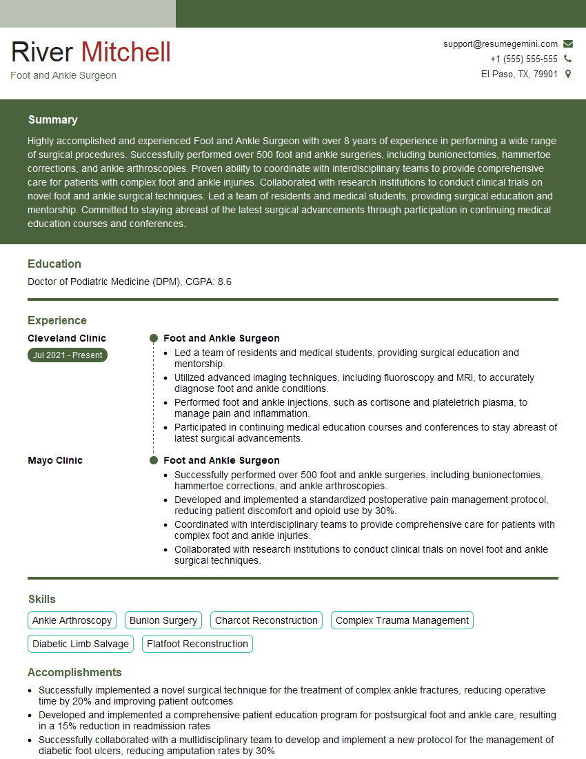
Mastering Flatfoot Reconstruction significantly enhances your career prospects in orthopedics and podiatry, opening doors to specialized practices and advanced roles. A strong resume is crucial for showcasing your expertise and securing your desired position. Creating an ATS-friendly resume is key to getting your application noticed. We highly recommend using ResumeGemini to build a professional and effective resume that highlights your skills and experience in Flatfoot Reconstruction. ResumeGemini provides examples of resumes tailored to this specific field, giving you a head start in crafting a compelling application.
Explore more articles
Users Rating of Our Blogs
Share Your Experience
We value your feedback! Please rate our content and share your thoughts (optional).
What Readers Say About Our Blog
This was kind of a unique content I found around the specialized skills. Very helpful questions and good detailed answers.
Very Helpful blog, thank you Interviewgemini team.
