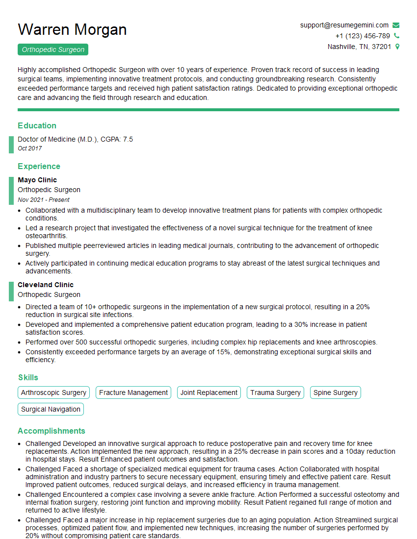
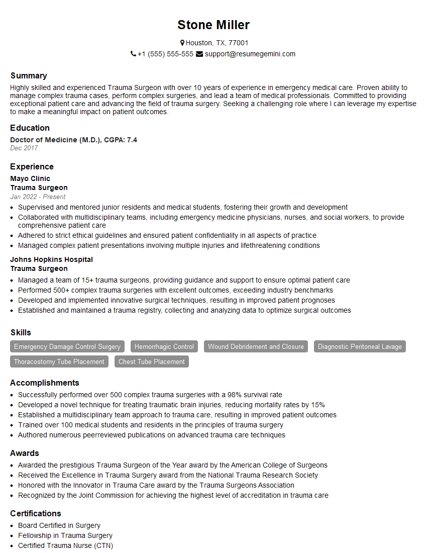
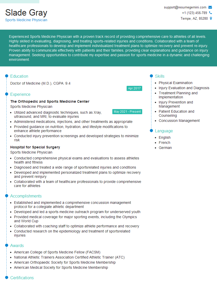
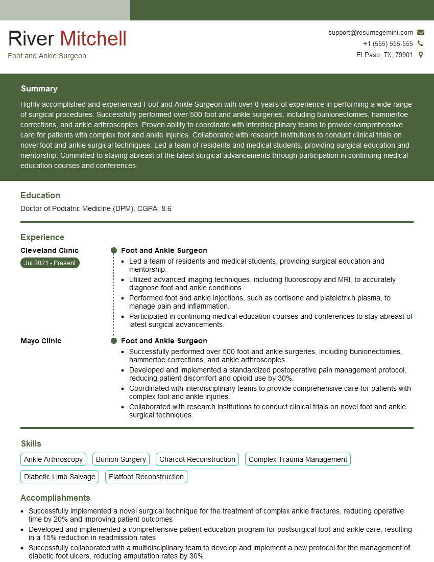
Cracking a skill-specific interview, like one for Foot and Ankle Fracture Repair, requires understanding the nuances of the role. In this blog, we present the questions you’re most likely to encounter, along with insights into how to answer them effectively. Let’s ensure you’re ready to make a strong impression.
Questions Asked in Foot and Ankle Fracture Repair Interview
Q 1. Describe the different classifications of ankle fractures.
Ankle fractures are complex, often involving multiple bones. We classify them based on the bones involved and the type of fracture. The most common classification system is the Lauge-Hansen classification, which describes the fracture based on the mechanism of injury and the resulting fracture pattern. It considers the initial direction of force (supination-adduction, supination-external rotation, pronation-abduction, pronation-external rotation) and the subsequent stage of injury. For example, a supination-adduction injury might result in a fracture of the medial malleolus, followed by a posterior malleolus fracture, potentially with a fibula fracture as well. Other classifications exist, such as the Weber classification, which focuses on the fibular fracture location relative to the syndesmosis (the strong ligamentous connection between the tibia and fibula). A Weber A fracture is below the syndesmosis, a Weber B fracture is at the syndesmosis, and a Weber C fracture is above the syndesmosis. These classifications guide treatment decisions based on the fracture’s severity and stability.
- Lauge-Hansen: Describes fracture patterns based on the mechanism of injury.
- Weber: Classifies fibular fractures based on their location relative to the syndesmosis.
Q 2. Explain the principles of open reduction and internal fixation (ORIF) for a tibial plafond fracture.
Open reduction and internal fixation (ORIF) for a tibial plafond fracture, a fracture of the distal tibia involving the articular surface, is a technically demanding procedure. The principles focus on achieving accurate articular reduction to restore joint congruity and stable fixation to allow early weight-bearing and prevent malunion or non-union. The process usually begins with meticulous open reduction under fluoroscopic guidance. This ensures perfect alignment of the articular fragments. Once the reduction is satisfactory, internal fixation is performed using plates and screws or potentially other techniques like intramedullary nails, depending on the fracture pattern and surgeon preference. The goal is to achieve rigid fixation that prevents motion at the fracture site, allowing for bone healing.
For example, a complex, comminuted (fragmented) tibial plafond fracture might require multiple plates and screws to achieve stable fixation, while a less complex fracture might be managed with a single plate. Post-operatively, careful monitoring for complications such as infection, malunion, or nonunion is crucial. The patient will typically require a period of non-weight-bearing followed by gradual weight-bearing as bone healing progresses.
Q 3. What are the indications for surgical versus non-surgical management of a Lisfranc fracture?
A Lisfranc fracture-dislocation involves the disruption of the tarsometatarsal joints. The decision to treat surgically or non-surgically depends primarily on the degree of displacement and instability. Non-surgical management, usually involving casting and non-weight bearing, is considered for minimally displaced fractures that are deemed stable on imaging. However, most Lisfranc injuries require surgical intervention. Surgical intervention (ORIF) is indicated for significant displacement, instability (as assessed clinically and radiographically), or articular incongruity. The reason for surgical intervention is to restore the normal alignment of the tarsometatarsal joints and maintain stability to prevent long-term problems like arthritis and instability. Unstable Lisfranc injuries, if not treated surgically, are prone to poor functional outcomes. Careful assessment through physical exam and imaging are critical to selecting the best management approach.
Q 4. How do you assess the stability of a fracture using imaging?
Assessing fracture stability relies heavily on imaging, primarily X-rays and computed tomography (CT) scans. X-rays provide a two-dimensional view and are essential for initial assessment. We look for the alignment of the bone fragments, the presence of displacement, and the integrity of the surrounding joint surfaces. However, X-rays alone may not adequately reveal the complexity of a fracture, especially in situations with comminution (bone fragments) or subtle displacement. CT scans offer superior visualization of bone fragments in three dimensions. This allows precise evaluation of the fracture pattern and its relationship to surrounding structures. Additional imaging modalities such as MRI can be used to assess soft-tissue structures like ligaments and tendons, critical for determining overall stability. Ultimately, determining the stability of a fracture involves a combination of assessing the fracture pattern on imaging and the patient’s physical exam findings.
For example, a displaced fracture with significant angulation or shortening is inherently unstable, while a minimally displaced fracture with intact supporting ligaments may be more stable.
Q 5. Describe the common complications associated with a calcaneal fracture.
Calcaneal fractures, fractures of the heel bone, are notoriously associated with significant complications. Common complications include:
- Subtalar arthritis: Damage to the subtalar joint surfaces often leads to post-traumatic osteoarthritis.
- Persistent pain: Many patients experience chronic pain despite successful fracture healing.
- Flattening of the heel: Loss of the normal heel architecture can result in gait abnormalities and further pain.
- Nerve injury: The surrounding nerves can be injured during the trauma or surgery.
- Infection: As with any open fracture or surgery, there is a risk of infection.
- Malunion/Nonunion: Poor healing of the fracture can lead to malalignment or nonunion of the bone fragments.
The severity of complications depends on several factors, including the severity of the fracture, the adequacy of treatment, and patient-specific factors.
Q 6. What are the different types of fixation devices used in foot and ankle fractures?
A variety of fixation devices are used in foot and ankle fracture repair, chosen based on the specific fracture pattern and location. These include:
- Plates and screws: Commonly used for fractures requiring rigid fixation, providing excellent stability. Different types of plates are used (e.g., anatomical plates, reconstruction plates).
- Intramedullary nails: These nails are inserted into the medullary canal of the bone, providing stable fixation for certain long bone fractures. They are less commonly used in the foot and ankle compared to plates and screws.
- Screws alone: Suitable for smaller fractures that don’t require extensive fixation.
- K-wires (Kirschner wires): Small pins used for temporary fixation or to aid in reduction before definitive fixation.
- External fixators: Used for complex or unstable fractures, these devices provide external support and stabilization of the fractured bones.
The choice of fixation is a crucial decision tailored to the individual patient and the unique features of the injury. The surgeon considers factors such as bone quality, fracture pattern, soft tissue injury, and the patient’s overall health in making this decision.
Q 7. How do you manage compartment syndrome in a patient with a foot or ankle fracture?
Compartment syndrome is a serious condition characterized by increased pressure within a confined anatomical space (compartment) that compromises blood supply to the muscles and nerves. In the context of foot and ankle fractures, it’s a potential complication that needs prompt recognition and treatment. Early signs include severe pain out of proportion to the injury, pain with passive stretch of the involved muscles, swelling, paresthesia (numbness or tingling), pallor (pale skin), and decreased pulses. Diagnosis involves measuring compartment pressures; if pressure exceeds a critical threshold, fasciotomy is often indicated.
Management of compartment syndrome involves immediate action. This includes:
- Elevation of the limb: To reduce swelling.
- Removing tight dressings or casts: To alleviate external compression.
- Measuring compartment pressures: Using a pressure gauge to determine the severity.
- Urgent fasciotomy: A surgical procedure to release the constricting fascia (tissue surrounding the muscle compartments) and relieve the pressure, restoring blood flow.
Delay in diagnosing and treating compartment syndrome can lead to permanent muscle damage, nerve damage, and limb loss. Hence, heightened awareness and prompt action are critical.
Q 8. Explain the principles of fracture healing.
Fracture healing is a complex process involving several stages. Think of it like building a house: you need a solid foundation, skilled labor, and the right materials. The initial stage is the inflammatory phase, where blood clots form at the fracture site, initiating the healing cascade. This is akin to laying the groundwork for your house. Next comes the reparative phase, characterized by the formation of a callus, a soft cartilage-like tissue that bridges the fracture gap. This is like building the frame of the house. Finally, in the remodeling phase, the callus gradually transforms into mature bone, strengthening and aligning the fracture. This is the finishing and strengthening phase. Factors affecting healing include the type of fracture, patient age, overall health, and proper alignment of the fracture fragments. A properly reduced and stabilized fracture heals faster and more reliably.
Q 9. Discuss the role of post-operative rehabilitation in foot and ankle fracture recovery.
Post-operative rehabilitation is crucial for optimal recovery after a foot and ankle fracture. It’s not just about physical therapy; it’s about regaining function and preventing long-term complications. Imagine a perfectly built house that isn’t properly furnished or maintained; it won’t function efficiently. The rehabilitation program is tailored to the specific fracture and the patient’s needs. It generally starts with controlled range-of-motion exercises to prevent stiffness, followed by strengthening exercises to rebuild muscle strength. Weight-bearing is gradually introduced as the fracture heals. Physical therapy plays a vital role, guiding patients through the process, providing support, and monitoring progress. Early mobilization, under guidance, is key to minimize stiffness and promotes blood flow to accelerate healing. Furthermore, patient education about proper footwear, activity modifications, and recognizing signs of complications is essential.
Q 10. How do you differentiate between a stress fracture and a fatigue fracture?
While both stress fractures and fatigue fractures involve bone breakage due to repetitive stress, there’s a key distinction. A stress fracture is a tiny crack in the bone caused by a single instance of high-impact trauma that surpasses the bone’s capacity. Imagine a sudden impact on already weakened bone. A fatigue fracture, on the other hand, results from repetitive low-impact stress over time, gradually weakening the bone until it fractures. Think of it like constantly bending a paperclip until it snaps. The key difference lies in the magnitude and duration of the stress applied. Diagnosing them involves a thorough history and physical exam, often aided by imaging studies like X-rays, bone scans, or MRI scans. Sometimes, stress fractures are initially subtle on X-rays and may require further imaging.
Q 11. Describe your experience with different surgical approaches to foot and ankle fractures.
My experience encompasses a wide array of surgical techniques for foot and ankle fractures. Open reduction and internal fixation (ORIF) is frequently employed, especially for displaced fractures. This involves surgically exposing the fracture, aligning the bone fragments, and securing them with plates, screws, or other internal implants. For example, a displaced ankle fracture often requires ORIF to restore the joint’s anatomy and ensure proper weight-bearing. In cases where less invasive approaches are appropriate, I utilize minimally invasive techniques, which often involve smaller incisions and less soft tissue damage. Arthroscopy can sometimes be used in specific ankle fractures to improve visualization and minimize tissue disruption. Finally, depending on the fracture pattern and patient factors, I may also employ external fixation, which uses pins and rods outside the skin to stabilize the fracture, providing an option for fractures that are challenging to fix internally or those with significant soft tissue damage.
Q 12. What are the indications for using external fixation in foot and ankle fractures?
External fixation is indicated in several scenarios involving foot and ankle fractures. It’s particularly useful when the fracture is severely comminuted (shattered into many pieces) or when significant soft tissue injury complicates internal fixation. Furthermore, it’s often preferred in open fractures where infection risk is high, as it allows for easy wound care and observation. In situations of severe swelling or compromised blood supply, external fixation offers the advantages of less invasive surgical intervention and allows for immediate management of soft tissue injuries. For fractures that require lengthening or distraction osteogenesis, external fixation provides the necessary adjustability. Essentially, it offers stability while minimizing risk to soft tissues in challenging situations.
Q 13. How do you manage a patient with a non-union fracture?
Managing a non-union fracture—where the bone fails to heal—requires a multi-faceted approach. First, the underlying cause needs to be addressed. This could involve surgical debridement (removal of infected or non-viable tissue), bone grafting (adding new bone to stimulate healing), or electrical stimulation. Sometimes, revision surgery may be necessary to improve fracture alignment or stability. In addition to surgical intervention, a well-structured rehabilitation program focused on restoring range of motion and strength is essential. Close monitoring and follow-up are crucial to ensure successful healing, including the use of imaging techniques to track progress and identify any complications. Sometimes, a combination of these approaches is necessary for optimal outcome. The goal is to restore anatomical alignment and functional capacity, and this may take time and patience.
Q 14. What are the common causes of delayed union?
Delayed union, where healing is slower than expected, can stem from several factors. Inadequate fracture reduction or stabilization is a common culprit; poorly aligned fragments hinder the healing process. Infection significantly impedes healing, as it creates an inflammatory environment hostile to bone formation. Insufficient blood supply to the fracture site can also delay union, as bone healing requires adequate nutrition and oxygen. Furthermore, systemic factors such as smoking, diabetes, malnutrition, and certain medications can negatively impact bone healing. Finally, patient compliance with post-operative instructions also plays a crucial role, ensuring proper immobilization and adherence to the prescribed rehabilitation program is essential.
Q 15. Describe your experience with post-operative complications, such as infection and malunion.
Post-operative complications following foot and ankle fracture repair are a significant concern. Infection, manifested as localized pain, swelling, redness, and potentially purulent drainage, is a major threat. We meticulously follow sterile surgical techniques and administer prophylactic antibiotics to minimize this risk. However, even with these precautions, infections can still occur, sometimes requiring surgical debridement (removal of infected tissue) and prolonged antibiotic therapy. Malunion, where the fracture heals in an unsatisfactory position, leading to pain, deformity, and functional limitations, is another challenge. This can arise from inadequate fracture reduction (alignment), insufficient immobilization, or patient non-compliance with post-operative instructions. I carefully assess the fracture during surgery and utilize appropriate fixation techniques – like plates and screws – to ensure optimal alignment. Post-operative imaging and careful clinical follow-up are crucial to detect and manage both infection and malunion promptly.
For example, I recently managed a patient who developed a deep infection after an open reduction internal fixation (ORIF) of a talus fracture. Immediate surgical debridement, intravenous antibiotics, and wound care were necessary to resolve the infection, followed by a prolonged course of oral antibiotics. In another case, a patient with a calcaneal fracture developed a malunion due to early weight-bearing against medical advice. We addressed this with corrective osteotomy (surgical bone cutting and realignment) and further immobilization.
Career Expert Tips:
- Ace those interviews! Prepare effectively by reviewing the Top 50 Most Common Interview Questions on ResumeGemini.
- Navigate your job search with confidence! Explore a wide range of Career Tips on ResumeGemini. Learn about common challenges and recommendations to overcome them.
- Craft the perfect resume! Master the Art of Resume Writing with ResumeGemini’s guide. Showcase your unique qualifications and achievements effectively.
- Don’t miss out on holiday savings! Build your dream resume with ResumeGemini’s ATS optimized templates.
Q 16. Explain your approach to pain management in a patient with a foot and ankle fracture.
Pain management is a crucial aspect of care following a foot and ankle fracture. My approach is multimodal and individualized, taking into account the patient’s pain tolerance, fracture type and severity, and any comorbidities. I typically start with a combination of analgesics such as nonsteroidal anti-inflammatory drugs (NSAIDs) like ibuprofen or naproxen, and acetaminophen for mild to moderate pain. For more severe pain, I might prescribe opioids, but I use them cautiously and strategically, focusing on minimizing the duration and potential for dependence. Regional nerve blocks, particularly ankle blocks, can provide excellent pain relief in the early post-operative period, reducing the need for systemic analgesics. I also emphasize non-pharmacological approaches like elevation, ice application, and physical therapy. Regular assessment of pain levels and adjustments to the pain management plan are essential to ensure the patient’s comfort and facilitate optimal healing.
For instance, a patient with a displaced ankle fracture might benefit from an ankle block immediately post-operatively, followed by a transition to oral NSAIDs and acetaminophen. Careful monitoring for opioid-related side effects is critical when these are prescribed. Regular physiotherapy will help maintain range of motion and reduce stiffness.
Q 17. What imaging modalities do you use to evaluate foot and ankle fractures?
Imaging plays a vital role in evaluating foot and ankle fractures. I routinely utilize several modalities:
- Plain radiographs (X-rays): These are the cornerstone of our initial assessment, providing excellent visualization of bone alignment, fracture lines, and the presence of any associated dislocations. We typically obtain anteroposterior (AP), lateral, and mortise views of the ankle, as well as oblique views as needed. For foot fractures, we adapt the views to best visualize the specific bone involved.
- Computed tomography (CT) scans: CT scans offer superior detail of bone anatomy, particularly in complex fractures involving multiple fragments or articular surfaces (joint surfaces). They are invaluable for pre-operative planning in ORIF cases.
- Magnetic resonance imaging (MRI): MRI excels in assessing soft tissue structures, such as ligaments, tendons, and cartilage. This is particularly helpful in evaluating associated injuries, such as ligament sprains or tears, which frequently accompany fractures.
The choice of modality depends on the clinical suspicion and the complexity of the fracture. For example, a simple, undisplaced fracture might only require X-rays, while a complex, comminuted (shattered) fracture would necessitate a CT scan for accurate assessment.
Q 18. How do you interpret radiographic findings of a foot and ankle fracture?
Interpreting radiographic findings of foot and ankle fractures requires a systematic approach. I start by assessing the alignment of bones, looking for displacement, angulation, and shortening. The fracture line itself is carefully analyzed to determine the type of fracture (e.g., transverse, oblique, comminuted). The involvement of articular surfaces is crucial, as intra-articular fractures often have worse prognoses and may require more aggressive treatment. I look for signs of associated injuries, such as joint dislocations or avulsion fractures (bone fragments pulled away by ligaments or tendons). Finally, I evaluate the quality of bone healing, assessing callus formation in the later stages of healing. This systematic review helps determine the appropriate treatment strategy.
For example, a displaced lateral malleolar fracture with significant displacement and widening of the mortise (ankle joint) on X-ray indicates a need for surgical intervention. A minimally displaced fracture of the fifth metatarsal, however, may be treated non-operatively with immobilization. The entire image analysis must be considered to provide an accurate diagnosis.
Q 19. What are the indications for bone grafting in foot and ankle fractures?
Bone grafting in foot and ankle fractures is indicated when there is a significant defect in bone or when there is insufficient bone stock for stable fixation. This situation commonly arises in comminuted fractures, segmental fractures (bone broken into multiple pieces), or fractures with substantial bone loss due to trauma or infection. The goal of bone grafting is to promote bony union, preventing non-union (failure of the fracture to heal) or malunion. Large bone defects or fractures that have already failed to heal (nonunion) often benefit from bone grafting in combination with other surgical techniques to restore the anatomy and stability of the foot and ankle.
For example, a patient with a severely comminuted calcaneal fracture with significant displacement and bone loss would likely require bone grafting along with internal fixation to achieve optimal healing and restoration of the hindfoot. Similarly, non-unions, as sometimes happen in navicular fractures, may be best addressed with bone grafting and possible fixation.
Q 20. Describe your experience with different types of bone grafts.
Various types of bone grafts are utilized, each with its own advantages and disadvantages:
- Autografts: These are grafts harvested from the patient’s own body, typically from the iliac crest (hip bone). Autografts have the advantage of being osteoinductive (stimulating bone formation) and osteoconductive (providing a scaffold for bone growth), minimizing the risk of rejection or disease transmission. However, harvesting autografts is an additional surgical procedure with its own associated morbidity (risks and complications).
- Allografts: These are grafts harvested from a deceased donor. They can be obtained readily and reduce the need for a secondary surgical site. However, there is a small risk of disease transmission, and they may not be as osteoinductive as autografts.
- Synthetic bone grafts: These are composed of various materials designed to mimic the properties of bone. They are readily available and easy to use but may not be as effective in promoting bone formation as autografts.
The choice of bone graft depends on several factors, including the size and location of the bone defect, the patient’s overall health, and the availability of resources. For example, a large defect in a tibial plafond fracture may require an iliac crest autograft, while a smaller defect in a metatarsal fracture might be addressed with a synthetic bone substitute.
Q 21. What are the different types of screws and plates used in ORIF?
Open reduction internal fixation (ORIF) utilizes various screws and plates to achieve stable fracture fixation. The choice depends on fracture type, location, and bone quality:
- Screws: Several types are available, including cancellous screws (for use in spongy bone), cortical screws (for use in dense bone), and cannulated screws (allowing the passage of a guide wire). Locking screws provide additional stability and are often preferred for osteoporotic bone or complex fractures.
- Plates: Various plate designs are used, including dynamic compression plates (DCP), reconstruction plates, and locking compression plates (LCP). LCPs are particularly useful in comminuted fractures and osteoporotic bone. Plate size and shape are chosen to match the anatomy of the bone being fixed.
The specific screw and plate combination is carefully selected during surgical planning to provide adequate stability and minimize the risk of complications. For example, a minimally displaced fracture of the fibula might be treated with a single screw, while a severely comminuted fracture of the tibia would require a larger plate and multiple screws for stable fixation. Bioabsorbable screws and plates are also available and are increasingly used to reduce the need for secondary removal surgeries.
Q 22. How do you select the appropriate fixation device for a specific fracture?
Selecting the right fixation device for a foot and ankle fracture is crucial for optimal healing and functional outcome. It’s a multi-factorial decision, depending on several key factors. Think of it like choosing the right tool for a job – a screwdriver for screws, a hammer for nails. In fractures, we consider the fracture pattern, bone quality, patient factors, and the desired stability.
- Fracture Pattern: A simple, stable fracture might only need a cast or a short-leg splint. However, a complex, unstable fracture – for example, a comminuted fracture (broken into multiple pieces) of the talus (ankle bone) – may require internal fixation with plates and screws for optimal alignment and stability.
- Bone Quality: Osteoporotic bone (brittle bone disease) is more prone to screw pullout. In such cases, we might choose larger screws, different screw types (like cancellous screws), or even consider alternative fixation methods like external fixation.
- Patient Factors: Age, activity level, overall health, and comorbidities (like diabetes) all influence the choice. A young, active patient might benefit from a more robust fixation allowing for early weight-bearing, while an elderly patient with comorbidities might need a less invasive approach to minimize risks.
- Desired Stability: The level of stability required depends on the fracture location and the forces acting on it. Some fractures require rigid fixation to allow immediate weight-bearing, while others allow for less rigid fixation with gradual weight-bearing.
For instance, a simple, undisplaced fracture of the fifth metatarsal (small toe bone) might be treated with a short-leg cast, while a displaced intra-articular fracture (a fracture involving a joint) of the tibia requires rigid internal fixation with plates and screws for anatomical reduction and stable fixation.
Q 23. Describe your experience with arthroscopy in foot and ankle surgery.
Arthroscopy plays a significant role in foot and ankle surgery, particularly in addressing articular (joint) injuries and assessing the extent of damage. It’s minimally invasive, allowing for a smaller incision and quicker recovery. I’ve extensively used arthroscopy for diagnosing and treating conditions like ankle impingement, osteochondral lesions (cartilage damage), and ligament tears.
For example, in a patient presenting with chronic ankle pain and limited range of motion, arthroscopy allows for a direct visualization of the joint, identification of any loose bodies, and treatment of cartilage defects through debridement or microfracture techniques. The minimally invasive nature significantly reduces post-operative pain and swelling, leading to faster rehabilitation compared to open surgery.
Moreover, I utilize arthroscopy in conjunction with other surgical techniques like open reduction and internal fixation (ORIF) for complex fractures involving the ankle joint. Arthroscopy helps ensure accurate reduction (realignment) of the fracture fragments and assess the integrity of surrounding ligaments and cartilage.
Q 24. What are the advantages and disadvantages of minimally invasive surgery for foot and ankle fractures?
Minimally invasive surgery (MIS) for foot and ankle fractures offers several advantages, but also has its limitations. Think of it as a trade-off between precision and invasiveness.
- Advantages: Smaller incisions lead to less pain, reduced scarring, faster healing, and quicker return to function. There’s also a lower risk of infection.
- Disadvantages: MIS might not be suitable for all fractures, especially complex ones requiring extensive manipulation or bone grafting. It can be technically more challenging, requiring specialized skills and equipment, and the visualization can be limited compared to open surgery.
For example, a minimally invasive approach might be ideal for a simple, undisplaced fracture of the fibula (outer ankle bone), while a severely comminuted fracture of the calcaneus (heel bone) may necessitate an open approach for accurate fragment reduction and stabilization.
The decision to use MIS versus open surgery is highly individualized and depends on the specific fracture characteristics, patient factors, and surgeon’s experience and expertise.
Q 25. How do you manage a patient with a complex fracture requiring multiple procedures?
Managing a patient with a complex fracture requiring multiple procedures necessitates a meticulous and phased approach. It’s like building a house – you lay the foundation first, then the walls, and finally the roof. We prioritize stabilization, infection prevention, and functional restoration.
The initial procedure usually focuses on stabilizing the most unstable fracture fragments. Subsequent procedures may address bone grafting, soft tissue reconstruction, or further fixation enhancement. Careful planning and timing are crucial to minimize the patient’s overall burden and maximize the chances of a successful outcome. Close monitoring for complications, such as infection or non-union (failure of the bones to heal), is paramount.
For example, a severely comminuted pilon fracture (fracture of the distal tibia involving the ankle joint) might require initial ORIF to stabilize the main fracture fragments. A later procedure might be needed for bone grafting to address any significant bone loss and achieve solid union. We would closely monitor the patient for infection, ensuring appropriate antibiotic prophylaxis and wound care.
Q 26. Explain your experience with managing patients with diabetes and foot and ankle fractures.
Managing patients with diabetes and foot and ankle fractures presents unique challenges due to their increased risk of infection, delayed healing, and peripheral neuropathy. Think of it as navigating a minefield – each step requires extra caution.
Pre-operative optimization of blood glucose control is crucial. We often collaborate with endocrinologists to ensure the patient’s glucose levels are within the optimal range. Prophylactic antibiotics are given to reduce the infection risk. Meticulous wound care and aggressive infection management are essential. In some cases, we might use advanced wound care techniques like negative pressure wound therapy.
Post-operatively, we closely monitor for signs of infection, delayed healing, and any neurovascular compromise. Regular follow-up appointments, meticulous wound care, and appropriate pain management are critical to achieving a favorable outcome. Patient education regarding foot care and risk factor management is also emphasized to minimize future complications.
Q 27. Describe your experience with managing patients with osteoporosis and foot and ankle fractures.
Patients with osteoporosis pose a significant challenge in foot and ankle fracture management. Their bones are fragile and prone to complications. It’s like working with delicate china – we need extra care to prevent further damage.
Our approach focuses on minimizing surgical trauma and using fixation methods that are less likely to cause further bone weakening. We might opt for less invasive techniques, smaller implants, or even consider using bone grafting material to enhance bone strength. Post-operative rehabilitation is carefully tailored to prevent further fractures and promote bone healing.
In addition to surgical management, we emphasize osteoporosis management, often recommending medication to improve bone mineral density. Patient education regarding fall prevention and diet is also emphasized to improve overall bone health.
Q 28. How do you counsel patients about the potential risks and complications of foot and ankle fracture surgery?
Counseling patients about the potential risks and complications of foot and ankle fracture surgery is a critical aspect of shared decision-making. It’s about equipping them with the knowledge they need to make an informed choice. This involves a transparent discussion encompassing both the benefits and potential drawbacks.
I explain the surgical procedure in detail, using clear and simple language, avoiding overly technical jargon. I discuss the potential complications, including infection, non-union, malunion (healing in a non-anatomical position), nerve injury, hardware failure, and chronic pain. I also outline the rehabilitation process and expected recovery time.
I emphasize the importance of realistic expectations and address any patient concerns or anxieties. The goal is to empower patients to participate actively in their treatment plan and understand what to expect before, during, and after surgery. It’s a collaborative partnership, and open communication is key.
Key Topics to Learn for Foot and Ankle Fracture Repair Interview
- Anatomy and Biomechanics: Detailed understanding of foot and ankle anatomy, including ligaments, tendons, and bone structures; analysis of biomechanical forces and their impact on fracture patterns.
- Fracture Classification Systems: Mastery of common classification systems (e.g., Lauge-Hansen, Weber) and their implications for treatment planning and prognosis.
- Imaging Interpretation: Proficient interpretation of radiographs, CT scans, and MRI scans to accurately diagnose fracture type, location, and displacement.
- Non-operative Management: Understanding of indications, techniques, and limitations of conservative treatment options, including casting, splinting, and bracing.
- Operative Techniques: Knowledge of various surgical approaches, including open reduction and internal fixation (ORIF), external fixation, and arthroscopy, for different fracture types.
- Implant Selection: Ability to choose appropriate implants based on fracture pattern, patient factors, and surgical approach.
- Post-operative Care and Rehabilitation: Understanding of post-operative management, including pain control, wound care, early mobilization protocols, and physical therapy regimens.
- Complications and Management: Knowledge of potential complications (e.g., infection, nonunion, malunion) and strategies for their prevention and management.
- Current Research and Trends: Awareness of advancements in surgical techniques, implant technology, and rehabilitation protocols.
- Problem-solving and Decision-Making: Ability to analyze complex cases, formulate appropriate treatment plans, and adapt to unforeseen circumstances during surgery or post-operative care.
Next Steps
Mastering Foot and Ankle Fracture Repair is crucial for career advancement in orthopedic surgery and related specialties. A strong foundation in these key areas will significantly enhance your interview performance and overall competitiveness in the job market. To maximize your job prospects, create an ATS-friendly resume that effectively showcases your skills and experience. ResumeGemini is a trusted resource that can help you build a professional and impactful resume tailored to your specific needs. Examples of resumes tailored to Foot and Ankle Fracture Repair are available to guide you through the process. Invest the time to craft a compelling resume—it’s your first impression on potential employers.
Explore more articles
Users Rating of Our Blogs
Share Your Experience
We value your feedback! Please rate our content and share your thoughts (optional).
What Readers Say About Our Blog
This was kind of a unique content I found around the specialized skills. Very helpful questions and good detailed answers.
Very Helpful blog, thank you Interviewgemini team.
