Preparation is the key to success in any interview. In this post, we’ll explore crucial Soft Tissue Reconstruction interview questions and equip you with strategies to craft impactful answers. Whether you’re a beginner or a pro, these tips will elevate your preparation.
Questions Asked in Soft Tissue Reconstruction Interview
Q 1. Describe the different types of skin grafts used in soft tissue reconstruction.
Skin grafts are crucial in soft tissue reconstruction, providing a cover for exposed tissue or filling defects. They are classified primarily by the depth of skin harvested:
- Split-thickness skin grafts (STSGs): These grafts include only the epidermis and a portion of the dermis. They are thinner, easier to harvest in larger quantities, and generally have a higher take rate. However, they lack the full thickness of the dermis, resulting in a less aesthetically pleasing outcome and potential for contracture. Think of it like peeling a thin layer of an apple; you’re taking a partial layer of skin.
- Full-thickness skin grafts (FTSGs): These include the entire epidermis and dermis. They provide a more aesthetically pleasing outcome, less contracture, and better texture and color match. However, they are more difficult to harvest, require a larger donor site, and have a lower take rate. This is akin to taking a complete “slice” of the apple; the donor site is more substantial.
- Mesh grafts: STSGs can be meshed, creating a larger surface area from the same amount of donor tissue. This allows for coverage of larger defects but results in a more porous appearance. Think of using a mesh to cover a hole—it expands the coverage but has a noticeable texture.
The choice between STSG and FTSG depends on the size and location of the defect, the quality of the recipient bed, and the cosmetic requirements.
Q 2. Explain the principles of microsurgical anastomosis.
Microsurgical anastomosis is the surgical connection of extremely small blood vessels (arteries and veins) under a surgical microscope. It’s the foundation of free flap surgery. The principles revolve around precision, meticulous technique, and attention to detail:
- Vessel identification and preparation: Accurate identification of the artery and vein is paramount. The vessels are meticulously dissected, cleaned of surrounding tissues, and prepared for anastomosis.
- Approximation: The ends of the vessels are carefully approximated (brought together) ensuring precise alignment of the lumen (inner space) and proper blood flow.
- Suture technique: Microsurgical sutures are incredibly fine, usually 8-0 to 11-0 nylon or polypropylene. These require specialized instruments and advanced skills. A continuous suture technique is often employed for arteries, while interrupted sutures are more typical for veins.
- Monitoring: Post-anastomosis, the flap is meticulously monitored for signs of patency (blood flow). This involves close observation, Doppler ultrasound, and sometimes intraoperative angiography.
Think of it like connecting two tiny garden hoses – incredibly precise work to ensure continuous flow.
Q 3. What are the common complications associated with free flap surgery?
Free flap surgery, while highly effective, carries inherent risks. Common complications include:
- Flap necrosis: This is the death of the flap tissue due to compromised blood supply. This can be partial or complete, requiring further surgery.
- Infection: Infection at the donor or recipient site is a significant concern. Prophylactic antibiotics and strict sterile technique are crucial.
- Hematoma/Seromas: Accumulation of blood (hematoma) or serous fluid (seroma) can compromise flap viability and increase the risk of infection.
- Lymphedema: Swelling in the limb due to lymphatic obstruction can occur, particularly with free flaps from the leg.
- Sensory deficits: Nerve injury during flap harvest or transfer can lead to altered sensation in the flap.
- Venous congestion: Insufficient venous drainage can lead to flap failure.
The incidence of these complications is reduced with meticulous surgical technique, appropriate patient selection, and meticulous postoperative care. For instance, careful monitoring of flap perfusion in the immediate post-operative period is paramount in preventing necrosis.
Q 4. How do you select the appropriate flap for a specific soft tissue defect?
Flap selection is a crucial decision in soft tissue reconstruction. It involves a multifactorial analysis, considering:
- Defect characteristics: Size, depth, location, and the surrounding tissues are analyzed.
- Patient factors: Overall health, comorbidities, and available donor sites influence flap choice.
- Functional requirements: The flap should provide not only aesthetic improvement but also restore function, such as sensation or movement.
- Flap availability: Location and suitability of potential donor sites are considered.
- Surgeon expertise: Certain flaps require specialized microsurgical skills.
For instance, a small defect on the face may be suitable for a local flap, while a large, complex defect in the head and neck might require a free radial forearm flap. The decision-making process often involves a team approach with the plastic surgeon, referring physician, and potentially a multidisciplinary team.
Q 5. Discuss the advantages and disadvantages of local, regional, and free flaps.
The choice between local, regional, and free flaps depends on the defect’s characteristics and the patient’s condition:
- Local flaps: These are harvested from the immediate vicinity of the defect. They are relatively simple to perform, with minimal donor-site morbidity and a high success rate. However, they may lack sufficient tissue bulk or length to cover large defects. Think of it like patching a hole with material from the nearby area.
- Regional flaps: These are based on larger vessels extending beyond the immediate area of the defect. They can cover larger defects and provide better tissue bulk than local flaps, but donor site morbidity can be slightly increased. This is like patching with material from the same room but further away.
- Free flaps: These are completely detached from their original blood supply and revascularized microsurgically. They can cover extensive defects, offering large volume and versatile tissue properties. However, they are complex procedures demanding specialized expertise and carry a higher risk of complications. This would be like bringing in material from an entirely separate building.
The decision involves weighing the advantages and disadvantages based on the specific surgical scenario.
Q 6. Describe your experience with different flap designs (e.g., random pattern, axial pattern).
My experience encompasses a wide range of flap designs. Understanding the vascular supply of the flap is crucial.
- Random pattern flaps: These rely on the diffuse subcutaneous blood supply and are suitable for smaller defects. Their reliability is limited by their size and distance from the blood supply. Examples include advancement flaps and rotation flaps often used for smaller facial defects.
- Axial pattern flaps: These are based on a named artery supplying the flap, which is a reliable blood supply. This allows for larger and more versatile flaps. Examples include the radial forearm flap or the latissimus dorsi flap which are often used for head and neck or lower extremity reconstruction.
I have extensive experience with both, tailoring the flap design to the specific anatomical location and defect characteristics. For instance, I’ve used a latissimus dorsi free flap for reconstructing a complex head and neck defect following tumor resection, and a local advancement flap for a smaller defect on the face.
Q 7. How do you manage complications such as flap necrosis or infection?
Managing complications like flap necrosis or infection requires a prompt and decisive approach.
- Flap necrosis: Early detection is crucial. Clinical signs include pallor, coolness, and loss of capillary refill in the flap. Immediate surgical exploration is usually necessary to debride non-viable tissue, improve blood flow, or consider a secondary reconstructive procedure. This could include revising the anastomosis, administering hyperbaric oxygen therapy, or even performing a secondary flap.
- Infection: This is managed with prompt surgical debridement of infected tissue, intravenous antibiotics targeting the identified pathogen, and supportive care. In severe cases, the flap might require removal, and the wound would need to be cleaned before a secondary reconstructive option can be considered.
In both scenarios, thorough wound care, close monitoring, and appropriate adjunctive therapies are critical for successful management.
Q 8. Explain the importance of pre-operative planning in soft tissue reconstruction.
Pre-operative planning in soft tissue reconstruction is paramount for achieving optimal outcomes. It’s like meticulously designing a blueprint before building a house – crucial for structural integrity and aesthetics. This phase involves a comprehensive assessment of the defect, including its size, depth, location, and surrounding tissues. We carefully analyze the patient’s medical history, including any comorbidities that might impact healing. Imaging studies such as CT scans and MRIs help visualize the underlying anatomy and vascular supply. Based on this detailed analysis, we select the most appropriate reconstruction technique, choosing between local flaps, regional flaps, or free flaps depending on the defect’s characteristics and the patient’s overall health.
For example, a small, superficial defect might be amenable to a local advancement flap, while a large, deep defect requiring substantial tissue volume may necessitate a free flap transfer. This planning stage also includes determining the necessary surgical instruments and preparing the operating room to ensure a smooth and efficient procedure. Careful planning significantly reduces complications and improves the chances of successful reconstruction.
Q 9. Describe your approach to assessing the vascularity of a recipient site.
Assessing the recipient site’s vascularity is critical for flap survival. Think of it as checking the soil’s fertility before planting a sapling – you wouldn’t plant in barren land, right? We utilize a multi-modal approach. Firstly, we conduct a thorough clinical examination, assessing the skin color, temperature, and capillary refill time. A pale, cool, and poorly perfused site suggests compromised vascularity. We then employ Doppler ultrasound to evaluate the blood flow in the underlying arteries and veins. This non-invasive technique provides real-time visualization of blood vessels and their patency. In cases of uncertainty or complex anatomy, we might use angiography, a more invasive technique that involves injecting contrast dye into the vessels to visualize them radiographically. The findings from these assessments guide our decision-making process, influencing the choice of flap and surgical technique to ensure adequate perfusion.
Q 10. What are the key considerations for post-operative care in soft tissue reconstruction?
Post-operative care is just as important as the surgery itself – it’s the crucial nurturing phase after planting the sapling. It focuses on optimizing the healing environment and minimizing complications. This includes meticulous wound dressing to protect the flap from trauma and infection. We regularly monitor vital signs, including blood pressure, heart rate, and oxygen saturation, to detect any signs of distress. Pain management is crucial, often employing a multimodal analgesic approach to ensure patient comfort. Regular assessment of flap viability, as discussed later, is vital. Early identification and management of complications such as infection, hematoma, or seroma are critical for preventing long-term sequelae. Finally, close follow-up appointments with the patient are scheduled to monitor healing progress and address any concerns. The goal is to facilitate optimal healing and minimize any potential functional or aesthetic deficits.
Q 11. How do you monitor flap viability post-operatively?
Monitoring flap viability post-operatively is crucial to ensure its survival. It’s like carefully observing a plant’s growth after planting – you look for signs of health or stress. Clinical assessment involves careful observation of skin color, temperature, and capillary refill. A pale, cool, or dusky flap suggests compromised perfusion. We also assess for signs of infection, such as increased tenderness, swelling, erythema, or purulent discharge. Doppler ultrasound can be used to evaluate blood flow within the flap. In cases of concern, we may obtain laser Doppler flowmetry measurements to quantify blood flow objectively. If clinical signs or imaging suggest compromised viability, surgical intervention might be necessary, such as revising the flap or performing a venous decompression procedure. Early detection of complications is key to prompt intervention and improving the chances of flap survival.
Q 12. Describe your experience with different types of wound closure techniques.
My experience encompasses a wide range of wound closure techniques. The choice depends on the defect’s characteristics and the surrounding tissues. Simple interrupted sutures are used for smaller, less tensioned wounds. Continuous sutures are efficient for longer wounds, but can lead to increased tension if not placed correctly. Subcuticular sutures provide a cosmetically superior closure with less visible scarring. For larger defects or those with significant tension, we often employ advanced techniques like undermining, releasing incisions, or skin grafts. In cases of significant tissue loss, skin flaps are utilized, ranging from local advancement flaps to more complex free flaps. The decision-making process always considers factors like tension, wound healing potential, and the patient’s overall health. For instance, a patient with poor healing capacity might benefit from a split-thickness skin graft, while a patient with a large defect might require a free flap reconstruction.
Q 13. How do you address skin tension during wound closure?
Addressing skin tension during wound closure is critical to preventing dehiscence (wound opening) and ensuring optimal aesthetic outcomes. Imagine trying to sew together two pieces of fabric that are too far apart – it wouldn’t hold. Techniques include undermining the surrounding tissues to create a laxity of the skin, allowing for easier closure without tension. This involves carefully dissecting the subcutaneous tissue to loosen the skin edges. Release incisions, strategically placed incisions that reduce tension, might be necessary for extensive defects. Z-plasties, surgical maneuvers that redistribute tension, are another option. Finally, skin grafts can be used in situations where there’s simply not enough skin to close the wound directly. The approach depends on the location, size, and depth of the defect, along with patient-specific factors. The ultimate goal is to achieve a tension-free closure that promotes optimal healing and minimizes scarring.
Q 14. What are the indications and contraindications for using a particular flap?
The choice of flap depends on various factors, including the size, location, and depth of the defect, the quality of the recipient site, and the patient’s overall health. For example, a local flap, such as a rotation flap, is ideal for smaller defects where there is sufficient adjacent tissue that can be mobilized. However, it’s unsuitable for large defects or areas with compromised vascularity. A regional flap, such as a musculocutaneous flap, offers a more robust blood supply and is suitable for larger defects but may require more extensive surgery and have a longer recovery time. Free flaps, which are harvested from a distant site and microsurgically revascularized, are the workhorses for reconstructing complex defects or situations with poor local tissue quality. However, they require specialized microsurgical expertise and involve higher risk. Contraindications for certain flaps could include inadequate recipient site vascularity, significant infection, or patient comorbidities that might impair healing. The decision-making process requires careful consideration of all these factors to select the most appropriate and safe flap choice.
Q 15. Explain the role of imaging (e.g., Doppler ultrasound) in soft tissue reconstruction.
Imaging plays a crucial role in soft tissue reconstruction, guiding surgical planning and monitoring post-operative outcomes. Doppler ultrasound, in particular, is invaluable for assessing the vascularity of potential flap donor sites. It allows us to visualize blood flow in real-time, helping determine the suitability of a flap based on its perfusion. For instance, we use Doppler to evaluate the superficial temporal artery and vein for a temporoparietal fascial flap before harvesting it for reconstruction of facial defects. A robust signal indicates a healthy vascular supply, making it a suitable option. Conversely, a weak or absent signal suggests a compromised vessel, prompting consideration of alternative flap options. Other imaging techniques like CT angiography and MRI provide additional information on the underlying anatomy, including bone structures, nerve pathways and the extent of soft tissue loss, further enhancing the precision of our surgical planning.
Career Expert Tips:
- Ace those interviews! Prepare effectively by reviewing the Top 50 Most Common Interview Questions on ResumeGemini.
- Navigate your job search with confidence! Explore a wide range of Career Tips on ResumeGemini. Learn about common challenges and recommendations to overcome them.
- Craft the perfect resume! Master the Art of Resume Writing with ResumeGemini’s guide. Showcase your unique qualifications and achievements effectively.
- Don’t miss out on holiday savings! Build your dream resume with ResumeGemini’s ATS optimized templates.
Q 16. How do you manage a compromised flap?
Managing a compromised flap is a critical situation requiring immediate action. The first step is to carefully assess the extent of compromise – is it partial or complete? What is the cause? Ischemia (lack of blood flow) is the usual culprit, potentially due to kinking, compression, or thrombosis (blood clot) in the supplying vessels. Treatment depends on the cause and severity. For partial compromise, we may attempt to improve blood flow by releasing any tension on the pedicle (the stalk of the flap containing the blood vessels). This might involve repositioning the flap or releasing any tight sutures. We may also administer medications to improve perfusion, such as vasodilators. However, for complete compromise where the flap is visibly necrotic (dying), surgical intervention is crucial. This often involves exploration of the pedicle to identify and correct the cause of occlusion, such as removing a thrombus or releasing a kink. If salvage is impossible, the compromised tissue is excised, and alternative reconstruction techniques are planned. Continuous monitoring, using Doppler ultrasound for example, and vigilant observation of flap viability are essential aspects of managing potential compromise during the post-operative period.
Q 17. Describe your experience with different types of mesh grafts.
My experience encompasses a wide range of mesh grafts, each with specific applications and advantages. For example, polypropylene meshes are commonly used for hernia repair and abdominal wall reconstruction due to their strength and biocompatibility. However, they can sometimes cause significant scarring and inflammation. I’ve also utilized expanded polytetrafluoroethylene (ePTFE) meshes, which are porous and allow for tissue ingrowth, leading to better integration and reduced scarring. These are particularly useful in areas requiring more flexibility. In situations where biointegration is paramount, I might consider using absorbable meshes that are gradually replaced by host tissue over time. The choice of mesh depends heavily on the location and nature of the defect, as well as individual patient factors such as risk of infection and potential for complications. The decision involves a careful evaluation of the pros and cons of each mesh type, considering both short-term and long-term outcomes.
Q 18. What are the challenges in reconstructing complex defects involving bone, nerve, and vessels?
Reconstructing complex defects involving bone, nerve, and vessels presents significant challenges demanding a multidisciplinary approach. The key lies in meticulous pre-operative planning and careful execution. Imaging techniques such as CT scans and MRI play an essential role in defining the extent of the defect and understanding the spatial relationships between bone fragments, nerve branches, and vascular structures. Often, these reconstructions necessitate a staged approach, starting with bone stabilization, followed by nerve repair (if feasible), and finally soft tissue coverage. Free tissue transfer, where a tissue flap is transferred with its own blood supply, is frequently required. Microsurgical techniques are employed for precise re-anastomosis of vessels and nerves. Moreover, we carefully consider the potential for complications, including infection, flap failure, nerve dysfunction, and non-union of bone. Close post-operative monitoring and appropriate management of complications are vital for optimal outcomes. A recent case involving a severe traumatic facial injury required a composite graft involving bone, nerve, and skin, successfully restoring facial form and function, but only after meticulous planning and a collaborative effort between plastic surgeons, neurosurgeons, and orthopedic surgeons.
Q 19. How do you incorporate patient expectations into your surgical planning?
Incorporating patient expectations is paramount to achieving successful surgical outcomes and patient satisfaction. The first step is a thorough discussion during the initial consultation. We engage in open dialogue about the nature of the defect, realistic expectations regarding the reconstruction, potential risks and complications, and the limitations of the procedure. I use photos, anatomical drawings and 3D models to visualize the planned surgical outcome. It’s crucial to manage expectations realistically; emphasizing what can be achieved while also acknowledging potential limitations. This process involves active listening, addressing the patient’s concerns and addressing any misconceptions. For instance, in a patient expecting perfect restoration of a severely damaged limb, we need to clearly explain the functional improvements achievable while acknowledging that complete cosmetic restoration might not be possible. Regular follow-up consultations also play a key role in managing expectations and addressing any post-operative concerns.
Q 20. Describe your experience with using biomaterials in soft tissue reconstruction.
Biomaterials are increasingly playing a significant role in soft tissue reconstruction, offering innovative solutions to complex challenges. I have experience using various biomaterials, including biodegradable scaffolds that provide a temporary matrix for tissue regeneration. These scaffolds can help guide tissue growth and improve the integration of grafts. Furthermore, I have used bioactive glass in bone augmentation procedures, to promote bone formation in conjunction with bone grafts. Another example is the use of collagen-based matrices to enhance wound healing and improve the quality of the scar tissue. The choice of biomaterial is guided by the specific clinical scenario and the desired outcome. We carefully evaluate the biocompatibility, degradation rate, and mechanical properties of the biomaterial to ensure its suitability and to minimize potential adverse effects. The field of biomaterials is constantly evolving, and I am actively exploring new advancements in this area.
Q 21. What is your experience with different types of microsurgical instruments?
My experience with microsurgical instruments is extensive, given their essential role in many reconstructive procedures. I am proficient in using a wide array of instruments, including various sizes and types of micro-forceps, micro-scissors, and micro-vascular clamps. The choice of instrument depends on the specific task, for example, delicate nerve repair requires finer instruments compared to vascular anastomosis. We utilize specialized microscopes with high magnification and illumination to facilitate precise handling and visualization during microsurgery. Maintaining the sharpness and sterility of the instruments is paramount for minimizing trauma and preventing infection. Regular maintenance and proper handling of these delicate instruments are crucial for successful microsurgical procedures. Expertise in handling these instruments requires years of training and practice, but the skills involved are incredibly rewarding in the context of complex reconstructive surgery.
Q 22. How do you address donor site morbidity?
Donor site morbidity, the complications arising from the area where tissue is harvested for reconstruction, is a major concern in soft tissue surgery. Minimizing this is crucial for patient well-being and optimal outcomes. We address this through several strategies:
- Careful Site Selection: We meticulously choose the donor site based on factors like tissue quality, minimal functional impact, and ease of closure. For example, for a large defect requiring a free flap, we might prioritize a less visible area like the inner thigh over the forearm if the functional consequences are comparable.
- Minimally Invasive Techniques: Where possible, we use minimally invasive techniques to harvest the graft, reducing the size and trauma to the donor site. This often translates to smaller scars and quicker healing.
- Advanced Closure Techniques: Careful wound closure techniques are employed, including layered closure, tension-relieving sutures, and the use of skin grafts or flaps to cover the wound in a timely manner. This reduces the chance of infection and promotes faster healing.
- Post-operative Care: Post-operative care is paramount, including meticulous wound dressing, pain management, and physical therapy to optimize healing and minimize scarring. Compression garments and regular wound assessments are also utilized.
- Early Intervention: Prompt identification and management of complications such as infection, hematoma, or seroma formation are crucial in mitigating long-term morbidity. This includes regular follow-up appointments and swift intervention if problems arise.
Ultimately, our aim is to balance the need for sufficient tissue for reconstruction with minimal disruption to the donor site, optimizing both functional and aesthetic outcomes for the patient.
Q 23. What is your experience with the use of expanders in soft tissue reconstruction?
Tissue expanders play a vital role in many soft tissue reconstruction cases, particularly in breast reconstruction after mastectomy. My experience involves using expanders to gradually stretch the skin and underlying tissues, creating space for a subsequent implant or autologous flap. This process minimizes the need for extensive tissue harvesting and reduces donor site morbidity.
- Pre-operative Planning: Careful preoperative planning is essential. We assess the patient’s skin elasticity and the size of the defect to determine the appropriate expander size and inflation schedule.
- Expander Placement: The expander is surgically placed under the skin, and the subcutaneous pocket is carefully created to ensure proper placement and prevent complications like capsular contracture.
- Gradual Inflation: Over several weeks, the expander is gradually inflated with saline under sterile conditions. This process is monitored closely through clinical examination and imaging.
- Expander Exchange: Once adequate tissue expansion is achieved, the expander is often exchanged for a permanent implant. Alternatively, the expanded tissue may be used as a recipient site for a flap.
- Addressing Complications: I have significant experience in managing potential complications like infection, extrusion, and seroma formation associated with tissue expanders. Prompt recognition and appropriate management are key for optimal outcomes.
Tissue expanders allow us to tailor reconstruction to the individual patient’s anatomy, achieving improved cosmetic and functional results. It’s a technique I utilize frequently and with great success.
Q 24. Describe your experience with the management of post-operative pain.
Post-operative pain management is a critical aspect of soft tissue reconstruction. A multimodal approach, incorporating various strategies, is essential for optimizing patient comfort and promoting a quicker recovery. My approach involves:
- Pre-emptive Analgesia: We begin pain management before surgery with appropriate pre-operative medications.
- Regional Anesthesia: Where applicable, regional anesthesia techniques like peripheral nerve blocks are used to reduce post-operative pain and minimize opioid requirements.
- Opioid Analgesics: Opioids are often necessary for the initial post-operative period, but we utilize a patient-controlled analgesia (PCA) pump to allow patients greater control over their pain. We carefully monitor patients for side effects and strive to reduce opioid use as quickly as possible.
- Non-Opioid Analgesics: We use non-opioid analgesics such as NSAIDs and acetaminophen to complement opioid therapy and reduce side effects.
- Adjuvant Analgesics: Adjuvant analgesics, such as gabapentinoids or antidepressants, can be very effective in managing neuropathic pain often associated with extensive tissue trauma.
- Physical Therapy: Early mobilization and physical therapy are crucial for promoting healing and restoring function. It helps reduce pain through improved circulation and range of motion.
Our goal is to provide comprehensive, individualized pain management that minimizes discomfort while ensuring patient safety. Regular monitoring and open communication with patients are key to adjusting the treatment plan as needed.
Q 25. Explain your understanding of the principles of tissue engineering in soft tissue reconstruction.
Tissue engineering holds immense promise for advancing soft tissue reconstruction. It involves using cells, biomaterials, and growth factors to create functional tissues and organs. My understanding centers around several key principles:
- Cell Sourcing: Identifying and culturing appropriate cell types, such as fibroblasts or stem cells, for the desired tissue is crucial. Sources can include the patient’s own cells (autologous) or from donors (allogeneic).
- Biomaterial Scaffolds: Biocompatible scaffolds provide structural support for cell growth and tissue formation. These materials are designed to be gradually degraded and replaced by newly formed tissue.
- Growth Factors: Growth factors and signaling molecules play critical roles in stimulating cell proliferation, differentiation, and tissue organization.
- Bioreactor Technology: Bioreactors create a controlled environment that mimics the physiological conditions necessary for tissue development, promoting optimal cell growth and differentiation.
While still evolving, tissue engineering offers the potential for generating tissues that are customized for individual patients, reducing the reliance on autologous grafts and improving the quality of reconstructed tissue. This technology is rapidly progressing and I actively follow its developments to stay informed of new applications in soft tissue reconstruction.
Q 26. How do you counsel patients regarding the risks and benefits of different reconstructive options?
Counseling patients about reconstructive options is a vital part of my practice. It involves a comprehensive discussion covering the risks, benefits, and limitations of each approach. This process includes:
- Thorough Assessment: I begin by thoroughly assessing the patient’s medical history, the nature of the defect, and their aesthetic goals.
- Option Explanation: I explain various options, including free flaps, local flaps, skin grafts, implants, and tissue expanders, in clear and understandable terms, using visual aids such as photographs or diagrams when helpful.
- Risk Discussion: I discuss the potential risks associated with each procedure, such as infection, hematoma formation, nerve injury, and poor cosmetic outcomes. I emphasize that no surgical procedure is risk-free.
- Benefit Explanation: I explain the potential benefits of each option, such as improved function, improved appearance, and enhanced quality of life.
- Shared Decision-Making: The final decision rests with the patient, but my role is to provide them with sufficient information to make an informed choice that aligns with their individual needs and preferences.
- Realistic Expectations: I manage expectations by setting realistic goals and emphasizing that the reconstruction may not perfectly replicate the original anatomy.
Open communication and empathy are essential throughout this process. The patient’s involvement in the decision-making process empowers them and contributes to a more positive outcome.
Q 27. Describe a challenging case in soft tissue reconstruction and how you overcame it.
One challenging case involved a patient with a massive, complex wound on their lower extremity after a severe motorcycle accident. The injury involved extensive soft tissue loss, exposed bone, and significant vascular damage. Initial attempts at wound closure with local flaps failed due to insufficient tissue coverage.
To overcome this, I devised a multi-staged reconstruction plan. The first stage involved debridement of non-viable tissue and stabilization of the bone. Then, a series of negative pressure wound therapy treatments were employed to stimulate granulation tissue formation. Once sufficient granulation tissue had developed, I performed a free fibular flap transfer, with the fibula providing a structural support, and the periosteal and muscle components providing soft tissue coverage. This was followed by meticulous wound care, physical therapy, and regular monitoring for complications.
Ultimately, the patient achieved a successful outcome with substantial wound healing and functional recovery. This case highlighted the importance of a staged, individualized approach in managing complex soft tissue defects, emphasizing adaptability and creativity in addressing significant surgical challenges. The successful outcome was a testament to the power of careful planning and teamwork.
Q 28. What are your plans for continuing education and professional development in the field of soft tissue reconstruction?
Continuing education is critical in the rapidly evolving field of soft tissue reconstruction. My plan includes several key aspects:
- Participation in Conferences and Workshops: I regularly attend national and international conferences and workshops to stay abreast of the latest advancements in surgical techniques, materials, and technologies.
- Mentorship and Collaboration: I actively participate in mentorship programs and collaborate with experienced colleagues to share knowledge and refine my surgical skills.
- Review of Scientific Literature: I consistently review relevant scientific literature, including peer-reviewed journals and textbooks, to remain updated on the latest research findings and evidence-based practices.
- Focus on Emerging Technologies: I am particularly interested in exploring emerging technologies like 3D-printed scaffolds and advanced biomaterials to enhance reconstruction techniques.
- Surgical Skill Refinement: I continue to refine my surgical skills through ongoing practice and the pursuit of advanced training courses to refine specific techniques such as microsurgery or complex flap designs.
Continuous learning and professional development are essential for providing the highest quality of care to my patients. Staying at the cutting edge ensures I can offer them the best available treatment options.
Key Topics to Learn for Soft Tissue Reconstruction Interview
- Wound Healing Principles: Understanding the phases of wound healing (inflammation, proliferation, maturation), factors influencing healing, and complications like infection or dehiscence.
- Tissue Grafting and Flaps: Mastering the principles of autografts, allografts, and xenografts; understanding different flap designs and their applications (e.g., random pattern flaps, axial pattern flaps).
- Microsurgery Techniques: Familiarity with microsurgical instrumentation, anastomosis techniques (arterial and venous), and postoperative management of microsurgical reconstructions.
- Soft Tissue Augmentation: Knowledge of various materials used for soft tissue augmentation (e.g., alloderm, fat grafting) and their indications and limitations.
- Burn Reconstruction: Understanding the specific challenges of burn wound healing and the various surgical techniques employed for scar revision, contracture release, and functional restoration.
- Craniofacial Reconstruction: Knowledge of techniques used in the reconstruction of facial soft tissues following trauma or congenital anomalies.
- Breast Reconstruction: Understanding different approaches to breast reconstruction after mastectomy (e.g., autologous tissue reconstruction, implant-based reconstruction).
- Problem-Solving Approaches: Developing the ability to analyze complex cases, identify potential complications, and devise appropriate surgical strategies.
- Advanced Imaging Techniques: Familiarity with relevant imaging modalities (e.g., CT, MRI) used in the preoperative planning and postoperative assessment of soft tissue reconstruction.
- Surgical Planning and Case Selection: Understanding the criteria for selecting appropriate surgical techniques based on patient factors, wound characteristics, and available resources.
Next Steps
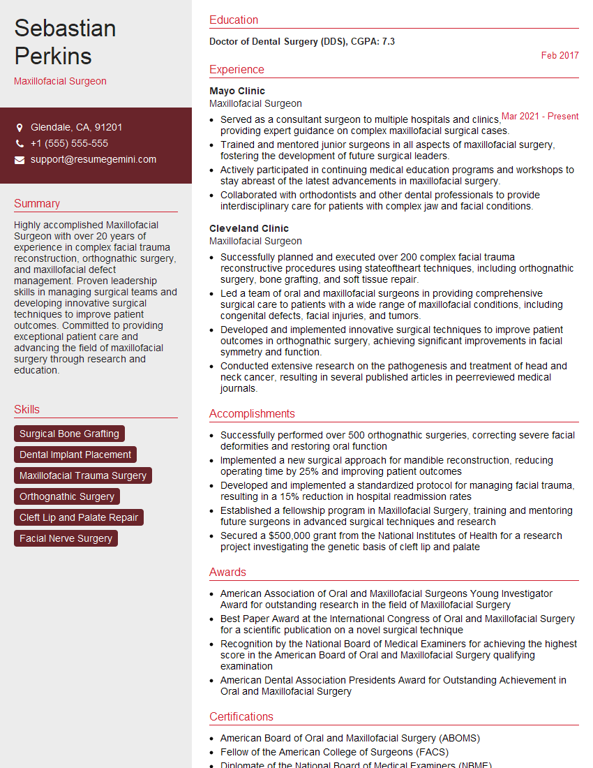
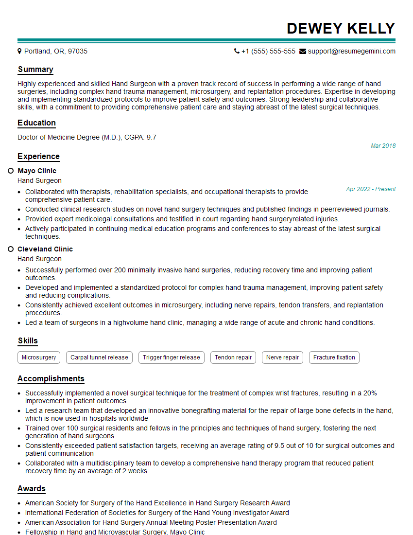
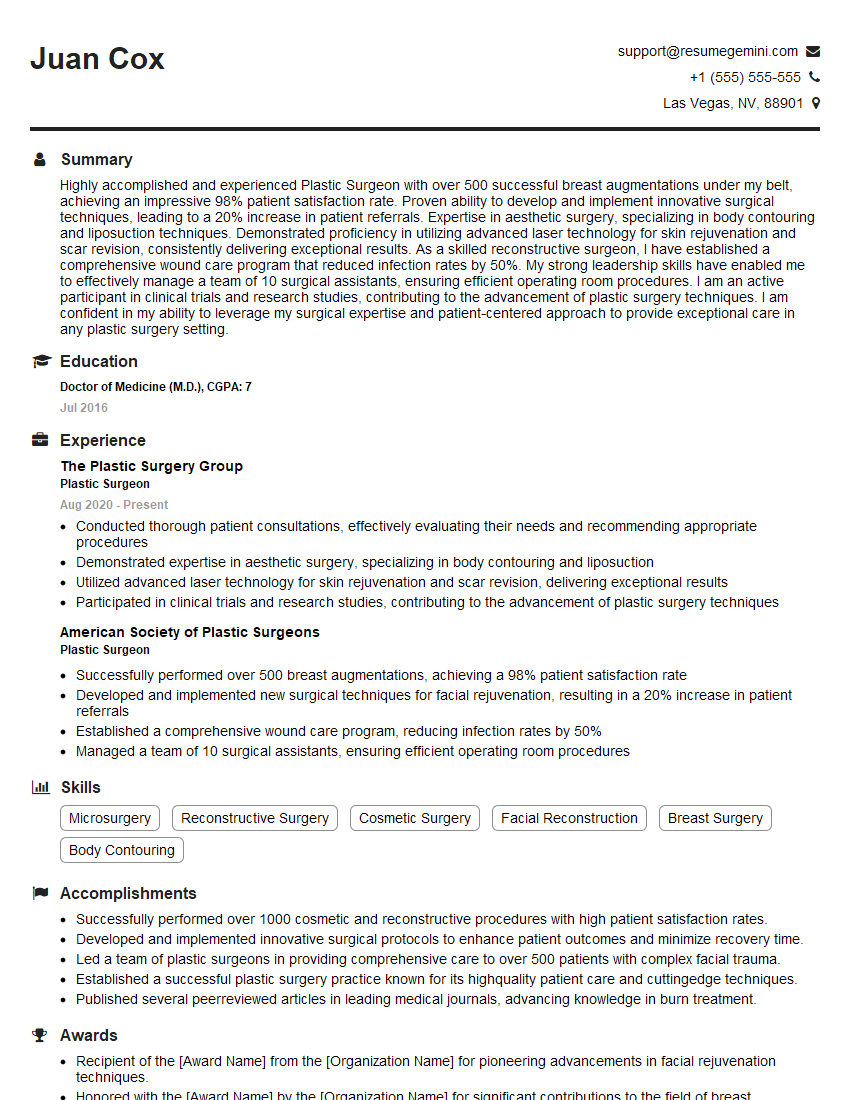
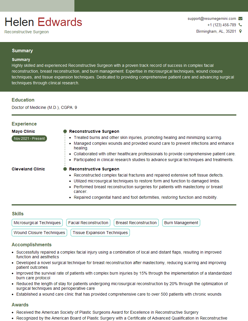
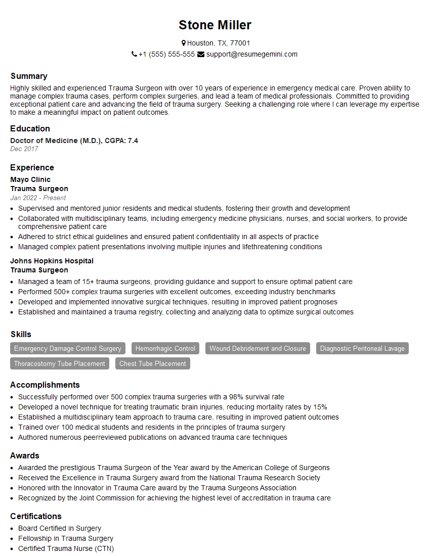
Mastering Soft Tissue Reconstruction opens doors to exciting career advancements, from specialized fellowships to leadership roles within renowned medical institutions. A strong resume is your key to unlocking these opportunities. Creating an ATS-friendly resume is crucial for getting your application noticed. To significantly enhance your resume and maximize your chances, we recommend using ResumeGemini. ResumeGemini provides a streamlined process to build a professional and impactful resume, ensuring your qualifications shine. Examples of resumes tailored to Soft Tissue Reconstruction are available to guide you.
Explore more articles
Users Rating of Our Blogs
Share Your Experience
We value your feedback! Please rate our content and share your thoughts (optional).
What Readers Say About Our Blog
This was kind of a unique content I found around the specialized skills. Very helpful questions and good detailed answers.
Very Helpful blog, thank you Interviewgemini team.
