Preparation is the key to success in any interview. In this post, we’ll explore crucial X-ray and Ultrasound Interpretation interview questions and equip you with strategies to craft impactful answers. Whether you’re a beginner or a pro, these tips will elevate your preparation.
Questions Asked in X-ray and Ultrasound Interpretation Interview
Q 1. Describe the difference between ionizing and non-ionizing radiation.
The key difference between ionizing and non-ionizing radiation lies in their ability to remove electrons from atoms. Ionizing radiation, like X-rays and gamma rays, carries enough energy to ionize atoms, meaning it can knock electrons out of their orbits. This creates charged particles (ions) that can damage DNA and other cellular components, potentially leading to health problems like cancer. Think of it like a powerful billiard ball striking other balls and scattering them wildly.
Non-ionizing radiation, such as ultrasound, radio waves, and microwaves, has less energy and doesn’t have the power to ionize atoms. Instead, it causes atoms to vibrate or rotate. While it can still cause heating effects at high intensities, it doesn’t directly damage DNA in the same way as ionizing radiation. This is analogous to gently nudging the billiard balls, causing some movement but not a disruptive scattering.
Q 2. Explain the principles of ultrasound image formation.
Ultrasound image formation relies on the principle of echolocation, similar to how bats navigate. A transducer, which is both a sender and receiver of sound waves, emits high-frequency sound pulses into the body. These pulses travel through the tissues, and whenever they encounter a boundary between tissues with different acoustic impedances (density and speed of sound), some of the sound is reflected back to the transducer as an echo.
The time it takes for the echo to return indicates the depth of the structure. The strength of the echo is related to the difference in acoustic impedance between the tissues. A computer then processes these echoes to create a grayscale image, where bright areas represent strong reflections (e.g., bone) and dark areas represent weak reflections (e.g., fluid).
Q 3. What are the common artifacts seen in ultrasound imaging and how are they addressed?
Several artifacts can affect ultrasound image quality. Acoustic shadowing occurs when a strongly reflective structure (like a stone) blocks sound waves, creating a shadow behind it. Acoustic enhancement is the opposite; sound waves passing through a fluid-filled structure (like a cyst) become amplified, appearing brighter behind the structure. Refraction bends sound waves as they pass through different tissues, distorting the image. Reverberation creates multiple reflections between two strong reflectors, appearing as parallel lines. Aliasing can cause wraparound effects with fast-moving structures like blood flow.
Addressing artifacts involves optimizing the ultrasound settings (e.g., frequency, gain), adjusting the transducer position and angle, and using different scanning techniques. Understanding the underlying cause of the artifact is crucial for correct interpretation.
Q 4. Differentiate between different types of X-ray imaging modalities (e.g., chest X-ray, CT, fluoroscopy).
These X-ray modalities differ significantly in their technique and application:
- Chest X-ray: A simple, quick, and widely used technique producing a two-dimensional image of the chest. It’s great for detecting pneumonia, pneumothorax, and bone fractures.
- Computed Tomography (CT): Uses X-rays and computer processing to create cross-sectional images of the body, providing much greater detail than a plain X-ray. It’s ideal for visualizing organs, blood vessels, and bone, making it valuable for diagnosing cancer, trauma, and internal bleeding.
- Fluoroscopy: Provides real-time X-ray imaging, like a live movie, showing the movement of structures. It’s used for guiding procedures like angiograms (viewing blood vessels) and swallowing studies (assessing swallowing function).
The key difference lies in the information obtained: a simple X-ray gives a broad overview, while CT provides detailed cross-sections, and fluoroscopy offers dynamic visualization.
Q 5. How do you identify and respond to a patient experiencing an adverse reaction during a procedure?
Responding to an adverse reaction requires immediate action. First, ensure patient safety – stop the procedure if possible. Then, assess the severity of the reaction: Is it mild (e.g., slight nausea), moderate (e.g., hives, vomiting), or severe (e.g., anaphylaxis, respiratory distress)?
For mild reactions, observation and supportive care may suffice. For moderate to severe reactions, initiate emergency protocols. This includes calling for help, administering oxygen, and potentially administering medications like epinephrine (for anaphylaxis). Closely monitor vital signs (heart rate, blood pressure, respiration). Once stabilized, document the event thoroughly, including symptoms, interventions, and the patient’s response. Appropriate follow-up care should be arranged.
Q 6. Explain the ALARA principle in relation to radiation safety.
ALARA stands for “As Low As Reasonably Achievable.” It’s a fundamental principle in radiation safety, emphasizing the minimization of radiation exposure to both patients and personnel. It doesn’t mean eliminating all radiation exposure, but rather using strategies to keep it as low as possible while still obtaining diagnostically useful images.
This involves optimizing imaging techniques (e.g., using the lowest possible radiation dose for a diagnostic image), using appropriate shielding (lead aprons for patients and staff), and implementing strict safety protocols. A balance must be struck between diagnostic benefit and radiation risk. For example, a chest X-ray delivers a relatively low radiation dose, providing valuable diagnostic information with an acceptable risk. On the other hand, a CT scan, providing much higher resolution, entails a higher radiation dose, requiring a thorough cost-benefit analysis.
Q 7. Describe the various ultrasound transducers and their applications.
Ultrasound transducers vary in frequency and design, each suitable for specific applications:
- Linear transducers (high frequency) are used for superficial structures like skin lesions and thyroid glands; they offer high resolution.
- Curvilinear transducers (lower frequency) penetrate deeper, suitable for abdominal and pelvic imaging, allowing visualization of larger organs and structures.
- Phased array transducers are often used for cardiac imaging because they provide a wide field of view and are suitable for accessing the heart through the intercostal spaces.
- Endocavity transducers are small and inserted into body cavities (e.g., vagina, rectum) for detailed internal visualization.
The choice of transducer depends on the depth and resolution needed for the specific examination. A high-frequency transducer provides better resolution but less penetration depth, while a low-frequency transducer allows deeper penetration but has lower resolution.
Q 8. What are the safety precautions you take when operating X-ray equipment?
Safety is paramount when operating X-ray equipment. My approach follows the ALARA principle – As Low As Reasonably Achievable. This means minimizing radiation exposure to both myself and the patient.
- Time: I minimize the duration of the X-ray exposure by using the shortest possible exposure time while maintaining image quality. Think of it like taking a quick photograph instead of a long exposure.
- Distance: I maintain a safe distance from the X-ray source during exposure, utilizing shielding such as lead aprons and barriers. The further you are from the radiation source, the lower the dose.
- Shielding: I ensure that both myself and any other personnel in the room are properly shielded with lead aprons and other protective equipment. This acts as a physical barrier against radiation.
- Regular Maintenance: I meticulously follow safety protocols and ensure the X-ray equipment undergoes regular maintenance checks to ensure its optimal functioning and radiation safety. Regular maintenance prevents malfunctions that might lead to increased exposure.
- Patient Protection: I always use appropriate shielding for the patient, such as lead gonadal shields, to protect organs from unnecessary radiation exposure. Protecting the patient is my top priority.
For example, during a chest X-ray, I’ll ensure the exposure time is as short as possible while still providing a clear image of the lungs. I’ll also ensure I’m behind a lead shield and the patient is properly positioned.
Q 9. How do you ensure proper patient positioning for optimal imaging?
Proper patient positioning is crucial for optimal imaging, as incorrect positioning can lead to artifacts, obscuring relevant anatomical structures, and requiring repeat exposures (increasing radiation exposure). My approach emphasizes precision and patient comfort.
- Anatomical Landmarks: I utilize anatomical landmarks to accurately position the patient. For instance, in a chest X-ray, I’ll make sure the patient’s shoulders are rolled forward to prevent obscuring the lung fields. For lateral views, I carefully position the patient so the central ray passes through the mid-axillary line.
- Immobilization: When necessary, I employ appropriate immobilization techniques to ensure the patient remains still during the exposure. This might involve sandbags or positioning aids to avoid blurring and artifacts.
- Communication: Clear communication with the patient is essential. I explain the procedure, ensuring the patient understands their role in remaining still and following instructions.
- Verification: Before each exposure, I verify the correct positioning through fluoroscopy (if applicable) or preview images, ensuring optimal image quality and avoiding repeat scans. This involves double-checking the position using the monitor.
For instance, in a lateral cervical spine X-ray, even a slight rotation can significantly affect the interpretation, so meticulous positioning is vital.
Q 10. Describe your experience with image acquisition and processing techniques.
My experience with image acquisition and processing spans various modalities and techniques. In X-ray, I’m proficient in using different settings (kVp, mAs) to optimize image contrast and reduce patient exposure, depending on the specific anatomical region and clinical question. For ultrasound, I have extensive experience adjusting parameters like gain, depth, frequency, and TGC to achieve optimal image quality.
- Digital Image Processing: I’m adept at post-processing techniques, such as windowing and leveling in X-ray, to enhance image visualization and feature identification. I utilize software features to improve image contrast and brightness for a clearer view.
- Image Quality Assurance: I regularly perform quality control checks to ensure optimal performance and consistent image quality. This includes regular calibration of equipment.
- Specific Modalities: My experience includes using various modalities, such as computed radiography (CR) and digital radiography (DR), along with diverse ultrasound transducers (linear, curvilinear, phased array) tailored for different applications (e.g., abdominal, cardiac).
For example, when faced with a suboptimal X-ray image due to patient movement, I might use post-processing techniques like edge enhancement to improve the visibility of subtle fractures. In ultrasound, I would adjust the gain to increase the brightness of a hypoechoic lesion (low reflection) for better visualization.
Q 11. What are the key differences between grayscale and Doppler ultrasound?
Both grayscale and Doppler ultrasound are used to visualize structures within the body, but they provide different types of information.
- Grayscale Ultrasound: This is the most basic type of ultrasound. It displays the anatomical structures based on their acoustic impedance – the resistance to the passage of sound waves. Different tissues reflect sound waves differently, creating varying shades of gray on the image. Brighter areas represent higher reflectivity (e.g., bone), while darker areas represent lower reflectivity (e.g., fluid).
- Doppler Ultrasound: This technique measures the velocity and direction of blood flow within vessels. It relies on the Doppler effect – the change in frequency of a wave in relation to an observer moving relative to the source. Doppler shifts are displayed as colors superimposed on the grayscale image. Red typically represents flow toward the transducer, while blue represents flow away.
Think of it like this: grayscale ultrasound shows you a static image of the roads, while Doppler ultrasound shows you the traffic flow on those roads.
Q 12. Explain the concept of acoustic impedance and its impact on ultrasound image quality.
Acoustic impedance is the resistance to the passage of sound waves through a tissue. It’s a crucial factor influencing ultrasound image quality. The greater the difference in acoustic impedance between two tissues, the stronger the reflected sound wave, resulting in a brighter echo on the image. Conversely, similar acoustic impedances result in weaker reflections and darker areas.
- Interface Reflections: Strong reflections occur at interfaces between tissues with significantly different acoustic impedances, such as the boundary between liver and kidney. These create strong echoes on the ultrasound image.
- Shadowing and Enhancement: High acoustic impedance tissues can cause shadowing (reduced signal behind the tissue) or enhancement (increased signal behind the tissue) artifacts, affecting image interpretation. For example, bone creates significant shadowing.
- Image Artifacts: Differences in acoustic impedance can lead to various artifacts, which can sometimes mimic pathology, highlighting the importance of understanding this concept.
For instance, the strong acoustic impedance difference between air and soft tissues creates a significant shadow behind air-filled structures (e.g., lungs), limiting visualization of underlying structures in conventional ultrasound.
Q 13. How do you interpret different density variations in X-ray images?
Density variations in X-ray images represent the varying ability of tissues to absorb X-rays. Different densities show up as different shades of gray.
- High Density (Bright White): Tissues that absorb X-rays well appear bright white, such as bone and metal. This means the X-rays are largely absorbed and don’t pass through to reach the detector.
- Low Density (Dark Gray/Black): Tissues that allow X-rays to pass through easily appear dark gray or black, such as air and fat. This means that the X-rays largely pass through with minimal absorption.
- Intermediate Density (Shades of Gray): Soft tissues like muscle, organs, and fluid have intermediate densities and appear as various shades of gray. Subtle differences in density can be crucial for diagnosis.
For example, a fracture will appear as a bright white line against the darker gray of the surrounding bone on an X-ray due to the disruption of bone density. Similarly, air in the lungs appears black, whereas consolidation (fluid buildup) in the lungs will appear brighter.
Q 14. Describe the different types of ultrasound contrast agents and their uses.
Ultrasound contrast agents are used to enhance the visibility of blood vessels or other structures within the body. They consist of microscopic bubbles that reflect ultrasound waves more strongly than surrounding tissues, improving image quality and allowing for better visualization.
- Gas-filled Microspheres: These are the most common type, usually made of albumin or other biocompatible materials encapsulating a gas such as air or perfluorocarbon. They improve visualization of blood flow and enhance lesion detection. They are typically administered intravenously.
- Liposomal Contrast Agents: These agents use liposomes (small, spherical vesicles) containing gas or other contrast-enhancing materials to improve image contrast and visualization.
The choice of contrast agent depends on the specific application. For example, gas-filled microspheres are frequently used in echocardiography to improve the visualization of myocardial perfusion. Their use allows better characterization of lesions in various organs such as the liver or kidneys.
Q 15. What are the limitations of ultrasound imaging?
Ultrasound, while a valuable non-invasive imaging modality, has several limitations. Its primary limitation stems from its dependence on sound waves, which interact differently with various tissues.
- Acoustic Shadowing and Enhancement: Bone and air significantly attenuate (reduce) ultrasound waves, creating shadowing behind these structures, obscuring underlying anatomy. Conversely, structures that transmit sound waves well, like fluid-filled cysts, can cause enhancement behind them, potentially masking pathology. Imagine trying to see through a brick wall with sound – difficult, right? That’s similar to what happens in ultrasound with bone.
- Operator Dependence: Image quality heavily relies on the skill and experience of the sonographer. Proper probe placement, angle, and technique are critical for optimal visualization. A poorly performed ultrasound can yield uninterpretable or misleading images.
- Limited Penetration Depth: Ultrasound waves don’t penetrate deep into the body as effectively as X-rays. This restricts its use in imaging deep-seated structures. For example, it might struggle to visualize structures deep within the abdomen or pelvis.
- Gas Interference: Air within the body, such as in the lungs or intestines, significantly scatters ultrasound waves, making visualization through these regions challenging. This often leads to poor image quality in areas with significant gas.
- Difficulty with Certain Tissues: Differentiating between certain tissues with similar acoustic impedance can be difficult. For instance, distinguishing between some solid masses and other tissue can be challenging.
Understanding these limitations is crucial for appropriate patient selection and interpretation of ultrasound findings. It’s essential to correlate ultrasound findings with clinical history and other imaging modalities when necessary.
Career Expert Tips:
- Ace those interviews! Prepare effectively by reviewing the Top 50 Most Common Interview Questions on ResumeGemini.
- Navigate your job search with confidence! Explore a wide range of Career Tips on ResumeGemini. Learn about common challenges and recommendations to overcome them.
- Craft the perfect resume! Master the Art of Resume Writing with ResumeGemini’s guide. Showcase your unique qualifications and achievements effectively.
- Don’t miss out on holiday savings! Build your dream resume with ResumeGemini’s ATS optimized templates.
Q 16. How do you handle a situation where the image quality is suboptimal?
Suboptimal image quality is a frequent challenge in both X-ray and ultrasound. My approach involves a systematic troubleshooting process.
- Identify the Cause: First, I carefully analyze the image to determine the source of the problem. This might involve considering patient factors (e.g., obesity causing acoustic shadowing in ultrasound or motion blur in X-rays), equipment malfunctions (e.g., improperly calibrated ultrasound transducer or faulty X-ray tube), or technical errors (e.g., incorrect ultrasound probe placement or improper X-ray exposure settings).
- Adjust Technique: Based on the identified cause, I adjust the imaging technique accordingly. For ultrasound, this might involve changing the probe, adjusting the depth, gain, or frequency settings, or modifying the scanning angle. For X-rays, this might involve adjusting the kilovoltage (kVp) and milliamperage (mA) settings, or changing the position of the patient or X-ray tube.
- Repeat the Image: Once adjustments are made, I repeat the imaging process to evaluate whether image quality has improved. If not, further investigation is needed.
- Consult with Colleagues: If troubleshooting is unsuccessful, I consult with experienced colleagues for their expertise. A second opinion can provide valuable insights and help identify subtle issues that might have been missed.
- Equipment Maintenance: If the problem stems from equipment malfunction, I would initiate a request for appropriate biomedical engineering support to ensure equipment is correctly calibrated and functioning properly. Regular quality control is crucial in preventing such situations.
In cases where image quality remains inadequate despite these measures, I would clearly document this in the report and clearly explain to the referring physician why certain findings couldn’t be evaluated and offer alternatives like a repeat study or different modality such as CT or MRI.
Q 17. Explain the concept of radiographic magnification.
Radiographic magnification refers to the enlargement of an object’s image on a radiograph compared to its actual size. This phenomenon occurs because the X-ray source and the image receptor (usually the film or digital detector) are at a distance from the object being imaged.
Imagine shining a flashlight on a small object onto a wall. The farther the object is from the wall, the larger its shadow appears. Similarly, in radiography, the farther the object is from the image receptor, the larger its image appears on the radiograph.
The degree of magnification is influenced by two factors:
- Object-to-Image Receptor Distance (OID): Increasing the OID increases magnification. Think of moving the object further from the wall in our flashlight analogy.
- Source-to-Image Receptor Distance (SID): Increasing the SID decreases magnification. Think of moving the flashlight farther from the wall. This reduces the size of the shadow.
The formula to calculate magnification is: Magnification = SID / (SID – OID)
Magnification can be a useful tool in specific radiographic techniques, such as magnification radiography of small bones. However, excessive magnification can reduce image sharpness and detail, so it’s important to carefully control these distances during image acquisition. Understanding magnification is critical for accurate interpretation of radiographic images.
Q 18. Describe your experience in quality assurance and quality control procedures for X-ray and ultrasound equipment.
Quality assurance (QA) and quality control (QC) are integral to ensuring accurate and reliable imaging results. My experience encompasses both routine and specialized procedures for both X-ray and ultrasound equipment.
- X-ray QA/QC: I’m familiar with performing and documenting annual QA tests on X-ray machines including, but not limited to, measurements of kVp accuracy, mAs linearity, half-value layer (HVL), and timer accuracy. These are done using standardized phantoms and quality control tools. We also perform regular checks on the image processing systems and image archiving systems to ensure image integrity.
- Ultrasound QA/QC: My ultrasound QA/QC experience involves routine testing of transducer output using test objects, verifying image uniformity and dynamic range. I’m trained in performing routine maintenance like cleaning and disinfection to avoid transducer damage and maintain optimal image quality. Additionally, I perform quality control by confirming the proper functioning of the ultrasound equipment’s various features before scanning each patient.
- Documentation: Thorough record-keeping of all QA/QC procedures is crucial. This documentation helps track equipment performance, identify trends, and ensure compliance with regulatory standards. These records are reviewed regularly, and any problems are reported immediately.
- Continuing Education: To stay abreast of the latest techniques and best practices, I actively participate in professional development activities and continuing medical education courses relating to image quality and equipment maintenance.
Proactive QA/QC minimizes the risk of producing suboptimal images and helps to ensure patient safety and the delivery of high-quality care.
Q 19. How do you maintain patient confidentiality and adhere to HIPAA regulations?
Patient confidentiality is paramount. I rigorously adhere to HIPAA regulations, which protect the privacy and security of Protected Health Information (PHI).
- Access Control: I only access patient information necessary for my work and utilize the strict access controls provided by our PACS and electronic health record systems. I never share patient information with unauthorized individuals.
- Secure Data Handling: I follow all protocols for secure data handling, including password protection, data encryption, and secure disposal of printed documents. I never leave patient information unattended.
- Privacy During Procedures: I ensure patient privacy during imaging procedures by draping and shielding them appropriately. Conversations about patient findings are conducted privately and never in public areas.
- HIPAA Training: I have completed comprehensive HIPAA training and remain updated on any revisions to the regulations.
- Reporting: When communicating findings, I only disclose information to authorized medical personnel and always use appropriate terminology. Patient identification is only ever done in secure electronic or paper systems.
Maintaining patient confidentiality is not just a regulatory requirement but an ethical obligation. I understand the potential consequences of a breach of privacy, and I am committed to safeguarding patient information at all times.
Q 20. Explain your understanding of image archiving and communication systems (PACS).
Picture Archiving and Communication Systems (PACS) are crucial for the storage, retrieval, and distribution of medical images. They’re essentially the central nervous system for radiology departments, revolutionizing how we handle and share images.
My understanding encompasses:
- Image Storage: PACS provides secure, long-term storage for all digital imaging studies (X-rays, ultrasounds, CTs, MRIs, etc.), eliminating the need for bulky film storage and improving access.
- Image Retrieval: Images can be quickly and easily accessed from various workstations within the hospital or remotely by authorized personnel, speeding up diagnosis and treatment.
- Image Distribution: PACS facilitates efficient distribution of images to referring physicians and other specialists, improving communication and collaboration.
- Image Manipulation: PACS allows for image enhancement, windowing (adjusting brightness and contrast), and measurements, improving image interpretation and diagnosis.
- Integration with EHR: Modern PACS systems are often seamlessly integrated with electronic health records (EHRs), allowing for easy access to patient information and a more comprehensive view of their medical history. This streamlined workflow eliminates duplicated effort and enhances patient care.
Experience with PACS is essential for efficient workflow in modern radiology. I have hands-on experience with several PACS platforms including image acquisition, quality control, and administration of various settings and protocols.
Q 21. How do you communicate findings to the physician in a clear and concise manner?
Communicating findings clearly and concisely to the referring physician is crucial for effective patient care. My approach emphasizes accuracy, brevity, and avoidance of jargon.
- Structured Reporting: I utilize a structured reporting format that organizes findings systematically, typically including a brief introduction, a description of the imaging technique used, and a comprehensive description of my findings with location, size, and relevant characteristics of any abnormalities.
- Precise Terminology: I employ precise medical terminology to ensure accuracy while avoiding unnecessary jargon. If complex terms are used, I provide clear definitions or explanations.
- Correlation with Clinical History: I always correlate my findings with the patient’s clinical history and the referring physician’s questions or concerns. This contextual information aids in the interpretation of images and improves communication.
- Visual Aids: I use annotations and measurements on the images themselves to highlight key findings, making them easy for the physician to locate and review. I also utilize image measurements and other quantitative data to support my description.
- Concise Summary: I provide a concise summary of my findings at the end of the report, highlighting the most important observations and recommendations.
- Accessibility: I ensure the report is easily accessible to the physician through electronic means and complies with all relevant standards for reporting.
Effective communication facilitates prompt and informed decision-making, ultimately contributing to improved patient outcomes. I regularly seek feedback from referring physicians to refine my communication style and ensure it meets their needs.
Q 22. Describe your experience working in a fast-paced, high-pressure environment.
My experience in emergency radiology has provided extensive training in high-pressure situations. Imagine a trauma bay – multiple patients arriving simultaneously, each requiring immediate imaging and interpretation. We’re talking seconds matter. In such an environment, prioritizing, efficient workflow, and accurate interpretation under pressure are paramount. I’ve honed my ability to rapidly assess situations, make critical decisions, and effectively communicate findings to the clinical team, often under intense time constraints. This includes managing competing priorities, delegating tasks when appropriate, and maintaining a calm demeanor even amidst chaos. I’m proficient in managing unexpected equipment malfunctions or workflow disruptions, ensuring minimal impact on patient care.
For example, during a major traffic accident, we received five critically injured patients within an hour. My ability to swiftly prioritize based on the severity of their injuries, quickly analyze the images, and communicate concisely with the surgical team was crucial to their successful treatment. This experience has instilled a deep sense of composure and efficiency that allows me to thrive in high-stress settings.
Q 23. Describe your experience with different types of medical imaging equipment.
My experience encompasses a wide range of medical imaging equipment, including various X-ray systems (conventional, fluoroscopy, computed tomography – CT), and ultrasound machines (both general purpose and specialized, such as transesophageal echocardiography – TEE). I’m proficient in operating these machines, optimizing image acquisition parameters (e.g., kVp, mAs in X-ray; frequency, gain in ultrasound), and troubleshooting technical issues. This includes understanding the intricacies of image reconstruction algorithms, particularly in CT, and being able to recognize artifacts and adjust settings to mitigate them. I’m familiar with PACS (Picture Archiving and Communication System) and other image management systems, allowing for efficient image retrieval and analysis.
Beyond the technical aspects, I have hands-on experience with different transducer types in ultrasound (linear, curvilinear, phased array) and their applications in various anatomical regions. My understanding extends to the physical principles behind image formation in both modalities, which allows me to better interpret the images and identify potential sources of error. I’ve also worked with specialized software for image post-processing and 3D reconstruction in both X-ray and ultrasound.
Q 24. What are the ethical considerations related to medical imaging?
Ethical considerations in medical imaging are crucial and multifaceted. They involve patient confidentiality (HIPAA compliance), radiation safety (ALARA principle – As Low As Reasonably Achievable), and responsible use of resources.
- Confidentiality: Protecting patient data is paramount. This includes ensuring secure storage and transmission of images and reports, adhering to strict access protocols, and only disclosing information to authorized personnel.
- Radiation Safety: Minimizing radiation exposure to patients is critical, particularly in X-ray and CT. This involves optimizing imaging protocols, utilizing appropriate shielding, and carefully weighing the benefits of the examination against the risks of radiation exposure.
- Resource Allocation: Ethical considerations involve judicious use of imaging resources, ensuring that tests are ordered appropriately and only when clinically indicated. Avoiding unnecessary examinations not only saves costs but also reduces the overall radiation burden on patients.
- Informed Consent: Obtaining informed consent from patients before any procedure is non-negotiable. This ensures patients understand the risks and benefits of the examination and make an informed decision.
A specific example is balancing the need for a detailed CT scan in a trauma patient with the risks of radiation exposure. In such cases, the ALARA principle must guide the decision-making process, using the lowest effective radiation dose while ensuring diagnostic quality.
Q 25. Describe your problem-solving skills related to diagnostic imaging.
Problem-solving in diagnostic imaging involves a systematic approach combining image analysis, clinical correlation, and critical thinking. I employ a structured process:
- Image Analysis: A thorough examination of the images, focusing on identifying key anatomical structures and any abnormalities (e.g., masses, fractures, fluid collections). This step requires attention to detail and familiarity with normal anatomy and variations.
- Clinical Correlation: This is a vital step. I always consider the patient’s clinical history, symptoms, and other relevant medical information. This context helps in differentiating normal variations from pathological findings.
- Differential Diagnosis: Based on the image findings and clinical information, I develop a differential diagnosis (a list of possible conditions), prioritizing the most likely diagnoses.
- Further Investigations: If needed, I will recommend further investigations such as additional imaging studies, blood tests, or consultations with specialists to refine the diagnosis.
- Reporting: I produce comprehensive reports that clearly communicate my findings, differential diagnoses, and recommendations to the referring physician.
For instance, I might encounter a chest X-ray showing an opacity in the lung. I wouldn’t simply label it an “opacity.” Instead, I’d assess the location, size, shape, and margins, correlate it with the patient’s symptoms (cough, fever, etc.), and formulate a differential diagnosis, which could include pneumonia, lung cancer, or a pulmonary embolism. Then, based on this differential, I might recommend further imaging (CT scan) or laboratory tests to arrive at a conclusive diagnosis.
Q 26. How do you stay updated with the latest advancements in X-ray and Ultrasound technology?
Staying current in X-ray and ultrasound is crucial. I actively engage in several strategies:
- Professional Organizations: Membership in organizations like the American College of Radiology (ACR) and the American Institute of Ultrasound in Medicine (AIUM) provides access to journals, conferences, and continuing medical education (CME) opportunities.
- Peer-Reviewed Journals: Regularly reading leading journals in radiology and ultrasound keeps me abreast of the latest research and technological advancements.
- Conferences and Workshops: Attending conferences and workshops allows for direct interaction with leading experts and exposure to cutting-edge technologies.
- Online Courses and Webinars: Many online platforms offer high-quality continuing education courses and webinars on imaging techniques and interpretation.
- Mentorship and Collaboration: Engaging in discussions with colleagues and mentors facilitates knowledge sharing and learning from others’ experiences.
For example, I recently completed a CME course on the latest advancements in contrast-enhanced ultrasound, significantly enhancing my diagnostic capabilities in liver and abdominal imaging.
Q 27. What are the common pathologies you are familiar with in various imaging modalities?
My experience covers a wide range of pathologies in both X-ray and ultrasound. Here are a few common examples:
- X-ray: Fractures (various types), pneumonia, pneumothorax (collapsed lung), atelectasis (lung collapse), pleural effusions (fluid in the lungs), congestive heart failure, foreign body aspiration.
- Ultrasound: Gallstones, kidney stones, liver disease (cirrhosis, hepatitis), abdominal masses, ovarian cysts, pregnancy complications, thyroid nodules, deep vein thrombosis (DVT).
The specific findings in imaging vary depending on the modality and the stage of the disease process. For instance, a simple fracture might appear as a clear line of discontinuity on an X-ray, whereas advanced liver cirrhosis might show characteristic changes in liver echogenicity and texture on ultrasound.
Q 28. Describe a challenging case you encountered and how you resolved it.
One challenging case involved a young female patient presenting with abdominal pain and a complex ultrasound image. Initial ultrasound showed a large, heterogeneous mass in the pelvis, but it was difficult to definitively characterize. It could have been a number of things, from a complicated ovarian cyst to something more sinister. The differential diagnosis was broad and concerning.
My approach was methodical. I reviewed the images carefully, focusing on subtle details like vascularity and internal echoes. I then correlated these findings with the patient’s clinical history, which included irregular menstrual cycles and elevated CA-125 levels (a tumor marker). I also consulted with a gynecologist, sharing my findings and differential diagnosis. Based on our collaborative assessment, we decided to perform a CT scan for better visualization of the mass and its relationship to surrounding structures. The CT scan revealed a large ovarian teratoma (a benign germ cell tumor), which was confirmed by subsequent surgical excision.
This case highlighted the importance of teamwork, meticulous image analysis, clinical correlation, and the judicious use of additional imaging modalities to arrive at an accurate diagnosis. It emphasized that a complex case requires not just expertise in interpreting images but also a broader clinical understanding and effective communication with other specialists.
Key Topics to Learn for X-ray and Ultrasound Interpretation Interview
- Image Acquisition and Optimization: Understanding the technical aspects of producing high-quality X-ray and ultrasound images, including patient positioning, transducer selection, and parameter adjustments. Practical application: Troubleshooting image artifacts and optimizing image quality for accurate interpretation.
- Anatomical Knowledge: Deep understanding of normal anatomy relevant to X-ray and ultrasound imaging. Practical application: Differentiating normal anatomical structures from pathology in various imaging modalities.
- Pathology Recognition: Identifying common pathologies and abnormalities visible on X-rays and ultrasound images. Practical application: Describing the characteristic findings of different diseases and conditions.
- Differential Diagnosis: Developing the ability to consider multiple possibilities when interpreting images and narrowing down the diagnosis based on the available information. Practical application: Formulating a differential diagnosis based on image findings and clinical history (if provided).
- Reporting and Communication: Structuring and writing clear, concise, and accurate reports that effectively communicate findings to referring physicians. Practical application: Using appropriate medical terminology and avoiding ambiguity in report writing.
- Radiation Safety and ALARA Principles: Understanding and adhering to radiation safety protocols and ALARA (As Low As Reasonably Achievable) principles. Practical application: Applying safety procedures to minimize radiation exposure to both patients and healthcare professionals.
- Ethical Considerations: Understanding the ethical implications of medical imaging and maintaining patient confidentiality. Practical application: Making informed decisions regarding imaging protocols and reporting.
- Image Processing and Enhancement Techniques: Familiarity with basic image processing techniques that can aid in interpretation. Practical application: Understanding the benefits and limitations of various image enhancement tools.
Next Steps
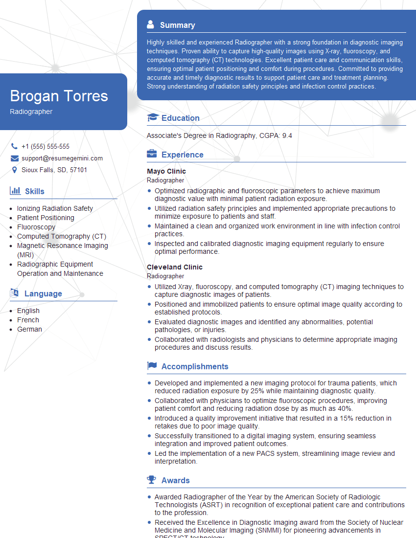
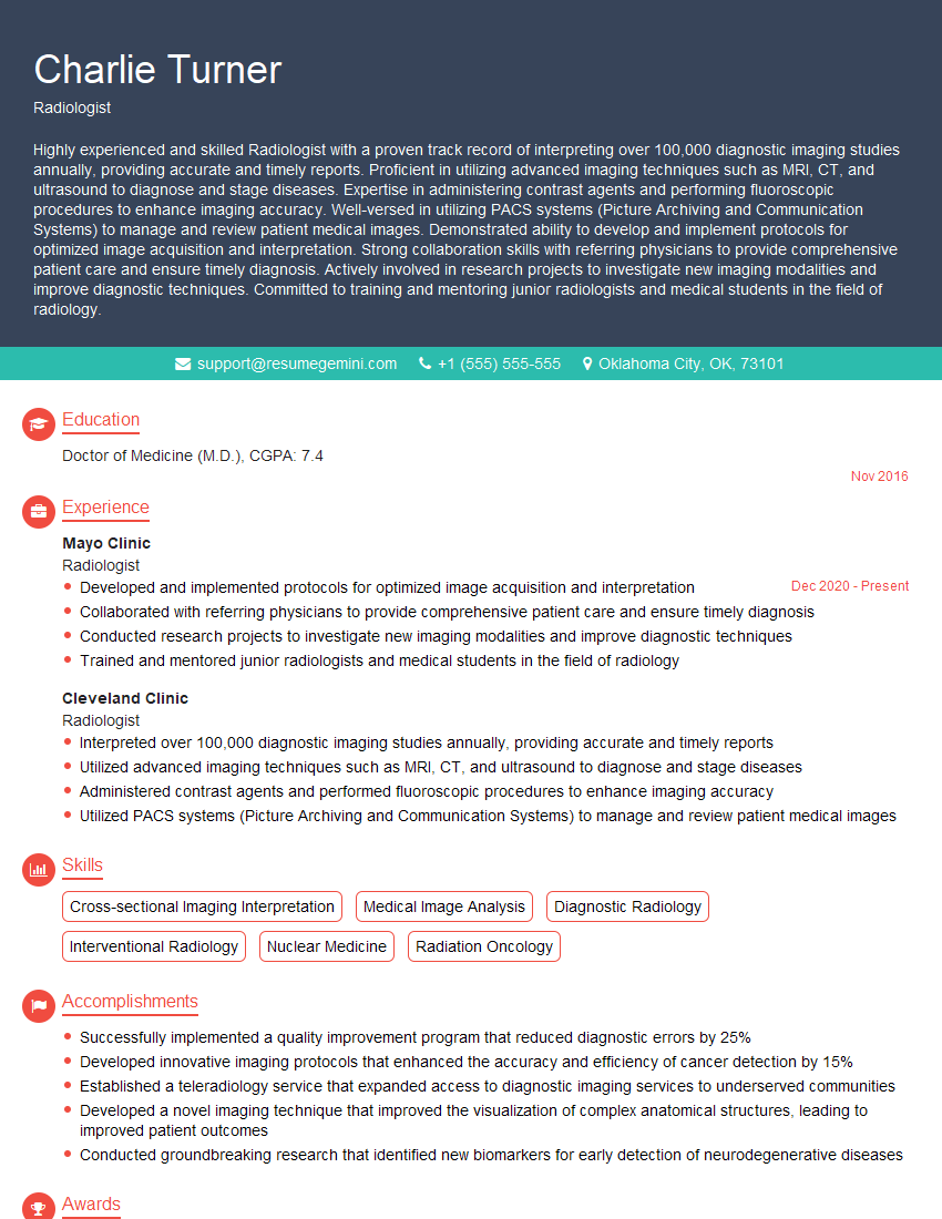
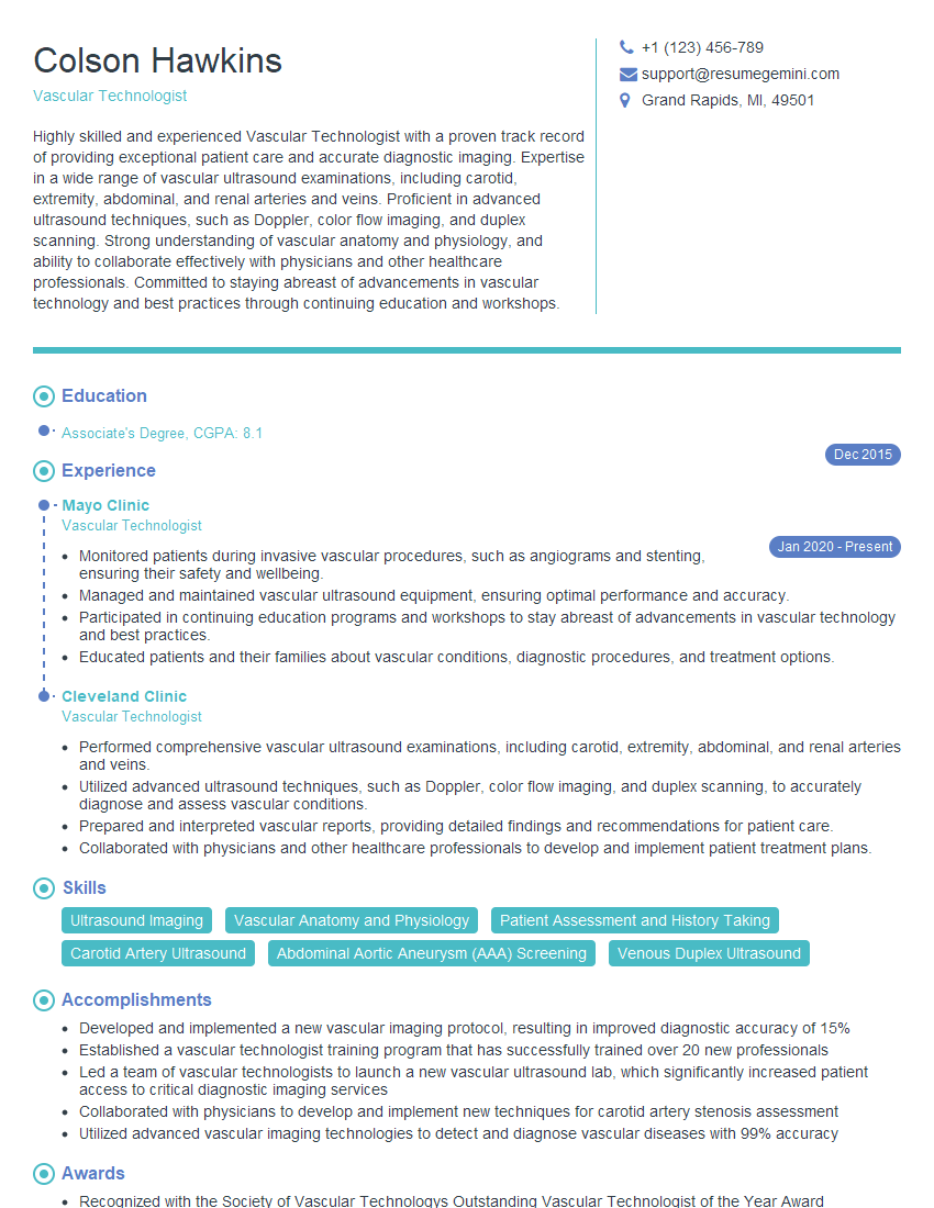
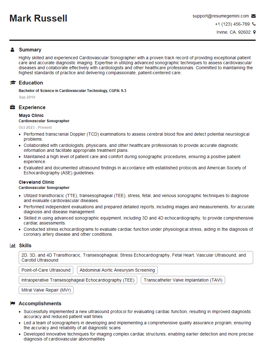
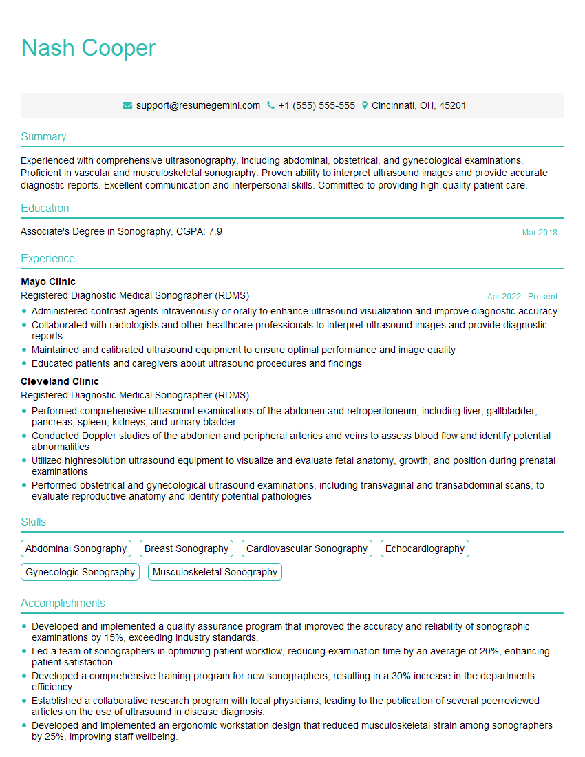
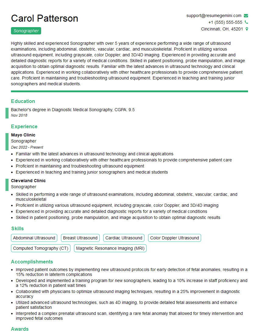
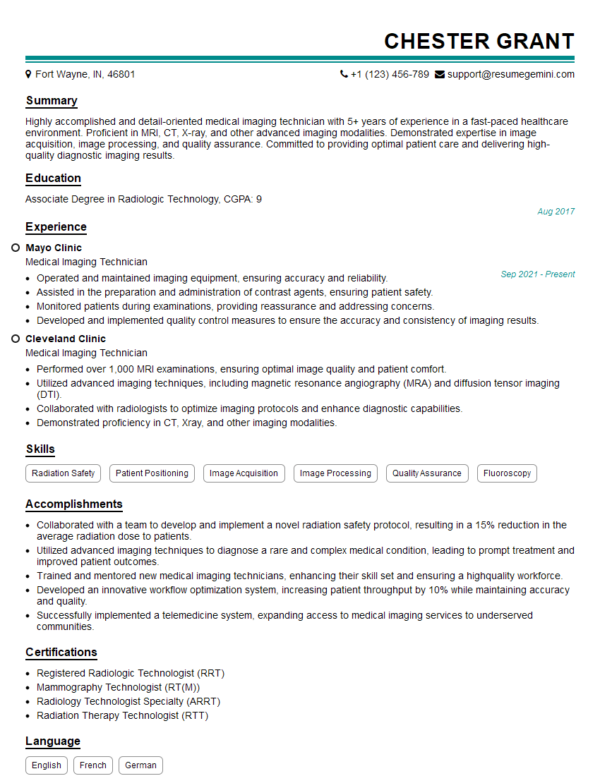
Mastering X-ray and ultrasound interpretation is crucial for career advancement in medical imaging, opening doors to specialized roles and increased responsibility. A strong resume is your key to unlocking these opportunities. Crafting an ATS-friendly resume is essential to get your application noticed by recruiters and hiring managers. We highly recommend using ResumeGemini to build a professional and impactful resume that highlights your skills and experience effectively. ResumeGemini provides examples of resumes tailored to X-ray and Ultrasound Interpretation roles, helping you create a document that truly showcases your qualifications.
Explore more articles
Users Rating of Our Blogs
Share Your Experience
We value your feedback! Please rate our content and share your thoughts (optional).
What Readers Say About Our Blog
This was kind of a unique content I found around the specialized skills. Very helpful questions and good detailed answers.
Very Helpful blog, thank you Interviewgemini team.
