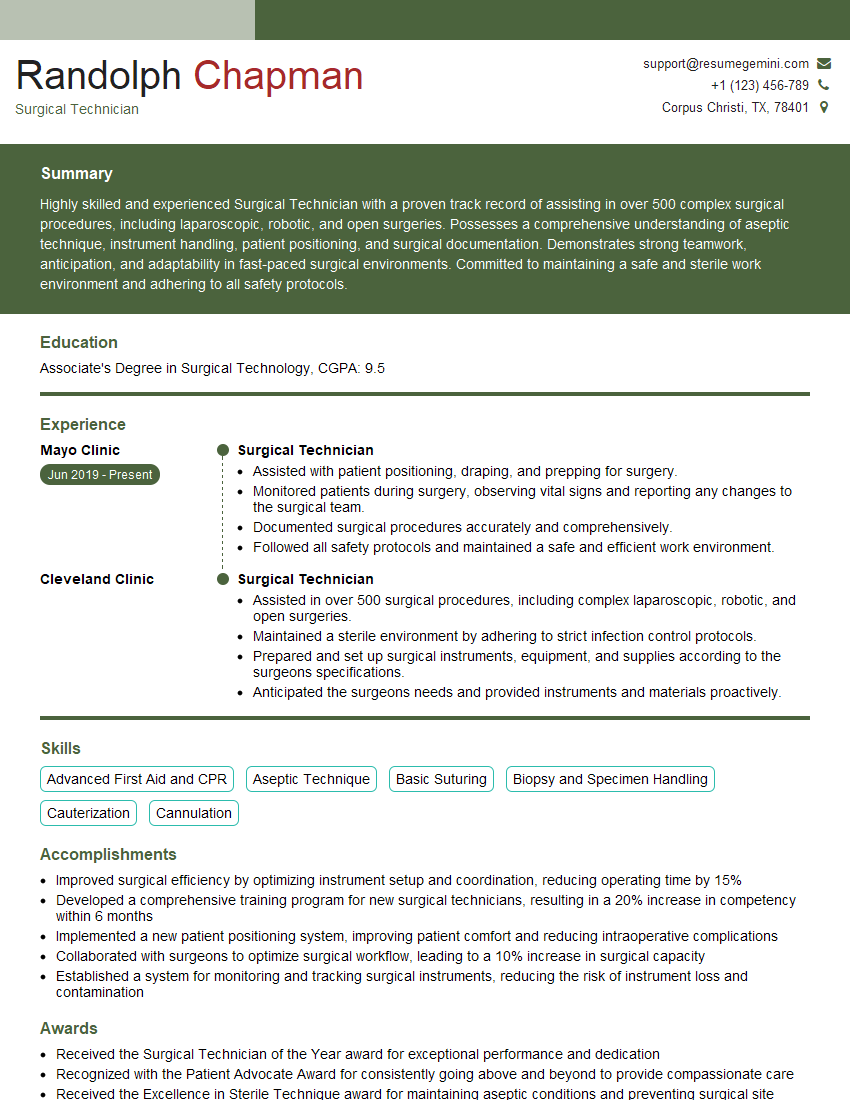
The right preparation can turn an interview into an opportunity to showcase your expertise. This guide to Preoperative and Postoperative Wound Care interview questions is your ultimate resource, providing key insights and tips to help you ace your responses and stand out as a top candidate.
Questions Asked in Preoperative and Postoperative Wound Care Interview
Q 1. Describe the different stages of wound healing.
Wound healing is a complex process typically divided into four overlapping phases: hemostasis, inflammation, proliferation, and maturation. Think of it like building a house; each stage is crucial for a strong and lasting structure.
- Hemostasis: This initial phase involves blood clotting to stop bleeding. It’s like laying the foundation – essential before construction begins. Platelets aggregate to form a clot, and vasoconstriction limits blood flow to the injury site.
- Inflammation: This phase, lasting approximately 3-21 days, involves the body’s immune response to clear debris and pathogens. This is like clearing the land and preparing the site – removing any obstacles for building. You’ll see redness, swelling, heat, and pain as the body fights off infection.
- Proliferation: This phase focuses on tissue regeneration and wound contraction. Fibroblasts create collagen, which forms the scaffolding for new tissue; this is like the framing and construction of the house. Granulation tissue, a pinkish-red tissue, indicates healthy healing.
- Maturation: This final phase, lasting months to years, involves collagen remodeling and scar tissue formation. It’s like the finishing touches – painting, landscaping, and making everything aesthetically pleasing and strong. The scar tissue becomes stronger and paler over time.
Understanding these stages is critical for effective wound management. For instance, prolonged inflammation suggests potential complications like infection, requiring timely intervention.
Q 2. Explain the assessment of a surgical wound.
Assessing a surgical wound involves a systematic approach, focusing on location, size, appearance, and surrounding tissue. Imagine you’re a detective investigating a crime scene; every detail matters.
- Location: Note the exact anatomical site of the incision.
- Size: Measure length, width, and depth in centimeters.
- Appearance: Observe the wound edges for approximation (how well they’re meeting), color (healthy tissue is pink or red, while pale tissue indicates poor perfusion), and presence of any drainage (serous, serosanguinous, purulent). The presence of purulent drainage (pus) is a major red flag.
- Surrounding Tissue: Assess for erythema (redness), edema (swelling), warmth, tenderness, and induration (hardening).
- Approximation: Are the wound edges well approximated or are there gaps? This is critical for assessing healing potential.
- Closure Method: Note if the wound was closed with sutures, staples, or adhesives.
Documentation is crucial. A detailed wound assessment helps track healing progress and identify potential problems early on. A photographic record can also be invaluable.
Q 3. What are the risk factors for postoperative wound complications?
Postoperative wound complications arise from a confluence of factors, increasing the risk for delayed healing or infection. Think of it as a recipe for disaster – if you have multiple ingredients, the likelihood of a bad outcome rises.
- Patient-related factors: Age (elderly patients heal slower), obesity (impaired blood supply to tissues), diabetes (impaired immune response and poor perfusion), smoking (vasoconstriction, reduced oxygenation), malnutrition (inadequate building blocks for healing), immunosuppression (chemotherapy, HIV), and pre-existing conditions.
- Surgical factors: Type of surgery (major vs. minor), duration of surgery (longer procedures increase risk), contamination during surgery, improper surgical technique, use of drains, and tension on the wound (poor approximation of wound edges).
- Postoperative factors: Infection at the surgical site, inadequate pain control, improper wound care, and poor hygiene.
Preoperative optimization of patient health and meticulous surgical technique are key preventative measures. Addressing risk factors before surgery is crucial.
Q 4. How do you identify and manage a wound infection?
Wound infection manifests as signs of local inflammation, often accompanied by systemic symptoms. Early detection is vital to prevent sepsis. Think of it like a fire alarm; you need to act swiftly before the fire spreads.
- Local signs: Increased pain, swelling, redness (erythema) extending beyond the wound edges, warmth, purulent drainage (pus), foul odor, and delayed healing.
- Systemic signs: Fever, chills, tachycardia (rapid heart rate), leukocytosis (increased white blood cell count), and malaise (general feeling of discomfort).
Management involves prompt treatment with antibiotics (after obtaining cultures to identify the infecting organism), wound debridement (removal of infected tissue), and appropriate wound dressings. In severe cases, surgical intervention might be necessary to drain an abscess or perform extensive debridement.
A simple example: If a wound displays increasing redness, warmth, and purulent drainage accompanied by fever, immediate action—including culture and antibiotics—is critical.
Q 5. What are the various types of wound dressings and their applications?
The choice of wound dressing depends on the type of wound, its stage of healing, and the presence of infection. Think of it like choosing tools for a specific job; each dressing has its own purpose.
- Alginate dressings: Highly absorbent, ideal for heavily exuding wounds (e.g., deep burns). They form a gel that helps manage drainage.
- Hydrocolloids: Maintain a moist wound environment, promoting autolytic debridement (self-cleaning) and reducing pain. Good for partial-thickness wounds and pressure ulcers.
- Hydrogels: Hydrating, soothing, and effective for dry or necrotic wounds. They help to rehydrate the wound bed.
- Foams: Highly absorbent, suitable for moderately to heavily exuding wounds. They provide protection and cushioning.
- Gauze dressings: Used for packing wounds and absorbing drainage; can be impregnated with various substances (e.g., antiseptic solutions).
- Transparent films: Allow for wound visualization while protecting from external contaminants. Ideal for superficial wounds with minimal exudate.
Incorrect dressing selection can hinder healing; for example, using a non-absorbent dressing on a heavily exuding wound will lead to maceration (softening) of the surrounding skin and potential infection.
Q 6. Discuss your experience with negative pressure wound therapy (NPWT).
Negative pressure wound therapy (NPWT) is a technique that uses a vacuum to remove excess wound fluid, promote granulation tissue formation, and reduce edema. I’ve extensively used it in managing complex wounds that weren’t healing adequately with conventional methods. Imagine it as a powerful vacuum cleaner for the wound, removing debris and promoting a healthy healing environment.
My experience shows that NPWT is particularly effective in treating chronic wounds, pressure ulcers, and surgical wounds with significant exudate or infection. It’s crucial to ensure proper application technique to avoid complications like tissue damage. I’ve observed faster healing times and reduced infection rates in many of my patients treated with NPWT, compared to those managed with traditional methods. The systematic removal of excess fluid creates space for granulation tissue formation, leading to accelerated healing.
However, NPWT isn’t without limitations. It’s not appropriate for all wounds, and careful patient selection is essential. For example, wounds with exposed vessels or organs are generally contraindicated.
Q 7. How do you manage a dehisced wound?
Wound dehiscence, the separation of wound edges, is a serious complication requiring prompt attention. Imagine a poorly constructed wall; the bricks start to fall apart. Immediate action prevents further complications like evisceration (organ protrusion).
Management involves:
- Assessment: The extent of separation needs careful evaluation. This includes assessing for signs of infection.
- Gentle Cleansing: The wound should be gently cleansed with saline solution.
- Dressing: A moist sterile dressing is applied to protect the wound and prevent further trauma.
- Positioning: The patient may be positioned to reduce tension on the wound.
- Surgical Repair: In most cases, surgical repair is necessary to close the wound. The patient will also require monitoring for signs of infection.
- Pain Management: Appropriate pain control is crucial for patient comfort and cooperation.
In cases of evisceration, immediate surgical intervention is life-saving. The aim is to prevent further complications such as infection and organ damage.
Q 8. Describe your experience with wound debridement techniques.
Wound debridement is the process of removing dead, damaged, or infected tissue from a wound to promote healing. I’m experienced in various techniques, selecting the most appropriate based on the wound type, depth, and the patient’s overall condition.
- Sharp debridement: This involves using sterile surgical instruments like scalpels or scissors to precisely remove necrotic tissue. It’s highly effective but requires surgical skill and precision. For example, I’ve used this technique successfully on a patient with a deep, infected leg ulcer, significantly improving the wound bed and allowing for healthy granulation tissue formation.
- Enzymatic debridement: This uses topical enzymes to break down necrotic tissue. It’s less invasive than sharp debridement, making it suitable for patients who are medically fragile. I’ve frequently used this approach on patients with pressure ulcers where aggressive debridement isn’t recommended.
- Autolytic debridement: This involves using the body’s own enzymes and moisture to break down necrotic tissue. It’s a slower process but less painful and is ideal for smaller, superficial wounds. I often use this as the primary method for smaller wounds, facilitating the body’s natural healing process.
- Mechanical debridement: This involves physically removing necrotic tissue using methods like wet-to-dry dressings or hydrotherapy. It’s relatively straightforward but can be uncomfortable and may damage healthy tissue if not performed carefully. I usually reserve this for wounds with large amounts of easily removable debris.
Choosing the right technique requires careful assessment of the wound and patient factors. It’s a dynamic process; the approach may need to be adjusted depending on the wound’s response to treatment.
Q 9. What are the indications for hyperbaric oxygen therapy?
Hyperbaric oxygen therapy (HBOT) involves breathing 100% oxygen in a pressurized chamber. It increases the amount of oxygen dissolved in the blood, improving oxygen delivery to tissues. Indications for HBOT include:
- Compromised wound healing: This is a common indication, particularly for wounds that are not healing due to infection, ischemia, or radiation damage. For example, diabetic foot ulcers that are unresponsive to conventional treatment can benefit significantly.
- Gas gangrene: HBOT is crucial in treating this life-threatening infection.
- Radiation necrosis: HBOT helps to improve blood supply to tissues damaged by radiation therapy.
- Crush injuries: In cases of severe crush injuries, HBOT can help reduce tissue damage and promote healing.
- Carbon monoxide poisoning: HBOT aids in the removal of carbon monoxide from the blood.
However, HBOT is not without potential risks and side effects, including barotrauma and oxygen toxicity. Therefore, careful patient selection and monitoring are crucial. It’s never a first-line treatment but an important adjunct therapy for specific, well-defined indications.
Q 10. How do you assess and manage a patient’s pain related to wound care?
Pain management is an integral part of wound care. I assess a patient’s pain using a multi-faceted approach:
- Pain scale: Using a standardized pain scale, like the 0-10 numerical rating scale, helps quantify pain levels consistently.
- Patient interview: I thoroughly discuss the patient’s pain experience, including location, intensity, quality, and duration. This helps to understand the nature of their discomfort better.
- Observation: Nonverbal cues, such as facial expressions and body language, can provide additional insight into pain levels.
Management strategies are tailored to each patient. Options include:
- Pharmacological interventions: Analgesics, such as opioids or NSAIDs, are prescribed based on the severity and type of pain. I frequently work with pain management specialists to optimize pain control.
- Non-pharmacological interventions: These include techniques like wound dressings that minimize trauma, repositioning to reduce pressure, and relaxation techniques. For instance, teaching deep breathing exercises can be highly effective.
- Wound care techniques: Careful, gentle wound care minimizes pain and discomfort during dressing changes. Using anesthetic creams or sprays before interventions is also effective.
Regular pain reassessment is critical to ensure the effectiveness of interventions and to adjust treatment as needed.
Q 11. Explain the importance of patient education in wound care.
Patient education is paramount in successful wound care. Informed patients are better equipped to participate actively in their healing process and are more likely to comply with treatment plans.
My approach involves:
- Explaining the wound: Clearly explaining the type of wound, its cause, and the expected healing timeline. Using visuals, like diagrams, helps patients understand better.
- Describing the treatment plan: Explaining all aspects of wound care, including dressing changes, medications, and follow-up appointments in simple, understandable terms. I use plain language, avoiding medical jargon whenever possible.
- Teaching self-care: Empowering patients with the knowledge and skills to perform necessary self-care, such as dressing changes and wound cleaning, when appropriate. Demonstrations are critical here.
- Identifying potential complications: Educating patients about potential complications like infection or delayed healing and how to recognize them. This empowers patients to seek timely help.
- Addressing concerns and questions: Providing ample opportunity for patients to ask questions and address their concerns. This fosters trust and ensures patient understanding.
Providing written materials and resources, such as brochures and websites, further supports patient education and promotes better outcomes.
Q 12. How do you document wound care interventions?
Accurate and detailed documentation of wound care interventions is crucial for effective communication and tracking progress. My documentation includes:
- Patient demographics: Name, date of birth, medical record number.
- Wound location and size: Precise location, dimensions (length, width, depth), and any undermining or tunneling.
- Wound appearance: Description of the wound bed (color, texture, presence of necrotic tissue or exudate).
- Interventions performed: Detailed description of all wound care interventions, including type of debridement, dressings used, and medications applied. I always record the amount of exudate removed.
- Pain assessment: Record the patient’s pain level before, during, and after interventions.
- Patient education provided: Documentation of any education provided to the patient and their family.
- Response to treatment: Observations regarding changes in wound appearance or patient’s condition. Progress notes are essential.
This information is documented in the patient’s electronic medical record using a standardized format to ensure consistency and facilitate efficient communication amongst the healthcare team. Clear, concise, and objective documentation is critical for optimal patient care and legal protection.
Q 13. What is your experience with different types of sutures and staples?
My experience encompasses a wide range of suture and staple materials, each with its own properties and applications. The choice depends on factors such as wound location, tension, and tissue type.
- Absorbable sutures: These sutures dissolve over time, eliminating the need for removal. Examples include Vicryl and PDS, commonly used for subcutaneous tissue closure. I select the type based on the rate of absorption required.
- Non-absorbable sutures: These need to be removed after the wound has healed. Nylon and silk are frequently used for skin closure. The selection here is often based on tissue fragility and the expected healing time.
- Staples: Metallic staples are often used for skin closure, especially in areas where rapid closure is needed, such as large surgical incisions. They are quicker to apply than sutures and are easily removed once healing is sufficient.
My proficiency extends to the proper technique of suture placement, staple application, and removal, minimizing patient discomfort and risk of infection. I always consider the patient’s individual needs and choose the most appropriate material and technique.
Q 14. How do you prevent pressure ulcers?
Pressure ulcers, also known as pressure sores or bedsores, are preventable. My approach to prevention involves a multi-pronged strategy focusing on minimizing pressure, improving tissue perfusion, and optimizing patient nutrition.
- Regular repositioning: Frequent turning and repositioning of patients in bed or wheelchairs is critical to reduce pressure points. A schedule should be followed, and the patient’s comfort should always be prioritized.
- Pressure-relieving surfaces: Using appropriate support surfaces, such as pressure-relieving mattresses, cushions, and overlays, can significantly reduce pressure on bony prominences.
- Skin care: Maintaining skin integrity through proper hygiene, moisturizing, and avoiding harsh soaps prevents damage. Prompt attention to skin breakdown is also necessary.
- Nutrition and hydration: Adequate nutrition and hydration are essential for wound healing and maintaining skin integrity. I work closely with dieticians to ensure appropriate nutrition plans.
- Mobility and exercise: Encouraging patients to engage in appropriate mobility and exercise programs, as tolerated, enhances tissue perfusion and reduces pressure points.
- Assessment and monitoring: Regularly assessing patients for risk factors for pressure ulcers, including mobility, nutrition, and sensory perception. This proactive approach is vital for early intervention. Using a standardized risk assessment tool, such as the Braden Scale, facilitates consistent monitoring.
Implementing these preventive measures reduces the incidence of pressure ulcers, improving patient comfort, reducing healthcare costs, and enhancing the overall quality of patient care.
Q 15. How do you manage a wound with excessive exudate?
Managing excessive wound exudate is crucial to prevent maceration (skin softening from prolonged moisture) and infection. The approach depends on the amount and type of exudate. We first assess the wound bed to determine the cause. Is it due to infection, inflammation, or simply a large wound?
Our strategy is multifaceted:
- Absorbent Dressings: For moderate exudate, highly absorbent dressings like alginate, foam, or hydrofiber are used. These wick away the fluid, maintaining a moist wound environment conducive to healing. For example, a patient with a large venous leg ulcer producing copious serosanguinous fluid might benefit from a calcium alginate dressing.
- Wound Vacuum Assisted Closure (VAC): In cases of significant exudate or where the wound bed is compromised, negative pressure wound therapy (NPWT), often called a VAC, is frequently employed. This system uses suction to remove excess fluid, stimulate granulation tissue formation, and reduce edema. I’ve seen remarkable results with VAC therapy in patients with traumatic wounds or dehisced surgical wounds.
- Compression Therapy: For wounds with excessive edema, like venous ulcers, compression bandages or stockings can assist in reducing fluid buildup and promoting lymphatic drainage. Correct application is critical to avoid compromising circulation. I always emphasize patient education about compression therapy, ensuring proper fit and application techniques.
- Frequent Dressing Changes: The frequency of dressing changes depends on the amount of exudate and the type of dressing. Some dressings require changes every day, while others can be left in place for several days. Regular monitoring prevents fluid build-up and reduces the risk of infection.
- Address Underlying Causes: Crucially, we must identify and treat the underlying cause of the excessive exudate, such as infection (requiring antibiotics) or inflammation (managed with appropriate measures).
Career Expert Tips:
- Ace those interviews! Prepare effectively by reviewing the Top 50 Most Common Interview Questions on ResumeGemini.
- Navigate your job search with confidence! Explore a wide range of Career Tips on ResumeGemini. Learn about common challenges and recommendations to overcome them.
- Craft the perfect resume! Master the Art of Resume Writing with ResumeGemini’s guide. Showcase your unique qualifications and achievements effectively.
- Don’t miss out on holiday savings! Build your dream resume with ResumeGemini’s ATS optimized templates.
Q 16. How do you manage a wound with insufficient granulation tissue?
Insufficient granulation tissue indicates a delay in the wound healing process. Granulation tissue is the pink, healthy tissue that fills the wound bed. Its absence suggests something is hindering the natural healing cascade.
Management strategies focus on optimizing the wound environment and addressing potential obstacles:
- Debridement: Removal of necrotic (dead) tissue or foreign material is vital. This can be achieved through sharp debridement (by a physician or specially trained nurse), autolytic debridement (using dressings to promote natural removal of dead tissue), or enzymatic debridement (using topical enzymes). I often find a combination of methods works best.
- Growth Factors: Topical growth factors, such as platelet-derived growth factor (PDGF), can stimulate cell proliferation and enhance granulation tissue formation. These are applied directly to the wound bed.
- Moist Wound Healing: Maintaining a moist wound environment is key for proper cell migration and collagen synthesis. Appropriate dressings, like hydrogels or hydrocolloids, help accomplish this. I make sure to educate patients on the importance of keeping the wound moist and covered.
- Addressing Underlying Conditions: Underlying conditions like diabetes, poor circulation, or malnutrition can significantly impact wound healing. Addressing these factors is critical for successful granulation tissue formation. For example, optimizing blood sugar control in a diabetic patient is paramount.
- Negative Pressure Wound Therapy (NPWT): As mentioned before, NPWT can also be beneficial in stimulating granulation tissue growth.
Q 17. What are the signs of a compromised surgical wound?
Recognizing signs of a compromised surgical wound is crucial for preventing serious complications. These signs can appear early or late in the post-operative period.
Key indicators include:
- Increased Pain: Significant or worsening pain at the incision site beyond what’s expected.
- Erythema (Redness): Increased redness beyond the immediate incision line, extending to the surrounding skin.
- Edema (Swelling): Significant swelling around the incision.
- Warmth: Increased warmth at the incision site, suggestive of inflammation or infection.
- Purulent Drainage: Presence of pus, indicating infection. The color and consistency of the drainage should be noted (yellow-green pus is a strong sign of infection).
- Fever: A systemic response, indicative of a possible infection.
- Dehiscence: Partial or complete separation of the wound edges.
- Evisceration: Protrusion of internal organs through the wound.
- Delayed Healing: Lack of progress in wound healing despite appropriate care.
Observing even subtle changes is crucial. Any combination of these signs warrants immediate evaluation and intervention by the surgical team.
Q 18. Describe your experience with ostomy care.
Ostomy care is a specialized area requiring comprehensive knowledge and skilled technique. My experience encompasses all aspects of care, from pre-operative patient education to post-operative assessment and ongoing management.
My responsibilities include:
- Patient Education: Thorough instruction on stoma anatomy, appliance selection, skin care, and potential complications.
- Stoma Assessment: Regular evaluation of the stoma’s size, color, and output.
- Appliance Application: Proper fitting and secure application of ostomy appliances to prevent leakage and skin irritation.
- Peristomal Skin Care: Preventing and managing skin breakdown around the stoma, using appropriate barrier products.
- Output Monitoring: Assessing the volume, color, and consistency of stoma output, identifying potential problems early.
- Troubleshooting: Addressing issues such as appliance leaks, skin irritation, or stoma complications.
- Psychological Support: Providing emotional support to patients adjusting to life with an ostomy.
I strive to empower patients to manage their ostomy effectively, promoting independence and improving their quality of life. For example, I remember one patient who was initially quite anxious about managing her ostomy. Through patient education and regular support, she gained confidence and was eventually able to manage her care independently, leading to a significant increase in her self-esteem.
Q 19. How do you manage a patient with a wound that is not healing?
A wound that is not healing requires a thorough investigation and a multi-pronged approach. This usually involves a collaborative effort between the healthcare team, the patient, and possibly specialists.
Management begins with:
- Comprehensive Wound Assessment: This involves evaluating the wound’s size, depth, location, exudate, and surrounding skin. We also check for signs of infection.
- Identifying Underlying Conditions: Diabetes, peripheral vascular disease, malnutrition, and immune deficiencies can all impede healing. We perform thorough assessments to identify these.
- Optimizing Patient Status: Addressing underlying conditions through medication, dietary changes, and other interventions is crucial.
- Wound Debridement: Removing necrotic tissue to promote healing is vital. The type of debridement method depends on the type and amount of dead tissue.
- Appropriate Wound Dressing: Selecting the right dressing to promote a moist healing environment is important. The choice depends on the wound’s characteristics.
- Advanced Wound Therapy: Options such as NPWT, growth factors, hyperbaric oxygen therapy, or skin grafts may be necessary in cases where standard care is ineffective. I’ve used hyperbaric therapy successfully in several cases of chronic wounds.
- Regular Monitoring and Re-assessment: The wound’s progress must be closely monitored, and the treatment plan should be adjusted as needed.
Many times, a non-healing wound is a symptom of a deeper medical issue. A holistic approach is essential for successful management.
Q 20. What are the different types of wound closures?
Wound closure techniques aim to approximate wound edges to facilitate healing. The choice depends on several factors including wound type, location, contamination, and patient factors.
Common methods include:
- Primary Closure: This involves directly approximating wound edges using sutures, staples, or adhesives. It’s typically used for clean, uninfected wounds with minimal tissue loss. Think of a cleanly incised surgical wound.
- Secondary Intention Healing: This involves allowing the wound to heal naturally from the bottom up, without closure. This method is used for wounds that are heavily contaminated, infected, or have significant tissue loss. A large pressure ulcer would usually heal this way.
- Tertiary Intention Healing: This technique involves initially leaving the wound open to allow for healing and then closing it later, once the infection is cleared and granulation tissue has formed. This might be used for a contaminated wound initially left open then sutured after a few days of antibiotic treatment.
- Sutures: Various types of sutures are available, selected based on the tissue strength and healing characteristics. Absorbable sutures dissolve over time, while non-absorbable sutures need to be removed.
- Staples: These are often used for larger wounds or skin incisions where quick closure is needed.
- Surgical Adhesive: Tissue adhesives are used for smaller, superficial wounds.
Q 21. What are the nursing interventions for preventing surgical site infections?
Preventing surgical site infections (SSIs) is paramount. A multi-pronged approach incorporating several nursing interventions is necessary.
Key interventions include:
- Preoperative Skin Preparation: Thorough skin cleansing with antiseptic solutions before surgery significantly reduces the bacterial load. We carefully follow established protocols to minimize the risk of contamination.
- Maintaining Sterile Technique During Surgery: Strict adherence to sterile technique during the surgical procedure is essential to prevent contamination of the surgical site. This includes appropriate gowning, gloving, and instrument handling.
- Postoperative Wound Care: Proper wound care includes dressing changes using aseptic techniques, monitoring for signs of infection, and maintaining a clean, dry wound environment. Regular observation and prompt treatment of any signs of infection are vital.
- Monitoring Vital Signs: Regular monitoring of vital signs helps detect early signs of infection, such as fever.
- Patient Education: Educating the patient about signs of infection and proper wound care helps in early detection and intervention. Empowering patients is crucial to the success of prevention measures.
- Appropriate Antibiotic Prophylaxis: Administering prophylactic antibiotics as prescribed by the surgeon further minimizes infection risk. This must be strictly according to the physician’s orders.
- Blood Glucose Control: In patients with diabetes, diligent control of blood glucose levels pre- and post-operatively is essential, as it significantly reduces SSI risk.
- Maintaining Normothermia: Maintaining normal body temperature helps prevent immune suppression which increases infection susceptibility.
SSIs can have significant consequences for patients, ranging from prolonged hospital stays to life-threatening complications. Therefore, proactive and consistent implementation of these interventions is critical.
Q 22. How do you select the appropriate wound dressing for a specific wound?
Selecting the right wound dressing is crucial for optimal healing. It’s not a one-size-fits-all approach; the choice depends on several factors, including the type of wound (e.g., acute, chronic, surgical), its depth and size, the presence of infection, the amount of exudate (drainage), and the patient’s overall condition.
- Acute wounds, like surgical incisions, often initially require dressings that protect the wound and absorb minimal drainage, such as a simple gauze dressing or a transparent film dressing.
- Chronic wounds, such as pressure ulcers or diabetic foot ulcers, may require dressings that manage heavy exudate (e.g., alginate dressings, hydrocolloids) or promote moisture balance (e.g., hydrogels).
- Infected wounds may need dressings with antimicrobial properties (e.g., silver-containing dressings) to help control infection.
For example, a patient with a clean surgical incision might be treated with a simple transparent film, allowing for easy visualization of the wound while protecting it from external contaminants. However, a patient with a heavily draining pressure ulcer might require an alginate dressing to absorb the excessive drainage and help maintain a moist wound environment, promoting healing.
Q 23. Describe your experience with wound culture and sensitivity testing.
Wound culture and sensitivity testing is essential when an infection is suspected or present. It involves collecting a sample from the wound bed using a sterile technique and sending it to a microbiology lab for analysis. The lab identifies the infecting organism(s) and determines their susceptibility to various antibiotics. This information is critical for guiding appropriate antibiotic therapy.
In my experience, I’ve seen how timely and accurate culture results can dramatically alter treatment plans. For instance, a patient with a non-healing wound initially treated with a broad-spectrum antibiotic might not show improvement. A culture could reveal a specific bacteria resistant to the initial antibiotic, allowing a change to a more effective treatment, speeding up healing and preventing complications.
I always emphasize meticulous sample collection to ensure the accuracy of the results. Proper technique minimizes contamination and maximizes the chances of isolating the actual causative organism, leading to effective treatment strategies.
Q 24. What is your experience with skin grafts?
Skin grafts are an important part of advanced wound care, used to cover large or deep wounds that aren’t healing properly. My experience encompasses assisting in the surgical grafting process, preparing the recipient site, and participating in post-operative wound care. I’ve worked with various types of grafts including split-thickness, full-thickness, and cultured skin substitutes.
Post-operative care is crucial for successful graft integration. It involves meticulous dressing changes to ensure the graft remains moist and free from infection. Monitoring for complications such as hematoma or seroma formation is vital. I’ve found that patient education on proper positioning and activity restrictions contributes greatly to successful graft take.
For example, I’ve been involved in the care of a burn patient who required multiple skin grafts. Through careful post-operative management, including meticulous dressing changes, infection control, and regular monitoring, we achieved excellent graft integration and the patient’s wounds healed successfully.
Q 25. How do you perform a wound assessment using the Braden scale?
The Braden Scale is a widely used tool for assessing a patient’s risk of developing pressure ulcers. It evaluates six factors: sensory perception, moisture, activity, mobility, nutrition, and friction/shear. Each factor is scored on a scale, and the total score indicates the level of risk.
Performing a Braden Scale assessment involves:
- Sensory Perception: Assessing the patient’s ability to feel pressure. For example, a patient with decreased sensation due to diabetes or nerve damage would score lower.
- Moisture: Evaluating the degree of skin moisture. Excessive moisture increases the risk of pressure ulcers.
- Activity: Assessing the patient’s activity level. Patients with limited mobility are at higher risk.
- Mobility: Evaluating the patient’s ability to change and control body position. Inability to reposition independently increases risk.
- Nutrition: Assessing the patient’s nutritional status. Poor nutrition weakens the skin and reduces its resistance to pressure.
- Friction and Shear: Assessing the risk of friction and shear forces on the skin. These forces can damage skin and lead to ulcer formation.
Each factor receives a score, and the total score determines the risk level (low, moderate, high). A lower score indicates a higher risk of pressure ulcer development, guiding preventive measures.
Q 26. What is your experience with advanced wound care modalities?
My experience with advanced wound care modalities includes the use of various technologies and therapies beyond standard dressings. This includes:
- Negative pressure wound therapy (NPWT): Applying sub-atmospheric pressure to a wound to promote healing by removing excess exudate, reducing edema, and stimulating tissue regeneration. I’ve used NPWT successfully in treating chronic wounds that failed to heal with conventional methods.
- Hyperbaric oxygen therapy (HBOT): Administering 100% oxygen at increased pressure to increase oxygen levels in the wound, improving healing and combating infection. I’ve seen positive results in patients with diabetic foot ulcers and compromised circulation.
- Growth factors and bioengineered skin substitutes: These help to stimulate cell growth and promote tissue regeneration in wounds that are slow to heal.
- Biofilms and debridement: The removal of necrotic tissue and biofilm is a crucial part of advanced wound care that enables effective healing
The choice of modality depends on the type and severity of the wound, the patient’s condition, and available resources. It’s essential to carefully assess each case and select the most appropriate treatment strategy for optimal outcomes.
Q 27. How do you maintain asepsis during wound care procedures?
Maintaining asepsis during wound care procedures is paramount to prevent infection. This involves a systematic approach using the principles of sterile technique.
- Hand hygiene: Thorough handwashing with soap and water or using an alcohol-based hand rub is the first and most crucial step.
- Sterile gloves and gown: Wearing sterile gloves and a gown protects both the patient and the healthcare provider from cross-contamination.
- Sterile instruments and supplies: Using sterile instruments, dressings, and solutions prevents the introduction of microorganisms into the wound.
- Proper draping: Creating a sterile field by draping the area around the wound prevents contamination.
- Careful technique: Maintaining a sterile field and avoiding touching non-sterile areas while performing the procedure is crucial.
Any breach of sterile technique can have serious consequences, leading to wound infection and potentially life-threatening complications. A methodical approach, coupled with vigilance and attention to detail, is essential for maintaining asepsis throughout the process.
Q 28. Describe your experience collaborating with other healthcare professionals on wound care
Collaboration is key in wound care. Effective management often requires a multidisciplinary approach involving physicians (surgeons, internists, infectious disease specialists), nurses, physical therapists, occupational therapists, dieticians, and sometimes even social workers.
I regularly collaborate with these professionals to develop and implement comprehensive care plans. For example, I work closely with surgeons to ensure proper wound closure and post-operative management. With physical therapists, we coordinate rehabilitation programs to improve mobility and prevent further injury. Nutritional support from dieticians is crucial for optimal healing, particularly in patients with compromised nutritional status.
Effective communication and shared decision-making are essential for optimal patient outcomes. Regular team meetings, detailed documentation, and a shared understanding of the patient’s condition and goals are vital to ensure seamless care coordination. I firmly believe this collaborative approach leads to better patient experiences and improved wound healing.
Key Topics to Learn for Preoperative and Postoperative Wound Care Interview
- Wound Assessment & Classification: Understanding different wound types (e.g., pressure ulcers, surgical wounds), their classifications (e.g., based on depth, contamination), and appropriate assessment techniques.
- Preoperative Wound Care: Preparing the surgical site, skin cleansing and prepping procedures, prophylactic measures to minimize infection risk, and the role of patient education.
- Postoperative Wound Management: Techniques for dressing changes, managing wound drainage, recognizing signs of infection (e.g., increased pain, swelling, redness, purulent drainage), and appropriate documentation.
- Wound Healing Principles: Understanding the phases of wound healing (inflammation, proliferation, maturation), factors that influence healing (e.g., nutrition, comorbidities, medications), and recognizing complications (e.g., dehiscence, infection).
- Infection Prevention & Control: Implementing sterile techniques, proper hand hygiene, appropriate use of personal protective equipment (PPE), and understanding infection control protocols.
- Pain Management: Strategies for assessing and managing wound-related pain, including pharmacological and non-pharmacological approaches.
- Advanced Wound Care Modalities: Familiarity with various wound care products (e.g., dressings, topical agents), negative pressure wound therapy (NPWT), and hyperbaric oxygen therapy (HBOT).
- Patient Education & Communication: Developing effective communication strategies to educate patients and families about wound care, emphasizing the importance of compliance and follow-up care.
- Legal & Ethical Considerations: Understanding the legal and ethical implications of wound care, including documentation, consent, and patient confidentiality.
- Problem-Solving in Wound Care: Developing critical thinking skills to identify and address challenges related to wound healing, such as delayed healing or infection.
Next Steps
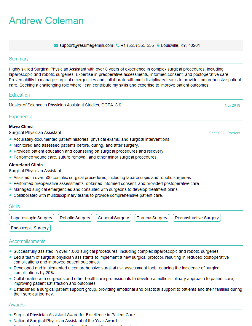
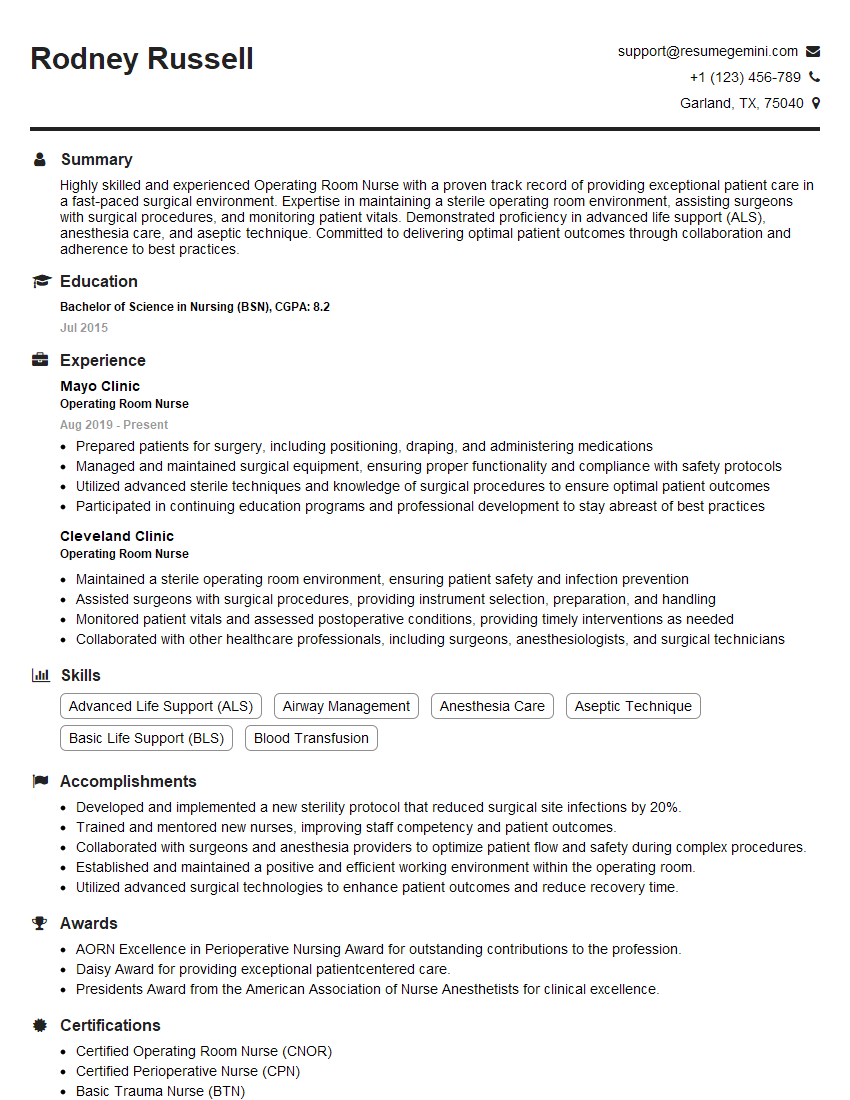
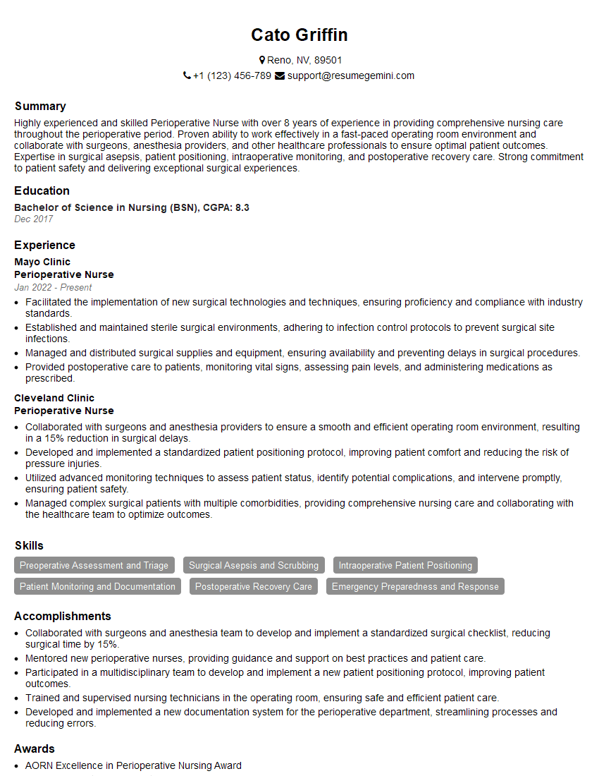
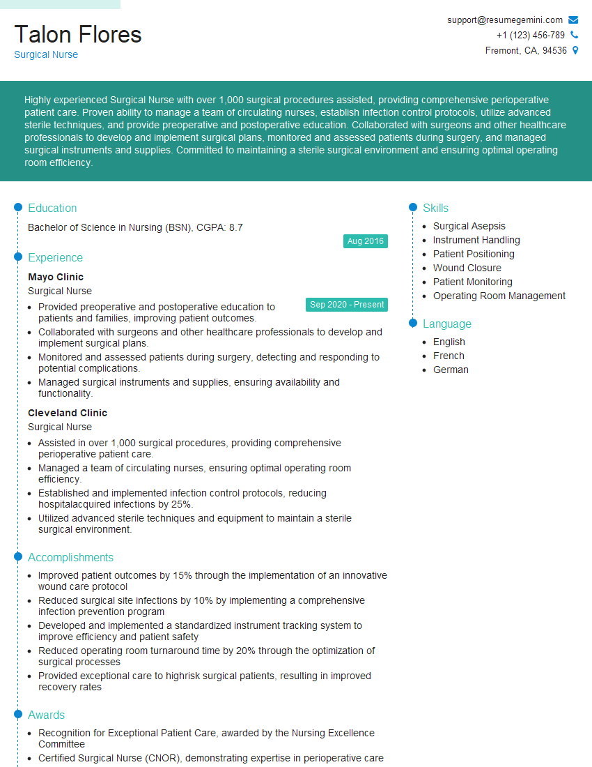
Mastering Preoperative and Postoperative Wound Care significantly enhances your career prospects in healthcare, opening doors to specialized roles and increased earning potential. To maximize your job search success, focus on building a strong, ATS-friendly resume that highlights your skills and experience effectively. ResumeGemini is a trusted resource to help you craft a professional and impactful resume that catches the eye of recruiters. Take advantage of their tools and resources – examples of resumes tailored to Preoperative and Postoperative Wound Care are available to guide you.
Explore more articles
Users Rating of Our Blogs
Share Your Experience
We value your feedback! Please rate our content and share your thoughts (optional).
What Readers Say About Our Blog
This was kind of a unique content I found around the specialized skills. Very helpful questions and good detailed answers.
Very Helpful blog, thank you Interviewgemini team.
