Are you ready to stand out in your next interview? Understanding and preparing for Jones Fracture Repair interview questions is a game-changer. In this blog, we’ve compiled key questions and expert advice to help you showcase your skills with confidence and precision. Let’s get started on your journey to acing the interview.
Questions Asked in Jones Fracture Repair Interview
Q 1. Describe the anatomical location of the Jones fracture.
A Jones fracture is a specific type of fracture affecting the base of the fifth metatarsal bone in the foot. This is the bone located on the outside of your foot, just below the little toe. More precisely, the fracture occurs at the metaphyseal-diaphyseal junction, which is the area where the wider, end part of the bone (metaphysis) meets the narrower, shaft-like portion (diaphysis). This location, being relatively poorly vascularized, makes it prone to delayed union or non-union (failure to heal properly).
Q 2. What are the common mechanisms of injury for a Jones fracture?
Jones fractures typically result from forceful plantarflexion and inversion of the foot. Imagine quickly pivoting on the outside of your foot while playing sports, or stepping awkwardly off a curb. These actions put a significant amount of stress on the fifth metatarsal, leading to a fracture at its weakest point. Other mechanisms can include a direct blow to the outside of the foot.
- Plantarflexion: Pointing your toes downwards.
- Inversion: Turning your foot inwards.
Athletes, particularly those participating in sports like basketball, soccer, and running, are at a higher risk due to the repetitive stress and sudden movements involved.
Q 3. Explain the difference between a Jones fracture and a fifth metatarsal avulsion fracture.
While both Jones fractures and fifth metatarsal avulsion fractures involve the fifth metatarsal, their location and mechanism differ significantly. A Jones fracture occurs at the metaphyseal-diaphyseal junction, as previously described. In contrast, a fifth metatarsal avulsion fracture happens at the tuberosity, the bony bump on the base of the fifth metatarsal. This tuberosity serves as an attachment point for several tendons and ligaments. An avulsion fracture occurs when these tendons or ligaments forcefully pull away from the bone, taking a small piece of bone with them. The avulsion fracture tends to be a less serious injury than a Jones fracture, generally healing quicker and with less risk of complications. Think of it this way: a Jones fracture is a break in the shaft of the bone, while an avulsion fracture is a small chip off of the bump at the end of the bone.
Q 4. Discuss the indications for surgical versus non-surgical management of a Jones fracture.
The decision between surgical and non-surgical management of a Jones fracture depends on several factors, including the patient’s age, activity level, fracture displacement, and overall health. Generally, undisplaced or minimally displaced Jones fractures in compliant patients with low activity demands may be treated non-surgically with immobilization in a cast or boot for 6-8 weeks, followed by gradual weight-bearing. However, displaced fractures, fractures in athletes requiring rapid return to activity, or those showing signs of non-union are typically treated surgically.
- Non-surgical: Cast immobilization, good patient compliance is critical.
- Surgical: Indicated for displaced fractures, high-demand patients, or signs of non-union.
Q 5. What are the advantages and disadvantages of open reduction and internal fixation (ORIF) for a Jones fracture?
Open reduction and internal fixation (ORIF) is a surgical procedure involving the direct visualization and realignment of the fractured bone fragments, followed by their stabilization with screws or plates.
- Advantages: ORIF provides stable fixation, allowing for early weight-bearing and faster return to activity. It significantly reduces the risk of non-union, which is a significant complication of Jones fractures.
- Disadvantages: ORIF carries the risks associated with any surgery, including infection, hardware complications (screw breakage, loosening), and potential nerve or tendon injury. It also involves a longer recovery period initially due to the surgery itself and potential post-operative discomfort.
Q 6. Describe the surgical technique for ORIF of a Jones fracture, including screw placement.
The surgical technique for ORIF of a Jones fracture typically involves a small incision over the fracture site. The surgeon then directly visualizes the fracture fragments, removing any interposed soft tissue. The fractured bone ends are meticulously reduced (realigned) and then stabilized using one or two screws. Screw placement is crucial. Ideally, the screws are placed obliquely, avoiding the precarious blood supply to the fracture site. This oblique screw placement helps to enhance fracture healing and mitigate the risk of complications. A small plate may be used for added stability in certain cases, particularly in highly comminuted (fragmented) fractures. The wound is then closed, and the foot immobilized in a post-operative cast or boot.
Q 7. What are the potential complications associated with surgical management of a Jones fracture?
Potential complications associated with the surgical management of a Jones fracture include, but are not limited to:
- Infection: A risk with any surgical procedure.
- Non-union: Failure of the fracture to heal, even with surgery, though significantly less common with ORIF.
- Malunion: Healing of the fracture in a malaligned position.
- Hardware complications: Screw breakage, loosening, or prominence.
- Nerve or tendon injury: Rare, but a potential risk during the surgical procedure.
- Delayed union: Slower than expected healing.
- Compartment syndrome: Rare but serious condition requiring immediate attention.
Careful surgical technique, meticulous postoperative care, and close patient follow-up are essential in minimizing these risks.
Q 8. Describe the non-operative management options for a Jones fracture.
Non-operative management of a Jones fracture, a fracture of the base of the fifth metatarsal, is primarily focused on immobilization and allowing the fracture to heal naturally. This approach is typically considered for less displaced fractures in otherwise healthy individuals.
The primary method involves using a short leg walking cast or boot, which provides stability to the foot and ankle, preventing movement at the fracture site. This immobilization is crucial for proper bone healing. The patient is typically non-weight-bearing initially, gradually progressing to weight-bearing as tolerated based on clinical assessment and imaging. The duration of immobilization typically ranges from 6-8 weeks, depending on the fracture healing progress monitored by radiographs.
Other supportive measures include using crutches to avoid stressing the injured foot, elevation of the foot to reduce swelling, and pain management with over-the-counter analgesics or prescription medication if necessary. Regular follow-up appointments with the physician are essential to monitor healing and address any complications that may arise.
Q 9. What are the factors that influence the choice of treatment modality for a Jones fracture?
The decision to manage a Jones fracture operatively or non-operatively hinges on several key factors. The fracture displacement is paramount; significantly displaced fractures are more likely to require surgery to achieve anatomical reduction and improve healing potential. Similarly, the patient’s overall health plays a role; patients with conditions like diabetes or those who are smokers may have impaired healing, making surgery a more favorable option.
- Fracture Displacement: Highly displaced fractures are more likely to need surgery.
- Patient Factors: Age, overall health, activity level, and compliance with non-operative treatment influence decision making.
- Comorbidities: Conditions like diabetes, smoking, or obesity can significantly impact healing and increase the risk of nonunion.
- Patient Preference: Patient wishes should be respected when formulating a treatment plan, provided they are based on informed consent.
For instance, a young, active athlete with a significantly displaced Jones fracture would likely benefit from surgical fixation to ensure rapid and complete healing and a faster return to sports. Conversely, an older, sedentary patient with a minimally displaced fracture may be a suitable candidate for non-operative management.
Q 10. How is the healing process of a Jones fracture monitored?
Monitoring the healing process of a Jones fracture involves a combination of clinical examination and imaging studies. Clinical examination focuses on assessing pain levels, swelling, and range of motion. The patient’s ability to bear weight is also closely monitored.
Radiographic imaging, specifically X-rays, is crucial for assessing fracture healing. Serial X-rays are typically taken at regular intervals (e.g., 4-6 weeks after the initial injury, and then again as needed) to evaluate callus formation and assess the progression of bony union. The radiographic appearance of bridging callus across the fracture line indicates progressive healing. Other imaging modalities such as CT scans or MRI may be utilized in specific cases for a more detailed assessment of the fracture, but X-rays remain the mainstay of fracture monitoring.
Q 11. What are the signs and symptoms of a nonunion in a Jones fracture?
A nonunion, the failure of a fracture to heal within a reasonable timeframe, can manifest in several ways in a Jones fracture. Clinically, persistent pain and swelling at the fracture site, despite adequate immobilization, are significant red flags. The patient may also experience persistent tenderness and limited range of motion in the foot and ankle. Functionally, the patient may struggle with weight-bearing on the affected foot.
Radiographically, a nonunion is characterized by the absence of bridging callus across the fracture line. The fracture fragments may appear sclerotic (increased bone density) with widening of the fracture gap. There may be evidence of reactive bone formation at the fracture ends, suggesting the persistent inflammatory process.
Q 12. How is a nonunion of a Jones fracture treated?
Treatment of a Jones fracture nonunion typically involves surgical intervention. The primary goal of surgery is to achieve stable fracture fixation and facilitate bone healing. This often involves open reduction and internal fixation (ORIF), where the fracture fragments are surgically realigned (reduced) and held in place with screws or plates. In some cases, bone grafting may be necessary to augment bone healing.
The surgical approach depends on several factors, including the degree of nonunion, the quality of the bone stock, and the patient’s overall health. In cases where significant bone loss is present, more complex procedures might be required, potentially including the use of bone grafts and/or specialized fixation devices. Post-operatively, a period of immobilization in a cast or boot is typically required, followed by a comprehensive rehabilitation program to restore function.
Q 13. What are the indications for bone grafting in a Jones fracture nonunion?
Bone grafting in a Jones fracture nonunion is indicated when there is a significant defect or gap at the fracture site hindering the natural healing process. This gap may be due to initial fracture displacement, bone loss from infection or avascular necrosis (death of bone tissue), or inadequate callus formation. Bone grafts provide a scaffold for new bone formation and promote healing. Several types of bone grafts can be used, including autografts (from the patient’s own body), allografts (from a donor), or synthetic bone graft substitutes.
The decision to use bone grafting is based on the size and characteristics of the fracture gap, the patient’s overall health, and the surgeon’s judgment. Adequate bone graft incorporation is crucial for successful union. Regular post-operative imaging is essential to monitor graft integration and fracture healing.
Q 14. Describe the role of physical therapy in the rehabilitation of a Jones fracture.
Physical therapy plays a critical role in the rehabilitation of a Jones fracture, both after non-operative and operative management. The goals of physical therapy are to reduce pain and swelling, restore range of motion in the ankle and foot, improve muscle strength and function, and ultimately, facilitate a return to normal activities. Early in the rehabilitation process, the focus is on range of motion exercises and pain management techniques such as ice and elevation.
As healing progresses, the emphasis shifts towards strengthening exercises for the muscles of the lower leg and foot, balance training, and proprioceptive exercises to improve coordination and stability. Once the patient is able to bear weight without pain, more advanced exercises such as gait training and functional activities will be introduced to help the patient regain their pre-injury activity levels. The duration of physical therapy varies depending on the individual patient’s needs and progress, but it typically lasts several weeks or months.
Q 15. What are the common complications of physical therapy following a Jones fracture repair?
Complications following physical therapy for a Jones fracture repair are thankfully uncommon but can be serious. The main concern revolves around inadequate healing or re-injury. This can manifest in several ways:
- Delayed Union or Non-union: The fracture doesn’t heal properly within the expected timeframe (delayed union) or fails to heal completely (non-union). This is often due to insufficient immobilization, early weight-bearing, or underlying medical conditions affecting bone healing. Imagine trying to mend a broken stick without properly splinting it; the break might not heal correctly.
- Re-fracture: Returning to activity too soon can lead to the fracture breaking again, setting back the healing process significantly. Think of it like trying to use a freshly mended bone too forcefully—it can easily break again.
- Chronic Pain and Stiffness: Improper rehabilitation or insufficient range-of-motion exercises can result in persistent pain and stiffness in the ankle and foot, significantly impacting long-term function. This is analogous to a muscle that’s been immobilized for too long becoming atrophied and stiff.
- Complex Regional Pain Syndrome (CRPS): Although rare, CRPS is a debilitating condition characterized by persistent pain, swelling, and changes in skin color and temperature. This is often triggered by injury and can significantly hamper recovery.
- Infection: While less common with modern surgical techniques, infection at the fracture site remains a possibility and can have severe consequences. Good sterile surgical techniques are vital.
Careful adherence to the physical therapist’s instructions, close monitoring of healing progress, and open communication with the healthcare team are crucial in minimizing these risks.
Career Expert Tips:
- Ace those interviews! Prepare effectively by reviewing the Top 50 Most Common Interview Questions on ResumeGemini.
- Navigate your job search with confidence! Explore a wide range of Career Tips on ResumeGemini. Learn about common challenges and recommendations to overcome them.
- Craft the perfect resume! Master the Art of Resume Writing with ResumeGemini’s guide. Showcase your unique qualifications and achievements effectively.
- Don’t miss out on holiday savings! Build your dream resume with ResumeGemini’s ATS optimized templates.
Q 16. What imaging modalities are used to diagnose a Jones fracture?
The primary imaging modality for diagnosing a Jones fracture is a lateral weight-bearing X-ray of the foot. This view allows visualization of the fracture line at the base of the fifth metatarsal. Sometimes additional views, such as an oblique X-ray, are needed for better visualization. In cases of unclear findings or suspected avulsion fractures, CT scans can provide more detailed images of the bone and surrounding soft tissues.
MRI scans are generally not needed for diagnosis but may be considered in cases where there’s a suspected avulsion fracture or if there’s a delay in healing to assess soft tissue involvement. A bone scan, using radioactive tracers to highlight areas of increased metabolic activity, may be employed to assess the healing process, particularly in cases of delayed union or non-union.
Q 17. How would you interpret an X-ray of a Jones fracture?
Interpreting an X-ray of a Jones fracture involves looking for a specific fracture pattern at the base of the fifth metatarsal, typically 1-1.5cm from the metatarsal head. The fracture line usually runs transversely, which distinguishes it from other fractures in that region.
Key features to look for include:
- Transverse or slightly oblique fracture line: The fracture line is usually not completely perpendicular but runs more across the bone.
- Fracture site: The fracture is located in the metaphyseal region of the fifth metatarsal (the junction of the shaft and the growth plate), often within 1-1.5cm of the tuberosity.
- Displaced or non-displaced: The bone fragments may be shifted (displaced) or aligned properly (non-displaced). Displaced fractures are more likely to require surgical intervention.
- Absence of other injuries: The X-ray should be evaluated for other fractures or injuries.
It’s crucial to remember that accurate interpretation requires experience. Often, subtle findings can make this a challenging diagnosis and a consultation with a radiologist is invaluable.
Q 18. What are the differential diagnoses for a Jones fracture?
Differential diagnoses for a Jones fracture are crucial because other injuries can mimic its symptoms. These include:
- Avulsion Fracture of the Fifth Metatarsal Tuberosity: This involves a small fragment of bone pulled away from the main bone, often caused by tendon or ligament injury. It typically presents differently on imaging.
- Stress Fracture of the Fifth Metatarsal: This is a fatigue fracture that occurs due to repetitive stress and often presents with less obvious imaging findings in the early stages.
- Fracture of the Base of the Fifth Metatarsal (non-Jones): Fractures in this area that don’t meet the specific criteria for a Jones fracture (e.g., different location, oblique fracture).
- Apophysitis (Sever’s Disease): In young athletes, this condition involves inflammation of the growth plate at the heel, sometimes mistaken for a fracture.
- Soft Tissue Injuries: Sprains, strains, and tendonitis in the foot and ankle can mimic the symptoms of a Jones fracture, although the imaging findings will be normal.
Careful history taking (mechanism of injury, specific symptoms), physical examination, and thorough image interpretation are necessary for accurate diagnosis.
Q 19. Discuss the importance of weight-bearing restrictions after a Jones fracture repair.
Weight-bearing restrictions after a Jones fracture repair are absolutely critical for successful healing. The fifth metatarsal, particularly in the Jones fracture location, receives substantial weight-bearing forces during activities. Premature weight-bearing can significantly increase the risk of non-union, delayed union, or re-fracture.
The duration and degree of weight-bearing restriction depend on several factors, including the fracture’s severity (displaced vs. non-displaced), the type of fixation used (non-operative vs. operative), and the patient’s overall health and compliance. Typically, initial weight-bearing is limited using crutches or a walking boot, gradually increasing weight-bearing as the fracture heals under the supervision of a healthcare professional.
Non-compliance with weight-bearing restrictions is a major reason for treatment failures. It’s vital for patients to understand and adhere to these instructions to ensure proper healing and avoid long-term complications.
Q 20. How long is the typical recovery time for a Jones fracture?
The typical recovery time for a Jones fracture varies greatly depending on several factors, including the severity of the fracture, the treatment method (surgical versus non-surgical), the patient’s age and overall health, and their adherence to the prescribed treatment plan. Recovery time often ranges from 6 to 12 weeks for non-operative management, while surgical intervention may extend the healing period.
However, a complete return to full activity, especially high-impact activities, may take considerably longer, potentially up to several months. This is because even if the bone heals, the surrounding soft tissues need time to regain their full strength and function. Each patient’s recovery is unique and guided by their individual progress and clinical assessments.
Q 21. Describe the use of different types of implants for Jones fracture fixation.
The choice of implant for Jones fracture fixation depends on various factors, including the fracture pattern, displacement, patient age, and surgeon preference. Here are some commonly used implants:
- Compression screws: These are frequently used in operative management to compress the fracture fragments and promote healing. The screws provide stability and prevent movement of the fractured bone pieces.
- Plates and screws: For complex or severely displaced fractures, plates and screws offer enhanced stability. A plate provides rigid support along the length of the bone, while screws further secure the fragments in place.
- Intramedullary (IM) nails: Although less common for Jones fractures, IM nails can be considered in certain circumstances to provide axial stability.
In some cases, particularly for minimally displaced or stable fractures, non-operative management, involving casting or splinting, can be successful. The decision of whether to use implants and which type to use is a complex one made by an orthopedic surgeon in consultation with the patient. Each option comes with its own set of advantages and disadvantages, with the primary goal being to obtain stable fracture reduction and facilitate bone healing.
Q 22. What are the advantages and disadvantages of different screw types in Jones fracture fixation?
The choice of screw type in Jones fracture fixation is crucial for optimal healing. Several factors influence this decision, including fracture characteristics (displacement, comminution), bone quality, and patient factors (age, activity level).
- Advantages and Disadvantages of Different Screw Types:
- Cancellous Screws: These are shorter screws ideal for less displaced fractures, offering good purchase in cancellous bone (spongy bone). Advantages: Easier insertion, less risk of over penetration. Disadvantages: May not provide sufficient stability for significantly displaced fractures, increased risk of screw loosening or pullout in osteoporotic bone.
- Cortical Screws: These are longer, stronger screws designed for cortical bone (compact bone). They offer superior fixation for displaced fractures. Advantages: Excellent stability, reduced risk of failure in high-demand patients. Disadvantages: More technically demanding to insert, greater risk of over-penetration and injury to adjacent structures.
- Herbert Screws: These are cannulated screws that offer a combination of cancellous and cortical screw characteristics. Advantages: Versatile, suitable for a range of fracture patterns. Disadvantages: Can be expensive.
The decision of which screw type to use is often guided by the surgeon’s experience and the specific fracture morphology, balancing the need for stability with the risks of complications.
Q 23. How do you address malunion after Jones fracture repair?
Malunion, where the fracture heals in an unsatisfactory position, can significantly impact function after a Jones fracture. Addressing malunion often requires surgery.
- Surgical Options:
- Osteotomy: This involves surgically cutting the bone to correct the malunion. A plate and screws are then used to stabilize the bone in the correct anatomical position.
- Revision Surgery: This might involve removing the existing hardware and re-fixating the fracture using a different technique (e.g., changing to a longer screw, adding additional fixation).
The decision on the most appropriate surgical approach is made on a case-by-case basis, considering the degree of malunion, the patient’s age and activity level, and the time elapsed since the initial injury. Non-operative management is rarely suitable for significant malunions.
Q 24. What are the risk factors for delayed union or nonunion in a Jones fracture?
Delayed union and nonunion (failure to heal) are serious complications of Jones fractures. Several factors increase the risk:
- Patient-Related Factors:
- Smoking: Nicotine significantly impairs bone healing.
- Diabetes: Poorly controlled blood sugar levels negatively impact bone healing.
- Obesity: Obesity is associated with decreased blood supply to the fracture site.
- Age: Older individuals tend to have slower bone healing.
- Fracture-Related Factors:
- Significant Displacement: Highly displaced fractures are more prone to complications.
- Comminution (fragmentation): Fractures with multiple bone fragments heal more slowly.
- Interruption of blood supply: The tenuous blood supply to the fifth metatarsal base contributes to a higher risk of non-union.
Minimizing these risk factors through pre-operative optimization (e.g., smoking cessation) and proper surgical technique improves the chances of successful fracture healing.
Q 25. How do you counsel a patient about the potential complications of Jones fracture repair?
Counseling patients about potential complications is a critical part of informed consent. I emphasize the importance of realistic expectations and discuss the potential for both common and less frequent complications in a clear and empathetic manner.
- Common Complications:
- Delayed Union/Nonunion: I explain that while most Jones fractures heal, some may heal more slowly or fail to heal entirely, requiring further surgery. I outline the potential need for bone grafting or other interventions.
- Infection: The risk of infection following surgery, although low, is explained.
- Hardware Problems: Potential issues with screw loosening or breakage are discussed.
- Malunion: The possibility of the fracture healing in an incorrect position is explained, highlighting its potential impact on function.
- Less Common Complications:
- Complex Regional Pain Syndrome (CRPS): This rare but debilitating condition can follow a fracture. I outline its symptoms and potential treatments.
- Nerve Injury: The risk of nerve damage during surgery is acknowledged, though infrequent.
I also discuss rehabilitation, the anticipated recovery time, and the importance of following post-operative instructions meticulously. Open communication throughout the process builds trust and ensures patient understanding.
Q 26. Explain your approach to managing a patient with a displaced Jones fracture.
My approach to a displaced Jones fracture typically involves operative fixation. Conservative management is generally not recommended due to the high risk of nonunion.
- Surgical Technique:
- Open Reduction and Internal Fixation (ORIF): This involves surgically exposing the fracture, reducing (realigning) the bone fragments, and securing them with screws. Fluoroscopic imaging is crucial during the procedure to ensure accurate reduction and screw placement.
- Screw Selection: The choice of screw type (Cancellous, Cortical, or Herbert) depends on factors such as fracture displacement, bone quality, and patient characteristics as previously discussed.
- Post-operative Management: This includes pain management, early mobilization with weight-bearing as tolerated, and physical therapy to restore full function. Weight-bearing restrictions are typically followed for a period of time, depending on fracture healing.
A meticulous surgical technique and close post-operative monitoring are essential to optimize the healing process and minimize complications.
Q 27. How would you manage a Jones fracture in a high-demand athlete?
Managing a Jones fracture in a high-demand athlete requires a more aggressive and tailored approach to ensure a swift and complete return to sport. The ultimate goal is a rapid, yet safe, return to optimal performance.
- Surgical Considerations:
- Early Surgical Intervention: Prompt surgical fixation, typically within the first few days after the injury, can reduce healing time.
- Rigid Fixation: Utilizing techniques that maximize stability and minimize motion at the fracture site (e.g., appropriately sized and positioned screws) is crucial for faster healing.
- Intensive Rehabilitation: A structured rehabilitation program focusing on strength, range of motion, and proprioception (body awareness) is essential. This program is personalized to the athlete’s sport and demands.
- Imaging Monitoring: Regular radiographic monitoring (X-rays) is essential to assess healing progress and identify potential complications early.
The collaboration between the surgeon, physical therapist, and athletic trainer is critical in helping high-demand athletes regain their full athletic potential.
Q 28. Discuss the current research and advancements in the management of Jones fractures.
Research on Jones fracture management is ongoing, focusing on improving healing rates and reducing complications.
- Areas of Current Research:
- Biological Augmentation: Research is exploring the use of bone growth factors and other biologics to enhance fracture healing. This could reduce the time to union, particularly in high-risk patients.
- Minimally Invasive Techniques: Studies are evaluating less invasive surgical techniques to reduce tissue trauma and improve recovery times. These may involve smaller incisions and different types of fixation.
- Biomechanical Analysis: Advanced biomechanical modeling and simulations are being used to optimize surgical techniques and improve implant designs.
- Improved Imaging Techniques: Newer imaging modalities can provide a better understanding of fracture healing dynamics, and aid in the earlier detection of complications.
These advancements offer promise for enhanced treatment of Jones fractures, leading to improved outcomes and a faster return to function for patients.
Key Topics to Learn for Jones Fracture Repair Interview
- Anatomy of the 5th Metatarsal: Understanding the specific anatomical location and biomechanics of the Jones fracture site is crucial.
- Fracture Classification and Types: Familiarize yourself with different classifications (e.g., based on location, displacement) and their implications for treatment.
- Diagnosis Techniques: Understand the role of X-rays, CT scans, and clinical examination in diagnosing Jones fractures and differentiating them from other injuries.
- Treatment Options: Explore the range of treatment options, including non-operative management (casting, bracing) and operative techniques (e.g., screw fixation, plate fixation).
- Surgical Approaches and Techniques: If focusing on surgical roles, delve into specific surgical approaches and the advantages/disadvantages of each fixation method.
- Post-operative Care and Rehabilitation: Gain a thorough understanding of post-operative protocols, including weight-bearing restrictions, physical therapy, and potential complications.
- Complications and Management: Learn to identify potential complications such as non-union, malunion, infection, and delayed union, and their management strategies.
- Biomechanics and Fracture Healing: Understand the principles of fracture healing and the biomechanical factors influencing the healing process in Jones fractures.
- Evidence-Based Practice: Stay updated on the latest research and clinical guidelines related to Jones fracture management.
- Case Studies and Problem Solving: Practice analyzing case scenarios and applying your knowledge to develop appropriate treatment plans.
Next Steps
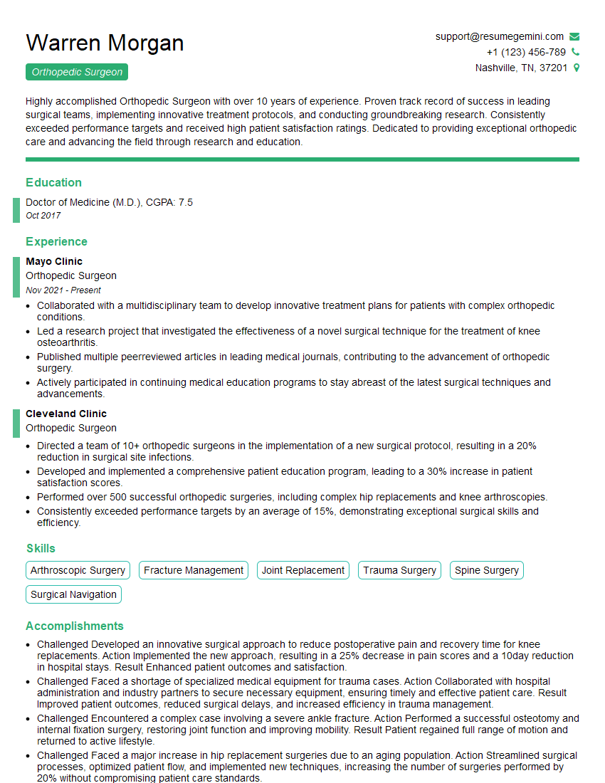
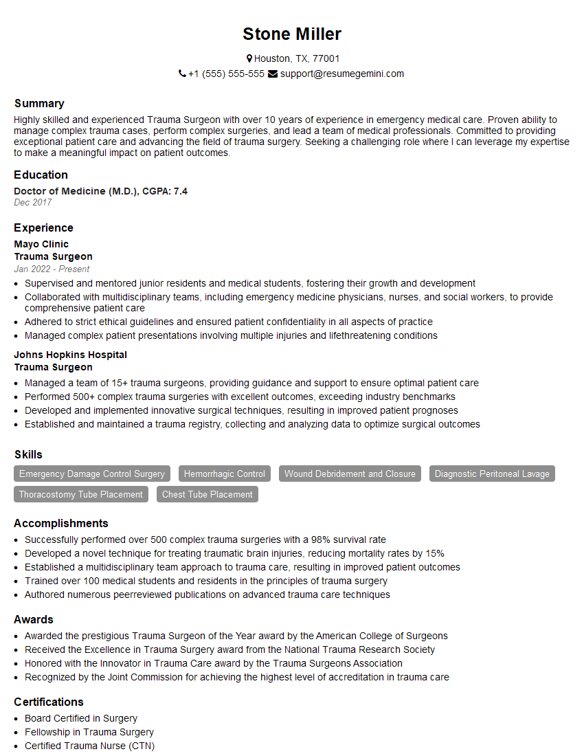
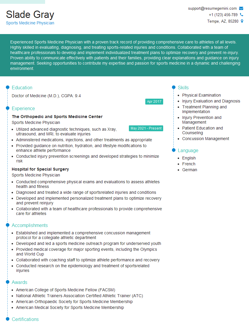
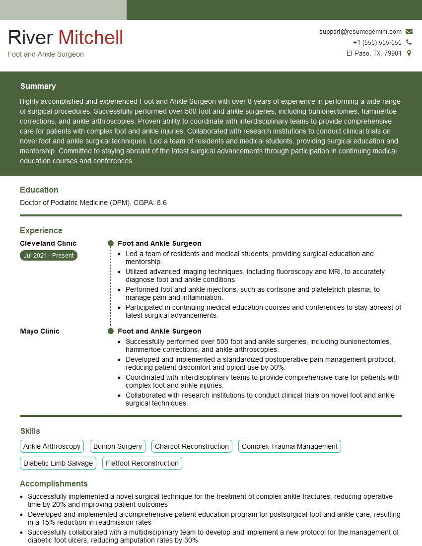
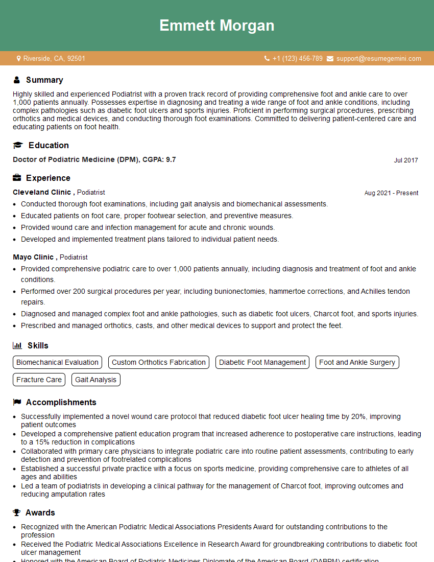
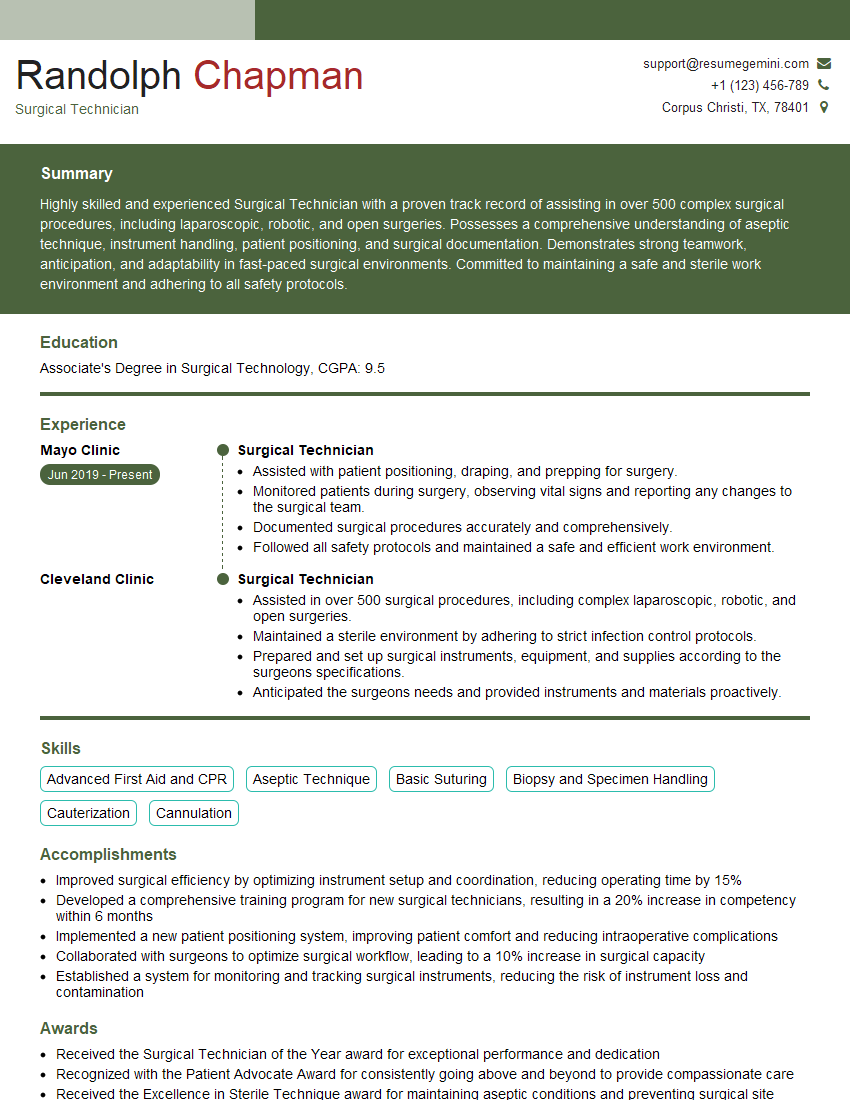
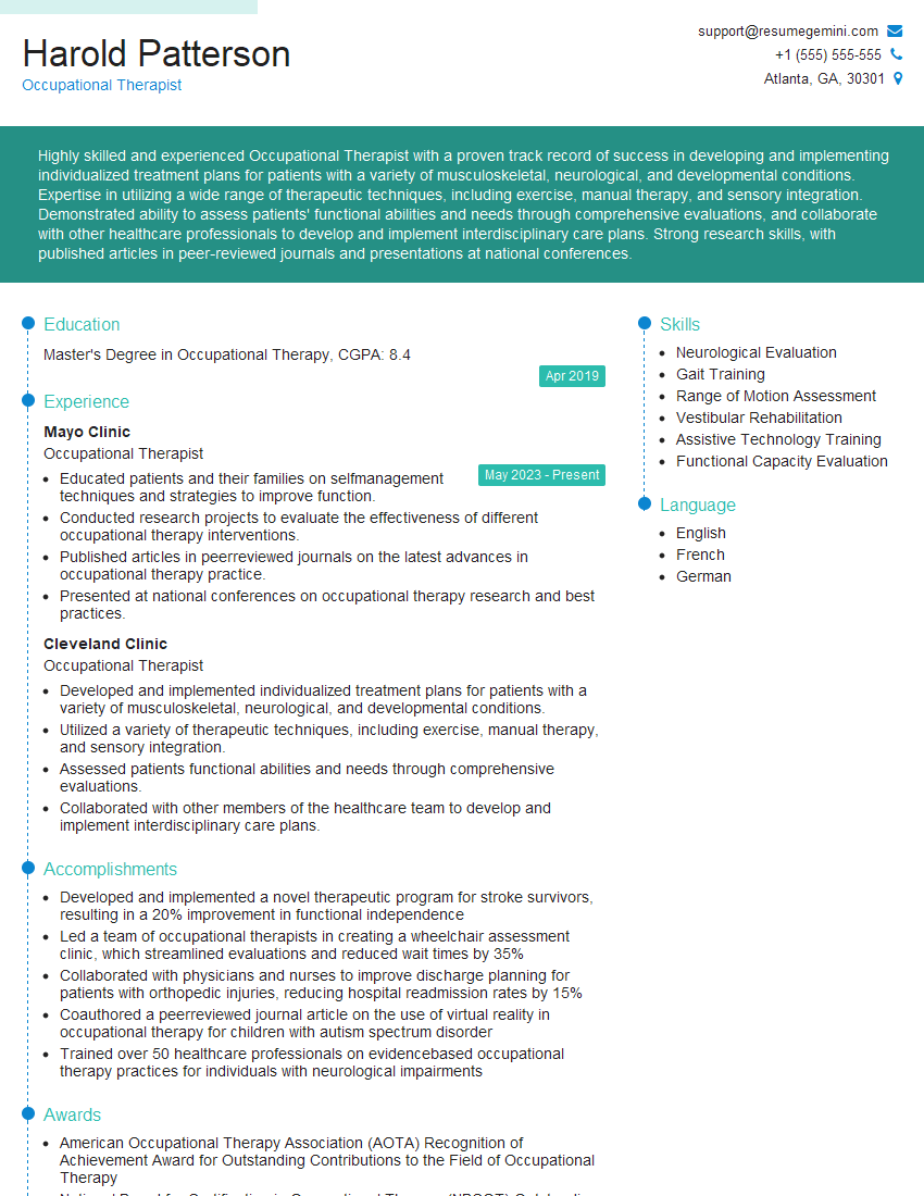
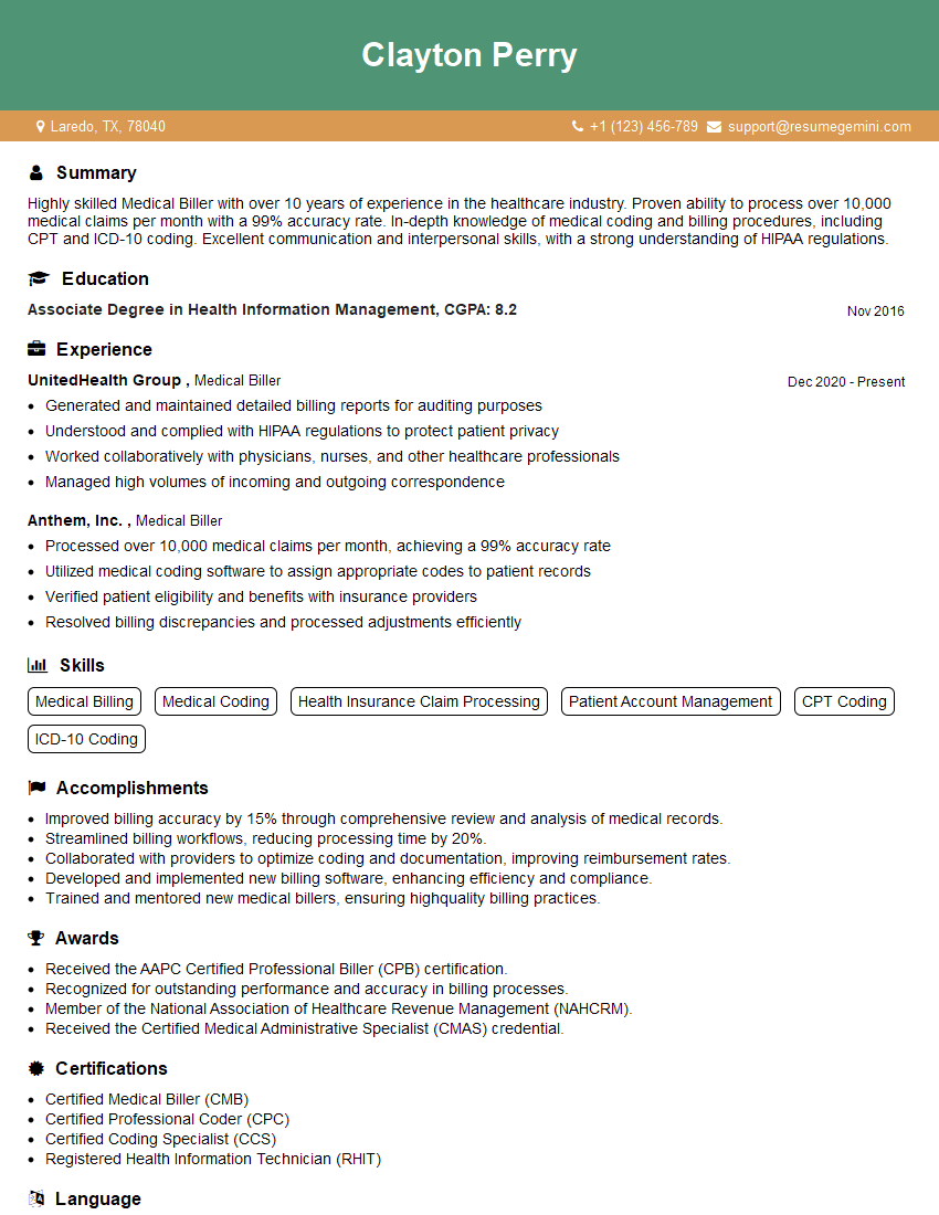
Mastering Jones Fracture Repair demonstrates a strong foundation in orthopedic surgery and trauma care, significantly enhancing your career prospects. A well-crafted resume is key to showcasing your expertise effectively to potential employers. Creating an ATS-friendly resume increases the likelihood of your application being noticed and considered. ResumeGemini is a trusted resource to help you build a professional and impactful resume, ensuring your skills and experience shine. Examples of resumes tailored to Jones Fracture Repair are available to help guide you.
Explore more articles
Users Rating of Our Blogs
Share Your Experience
We value your feedback! Please rate our content and share your thoughts (optional).
What Readers Say About Our Blog
This was kind of a unique content I found around the specialized skills. Very helpful questions and good detailed answers.
Very Helpful blog, thank you Interviewgemini team.
