Are you ready to stand out in your next interview? Understanding and preparing for Lisfranc Fracture Repair interview questions is a game-changer. In this blog, we’ve compiled key questions and expert advice to help you showcase your skills with confidence and precision. Let’s get started on your journey to acing the interview.
Questions Asked in Lisfranc Fracture Repair Interview
Q 1. Describe the anatomy of the Lisfranc joint.
The Lisfranc joint is a complex articulation in the midfoot, crucial for stability and mobility. It’s not a single joint, but rather a series of articulations between the medial cuneiform, intermediate cuneiform, lateral cuneiform, and cuboid bones of the midfoot and the bases of the first through fifth metatarsals. Think of it as a keystone arch – each bone contributes to the overall stability. The critical ligaments connecting these bones, particularly the Lisfranc ligament (connecting the medial cuneiform to the base of the second metatarsal), are essential for maintaining the integrity of the joint.
Imagine a stack of building blocks; the Lisfranc joint is where these blocks (bones) interlock. The ligaments act as the mortar, holding the blocks firmly together. Disruption of this intricate structure leads to the instability and pain characteristic of a Lisfranc injury.
Q 2. What are the common mechanisms of injury for a Lisfranc fracture-dislocation?
Lisfranc injuries typically result from high-energy trauma, such as a fall from a significant height or a motor vehicle accident, where a direct force is applied to the midfoot. These forces can cause a fracture, a dislocation, or a combination of both. Common mechanisms include:
- Axial loading: A direct force applied along the long axis of the foot, such as landing on the foot after a fall.
- Rotation: A twisting injury, often seen in sports, can shear the Lisfranc joint apart.
- Lateral compression: A force pushing the foot inward, which can lead to a disruption of the joint.
For instance, a football player who gets his foot stuck in the turf while twisting could easily suffer a Lisfranc injury. It’s crucial to understand that even seemingly minor trauma can, in some cases, result in significant Lisfranc instability.
Q 3. Explain the different classification systems for Lisfranc injuries.
Several classification systems exist for Lisfranc injuries, each with its own strengths and weaknesses. They generally categorize injuries based on the degree of involvement of the metatarsals and the associated ligaments and fractures.
- Myerson Classification: This system categorizes injuries based on the degree of displacement and articular involvement. Type I involves ligamentous injuries without displacement, while Types II–IV describe increasing degrees of fracture and displacement.
- Hardcastle Classification: This system focuses on the specific bones and ligaments involved, offering a more detailed anatomical description of the injury.
- Others: Numerous other classifications exist, using combinations of radiographic findings and surgical approaches.
These systems are crucial for guiding treatment planning. A simple ligamentous sprain might only need conservative management, whereas a complex fracture-dislocation will almost certainly require surgery.
Q 4. How do you differentiate a Lisfranc injury from other midfoot injuries?
Differentiating a Lisfranc injury from other midfoot injuries requires careful clinical examination and imaging. Key features that suggest a Lisfranc injury include:
- Pain and swelling precisely localized to the midfoot, often over the Lisfranc joint itself
- Deformity of the midfoot
- Tenderness specifically at the Lisfranc joint
- Pain on weight-bearing
- Difficulties with range of motion in the midfoot
Conditions such as navicular fractures, cuboid fractures, and other midfoot ligament sprains may present similarly. A thorough evaluation, including careful palpation of the midfoot and detailed imaging, is crucial for accurate diagnosis.
Q 5. What are the key findings on physical exam for a suspected Lisfranc injury?
Physical examination is a critical first step in evaluating a suspected Lisfranc injury. Key findings include:
- Pain and swelling in the midfoot region: This is often the presenting complaint.
- Tenderness to palpation at the Lisfranc joint: Careful palpation, feeling for tenderness over each articulation, is crucial.
- Midfoot instability: Assess for abnormal movement or widening of the joint space by comparing it to the uninjured side. A positive “piano key” sign, where the metatarsals can be shifted medially and laterally relative to the cuneiform bones, is a strong indicator of instability.
- Shortening or dorsal displacement of the metatarsals: A visible difference in length or position compared to the other foot suggests instability.
- Foot alignment: Examine for any rotational or angular deformities.
Remember, a high index of suspicion is necessary because subtle findings may be present.
Q 6. What imaging modalities are used to diagnose a Lisfranc injury, and what are their limitations?
Imaging is essential for definitive diagnosis. The following modalities are typically used:
- Plain radiographs: These are the initial imaging modality, providing valuable information on the alignment and presence of fractures. Weight-bearing radiographs are particularly useful in assessing joint stability. However, subtle ligamentous injuries might be missed.
- Computed tomography (CT): Provides detailed bone visualization and allows for better assessment of fracture patterns and joint alignment. It is better for detecting subtle fractures than plain radiographs.
- Magnetic resonance imaging (MRI): Excellent for evaluating ligamentous injuries and soft tissue damage. It’s particularly helpful in cases where radiographs are inconclusive.
Limitations: Plain radiographs may miss subtle ligamentous injuries or small fractures. CT scans are less sensitive to soft tissue injuries, and MRI can be affected by motion artifacts, making it sometimes less reliable for visualizing fractures.
Q 7. Describe the indications for non-operative management of Lisfranc injuries.
Non-operative management of Lisfranc injuries is typically reserved for minimally displaced injuries with intact articular surfaces and no significant instability. These cases usually involve:
- Stable fractures without significant displacement
- Ligamentous injuries without instability
- Patients with significant comorbidities preventing surgery
Treatment includes immobilization in a non-weight-bearing cast or boot for several weeks, followed by gradual weight-bearing as tolerated. Physical therapy plays a vital role in restoring range of motion and strength. However, the success rate is lower than surgical intervention, and careful monitoring is paramount to detect any signs of instability or non-union.
Q 8. What are the contraindications for non-operative management of Lisfranc injuries?
Non-operative management of Lisfranc injuries, while sometimes considered, is generally reserved for very specific circumstances. It’s contraindicated in cases where there’s significant displacement of the Lisfranc joint, involving any instability or disruption of the articular surfaces. Think of it like this: if you have a badly broken bone, you wouldn’t simply splint it and hope it heals correctly – you need proper realignment. Similarly, with a significantly displaced Lisfranc injury, the chances of a poor outcome, including chronic pain, arthritis, and instability, are significantly higher without surgery.
- Significant displacement of the tarsometatarsal joints: A fracture-dislocation where the bones are not properly aligned.
- Articular incongruity: The joint surfaces don’t fit together smoothly, leading to abnormal stress and wear.
- Open fracture: The bone breaks through the skin, increasing the risk of infection.
- Associated injuries: Other injuries, such as nerve or tendon damage, that require surgical intervention.
- Failure of closed reduction: If we attempt to realign the bones without surgery (closed reduction) and it’s unsuccessful, surgery becomes necessary.
Essentially, non-operative management is only considered for minimally displaced injuries in very select patients, and close follow-up with imaging is crucial to monitor healing and stability.
Q 9. Describe the different surgical approaches to Lisfranc fracture repair.
Surgical approaches to Lisfranc fracture repair aim to achieve anatomical reduction and stable fixation. Several approaches exist, each with its advantages and disadvantages depending on the specific injury pattern:
- Dorsal approach: This is the most common approach, providing excellent exposure to the dorsal aspect of the Lisfranc joint. It allows for precise reduction and fixation of the involved metatarsals and cuneiforms.
- Plantar approach: This approach is often used in conjunction with a dorsal approach, particularly when addressing plantar ligamentous injuries. It offers visualization of the plantar structures crucial to stability.
- Combined dorsal and plantar approaches: This is frequently employed for complex injuries requiring a multi-faceted approach. This provides access to the entire joint complex for comprehensive repair.
- Lateral approach: A less commonly used approach, usually reserved for access to specific fractures or for cases where other approaches are unsuitable.
The choice of surgical approach depends on factors like the location and extent of the fracture-dislocation, the surgeon’s experience, and any associated injuries. The goal in all cases remains precise reduction and stable fixation.
Q 10. Explain the principles of anatomical reduction in Lisfranc fracture repair.
Anatomical reduction in Lisfranc fracture repair is paramount. It means restoring the bones of the midfoot to their precise, pre-injury anatomical alignment. Think of it like meticulously assembling a complex jigsaw puzzle where each piece (bone) must fit perfectly into its designated spot. Imprecise reduction leads to suboptimal healing and long-term complications.
The principles involve:
- Restoration of articular congruity: Ensuring the joint surfaces of the metatarsals and cuneiforms are in perfect contact. This is essential to prevent premature wear and arthritis.
- Accurate reduction of all fracture fragments: Each fracture fragment, regardless of size, must be reduced and stabilized to its original position.
- Restoration of normal joint width: The spaces between the bones should be restored to their pre-injury widths, preventing instability.
- Addressing associated ligamentous injuries: Repair or reconstruction of any torn ligaments is crucial for stability.
Achieving anatomical reduction is often aided by intraoperative fluoroscopy (real-time X-ray imaging) to verify the accuracy of the reduction before fixation.
Q 11. What fixation methods are commonly used in Lisfranc fracture repair?
Various fixation methods are available for Lisfranc fracture repair, each with its own strengths and weaknesses. The goal is to provide rigid stabilization allowing for bone healing.
- Screws: These provide strong fixation and are commonly used to stabilize fractures of the metatarsals and cuneiforms. Different screw types, like cannulated screws and Herbert screws, offer unique benefits in specific situations.
- Plates: Plates offer more extensive stabilization, especially in comminuted fractures (fractures with multiple fragments). They can bridge gaps and provide structural support for the joint.
- K-wires (Kirschner wires): These smaller wires are often used as supplementary fixation, particularly in conjunction with screws or plates, offering additional stability.
- External fixation: This involves pins placed through the skin and bone, connected to an external frame. It’s commonly used in severely comminuted fractures or cases of infection. It offers external stabilization and allows for soft tissue management.
The chosen fixation method is tailored to the individual injury pattern and patient-specific factors.
Q 12. Discuss the advantages and disadvantages of different fixation methods.
The choice of fixation method is a critical decision, influenced by the specifics of each case. Here’s a comparative look:
- Screws: Advantages include minimal invasiveness, strong fixation for simple fractures, and relatively easy application. Disadvantages include potential for screw loosening or breakage, and limited ability to manage complex fracture patterns.
- Plates: Advantages include strong fixation, ability to manage complex fracture patterns, and better management of instability. Disadvantages include increased invasiveness, potential for plate breakage, and increased risk of infection.
- K-wires: Advantages include simplicity and ease of placement, useful in supplementary stabilization. Disadvantages include relatively weak fixation on their own and risk of migration or irritation.
- External fixation: Advantages include management of severe comminution or infection, excellent for soft tissue management, and good for unstable cases. Disadvantages include increased risk of pin-tract infection, less cosmetically appealing, and potential for reduced functionality during early recovery.
In practice, a combined approach, perhaps using screws and plates together, is frequently the optimal strategy. The decision often comes down to balancing the benefits of strong fixation with the need to minimize surgical invasiveness and potential complications.
Q 13. How do you address articular incongruity during Lisfranc repair?
Addressing articular incongruity is crucial for a successful outcome in Lisfranc repair. It’s like ensuring that all the gears in a complex machine mesh perfectly to function smoothly. Failure to address this leads to early osteoarthritis and long-term pain.
Techniques to manage articular incongruity include:
- Precise reduction and fixation: As previously mentioned, meticulously restoring the normal anatomical alignment of the joint surfaces is fundamental. This often requires meticulous manipulation and image guidance.
- Bone grafting: In cases where significant bone loss has occurred, bone grafting can be used to restore the joint’s architecture and ensure proper contact between the articular surfaces.
- Arthrodesis (fusion): In cases with extensive articular damage or where anatomical reduction is impossible, arthrodesis may be necessary. This involves surgically fusing the bones of the Lisfranc joint to create a stable, albeit less mobile, joint.
The optimal approach is carefully determined based on the severity of the articular incongruity and the overall condition of the joint.
Q 14. Describe your post-operative management protocol for Lisfranc fracture repair.
Post-operative management of Lisfranc fractures is crucial for ensuring proper healing and a good functional outcome. It’s a multi-stage process, meticulously planned to support the healing process.
The protocol generally includes:
- Immediate post-operative care: Pain management, elevation of the foot to minimize swelling, and assessment for neurological and vascular compromise.
- Immobilization: A non-weight-bearing period, typically using a cast or splint, is essential to protect the repair site and allow for bone healing. The duration varies depending on the extent of the injury and the fixation method employed.
- Physical therapy: A structured physical therapy program, beginning once the immobilization period is over, is key for regaining range of motion, strength, and function. This is a gradual process, starting with passive range of motion exercises and progressing to weight-bearing exercises.
- Weight-bearing: The transition to weight-bearing is gradual and carefully monitored, initially with partial weight-bearing and gradually increasing the load as healing progresses. This is frequently guided by radiographic assessment.
- Long-term follow-up: Regular clinical and radiographic follow-up is vital to monitor healing, detect potential complications, and assess functional outcome.
The success of post-operative management relies on close collaboration between the surgeon, the patient, and the physical therapist. Consistent adherence to the plan is critical to achieve optimal results.
Q 15. What are the common complications associated with Lisfranc fracture repair?
Lisfranc fracture repair, while often successful, carries several potential complications. These can broadly be categorized into early and late complications. Early complications, arising within the first few months post-surgery, often include infection, malunion (where the bones heal in a misaligned position), nonunion (where the bones fail to heal), and complex regional pain syndrome (CRPS), a chronic pain condition. Late complications, developing months or even years later, can encompass arthritis (degenerative joint disease), avascular necrosis (bone death due to lack of blood supply), and persistent pain or stiffness. The risk of these complications varies based on factors such as the severity of the initial injury, the surgical technique employed, and the patient’s overall health.
- Infection: A serious complication requiring prompt antibiotic treatment and potentially surgical debridement (removal of infected tissue).
- Malunion/Nonunion: Requiring revision surgery to correct the alignment or stimulate healing.
- CRPS: A debilitating condition requiring specialized pain management strategies.
- Arthritis: Can lead to long-term pain and reduced joint function, potentially requiring further intervention.
Career Expert Tips:
- Ace those interviews! Prepare effectively by reviewing the Top 50 Most Common Interview Questions on ResumeGemini.
- Navigate your job search with confidence! Explore a wide range of Career Tips on ResumeGemini. Learn about common challenges and recommendations to overcome them.
- Craft the perfect resume! Master the Art of Resume Writing with ResumeGemini’s guide. Showcase your unique qualifications and achievements effectively.
- Don’t miss out on holiday savings! Build your dream resume with ResumeGemini’s ATS optimized templates.
Q 16. How do you manage post-operative complications such as infection or non-union?
Managing post-operative complications requires a multi-faceted approach. Infection is tackled aggressively with intravenous antibiotics, surgical debridement if necessary, and close monitoring. Nonunion is a more challenging complication, often requiring revision surgery. This might involve bone grafting (using bone from another site or a bone graft substitute) to stimulate healing, the use of internal fixation devices (like plates and screws) for better stabilization, or even bone stimulators in some cases. For malunion, corrective osteotomy (surgical bone cutting and realignment) may be necessary. CRPS management involves a combination of medication, physical therapy, and sometimes nerve blocks or sympathetic nerve surgery. Regular follow-up appointments are crucial for early detection and timely intervention of complications.
For instance, I recently managed a patient with a Lisfranc nonunion. After careful assessment, we performed a revision surgery involving bone grafting and the application of a specialized plate for enhanced stability. He responded well to the treatment, and his fracture eventually healed.
Q 17. What are the criteria for return to activity after Lisfranc fracture repair?
Return to activity after Lisfranc fracture repair is a gradual process, tailored to each patient’s individual healing progress and the specific surgical technique used. It’s crucial to avoid premature weight-bearing, which could disrupt the healing process. Initial stages focus on controlled range-of-motion exercises and pain management. As healing progresses, patients gradually increase weight-bearing under the guidance of a physical therapist. Full weight-bearing and a return to sports or high-impact activities are generally not allowed for at least 4-6 months, and often much longer, depending on the complexity of the fracture and the patient’s response to treatment. Radiographic imaging plays a vital role in monitoring healing and determining readiness for increased activity levels. Clear communication and adherence to the prescribed rehabilitation protocol are essential for optimal outcomes.
Q 18. How do you counsel patients regarding the prognosis of Lisfranc injuries?
Counseling patients about the prognosis of Lisfranc injuries involves a realistic and empathetic approach. While many patients achieve excellent outcomes, it’s important to highlight the possibility of complications. I always explain that recovery is a long process, often requiring months or even years. I discuss the potential for persistent pain, stiffness, and long-term limitations in activity levels, especially in cases of severe injuries or complications. I emphasize the importance of adherence to the rehabilitation plan and the need for regular follow-up appointments. I also reassure patients that with proper care and rehabilitation, many patients regain a significant degree of function and can return to many of their activities, but perhaps not at the same level as before the injury.
For example, I explain that, while the goal is to achieve a pain-free, functional foot, perfect restoration of pre-injury function is not always guaranteed. This honest communication helps manage patient expectations and fosters collaboration during the recovery journey.
Q 19. What are the long-term outcomes associated with Lisfranc fracture repair?
Long-term outcomes after Lisfranc fracture repair vary widely depending on several factors, such as the severity of the initial injury, the surgical technique, the presence of complications, and patient compliance with rehabilitation. In many cases, patients achieve good functional outcomes with minimal pain and return to a satisfactory level of activity. However, some patients experience persistent pain, stiffness, and limitations in activity levels. The development of post-traumatic arthritis is a significant long-term concern, which can necessitate further interventions, such as joint fusion or arthrodesis.
Longitudinal studies indicate that a considerable number of patients have ongoing pain and functional deficits even years after surgery. Therefore, ongoing monitoring and management are essential for optimizing long-term outcomes. Successful outcomes are often tied to early diagnosis, appropriate surgical management, and meticulous adherence to the rehabilitation program.
Q 20. Discuss the role of weight-bearing in the rehabilitation process.
Weight-bearing plays a crucial, yet carefully managed, role in Lisfranc fracture repair rehabilitation. Early weight-bearing is generally avoided to prevent disruption of the fracture healing process. The initial phase typically involves non-weight-bearing or partial weight-bearing with the assistance of crutches or a walker. As healing progresses, under the guidance of the physician and physical therapist, the patient gradually increases weight-bearing, progressing from touch-down weight-bearing to partial, then full weight-bearing. The transition to full weight-bearing usually happens over several weeks or months and is guided by radiographic evidence of fracture healing. Premature weight-bearing can lead to nonunion or malunion, necessitating further surgery. A carefully planned and individualized weight-bearing protocol is critical for optimal outcomes.
Q 21. What are the common causes of failure in Lisfranc fracture repair?
Failure in Lisfranc fracture repair can stem from various factors. Inadequate reduction (misalignment of the fractured bones) during surgery is a major culprit. This can lead to malunion or nonunion. Inadequate fixation, where the surgical implants fail to provide sufficient stability, also contributes to failure. Other factors include infections, patient non-compliance with the rehabilitation protocol (leading to premature weight-bearing), and the presence of certain comorbidities (like diabetes, which can impair healing). In addition, the severity of the initial injury, the patient’s age, and overall health can influence the likelihood of failure. Preventing failure hinges on precise surgical technique, robust fixation, meticulous post-operative care, and strict adherence to the rehabilitation protocol.
Q 22. How do you address malunion or non-union in a Lisfranc fracture?
Malunion and nonunion in Lisfranc injuries represent significant challenges, often requiring revision surgery. A malunion is when the bones heal in a misaligned position, leading to pain, instability, and arthritis. A nonunion is when the fracture fails to heal completely. Addressing these complications requires a thorough assessment of the deformity, including radiographic analysis (X-rays, CT scans) to determine the precise location and degree of malalignment or non-union.
Treatment strategies vary depending on the severity. For malunions, corrective osteotomy (surgical bone cutting and realignment) may be necessary, followed by internal fixation (plates and screws) to maintain the corrected alignment. Bone grafting may be used to stimulate healing in areas of significant bone loss. For nonunions, debridement (removal of non-viable tissue), bone grafting, and the use of electrical bone stimulation may be employed to promote healing. In some cases, external fixation may be used temporarily to stabilize the joint while allowing healing to occur. The choice of surgical technique is highly individualized based on factors such as the patient’s age, activity level, and the specifics of their injury.
Post-operative rehabilitation plays a critical role in achieving successful outcomes after revision surgery for malunion or nonunion. A structured program of physical therapy focusing on range of motion, strength, and functional activities is crucial for restoring optimal joint mechanics and function.
Q 23. Describe your experience with minimally invasive techniques for Lisfranc repair.
Minimally invasive techniques for Lisfranc repair offer several advantages, including reduced surgical trauma, decreased blood loss, faster recovery times, and smaller scars. I have significant experience with these techniques, employing them whenever anatomically feasible. These typically involve the use of smaller incisions and specialized instruments to access and repair the injured structures. Fluoroscopic guidance (real-time X-ray imaging) is often used to ensure accurate reduction and fixation.
One example is the use of minimally invasive percutaneous screws to stabilize the Lisfranc joint. This method avoids extensive dissection of soft tissues, reducing the risk of complications such as infection and nerve or tendon injury. Another example is the use of small incisions to access the joint and perform an open reduction and internal fixation, followed by a limited incision closure.
The decision to use minimally invasive techniques depends on several factors. The severity of the injury, the presence of any associated injuries (such as ligamentous damage or fractures of other bones), the patient’s bone quality, and surgeon experience all play a role. It is important to note that while minimally invasive surgery offers benefits, there are situations where an open approach might be more appropriate to thoroughly address the injury.
Q 24. What is your preferred method for assessing the stability of a Lisfranc repair?
Assessing the stability of a Lisfranc repair is crucial for ensuring a successful outcome. My preferred method incorporates a multi-modal approach. I begin with a thorough clinical examination, assessing range of motion, pain, and tenderness. This is followed by a comprehensive imaging evaluation, including weight-bearing X-rays to assess the alignment and reduction of the fracture fragments. Computed tomography (CT) scans provide detailed three-dimensional images, which are essential for detecting subtle instabilities.
Fluoroscopy during the surgery is paramount for real-time confirmation of reduction and implant placement. Postoperatively, I use stress X-rays to assess stability under load, applying controlled stresses to the Lisfranc joint to visualize any signs of instability or displacement of the fracture fragments. In selected cases, I may utilize other advanced imaging such as MRI to evaluate ligamentous injuries. The combination of clinical, radiological and intraoperative assessment provides the most reliable measure of stability.
Q 25. What is your experience with different types of implants for Lisfranc repair?
My experience encompasses a wide range of implants used in Lisfranc repair, including plates, screws, and occasionally, external fixators. The selection of the implant is dictated by the specific fracture pattern, bone quality, and patient-specific factors. I find that small fragment screws and plates offer excellent stability for many Lisfranc injuries. The use of minimally invasive techniques often favors smaller screws, minimizing tissue disruption.
For complex fractures with significant comminution (fragmentation) or instability, I may use larger plates to provide greater stability. External fixators are reserved for particularly challenging cases, such as those with significant soft tissue injury or instability where the use of internal fixation might be compromised. I carefully weigh the benefits and risks associated with each implant type to select the most appropriate option for each individual case. The choice of material (e.g., titanium, stainless steel) is also considered, and it depends on patient factors and implant characteristics.
Q 26. Describe a challenging Lisfranc fracture case you have managed and how you approached it.
One particularly challenging case involved a young, active male who presented with a severely comminuted Lisfranc fracture, associated with significant ligamentous disruption and soft tissue damage. The initial X-rays showed multiple fracture fragments and dislocation of the Lisfranc joint. Traditional techniques would have involved extensive open surgery, increasing the risk of complications.
I opted for a minimally invasive approach utilizing small incisions and fluoroscopic guidance. I carefully reduced the fracture fragments and stabilized the joint with small screws, meticulously preserving as much soft tissue as possible. However, due to the significant ligamentous injury, I also incorporated a temporary external fixator for additional stability. After several weeks of careful monitoring, the external fixator was removed, allowing for a focused rehabilitation program. His post-operative course was complicated by some persistent pain, but through diligent physical therapy, he ultimately regained excellent function and returned to his pre-injury activity level. This case highlighted the importance of careful planning and adaptability in managing complex Lisfranc injuries.
Q 27. How do you stay updated on the latest advancements in Lisfranc fracture repair?
Staying updated on the latest advancements in Lisfranc fracture repair is a continuous process. I actively participate in professional organizations such as the American Academy of Orthopaedic Surgeons (AAOS) and attend relevant conferences and workshops to learn about new techniques, implants, and research findings. I regularly review peer-reviewed medical journals and publications, focusing on high-impact journals specializing in foot and ankle surgery. I also participate in continuing medical education (CME) courses related to foot and ankle surgery. This multi-faceted approach ensures I remain knowledgeable about the most current evidence-based practices in Lisfranc fracture management.
Q 28. What are your research interests related to Lisfranc fracture repair?
My research interests currently center on improving the long-term outcomes of Lisfranc fracture repair. I am particularly interested in exploring innovative surgical techniques and implant designs that minimize complications and optimize functional recovery. I am also interested in investigating the role of advanced imaging modalities in guiding surgical decision-making and improving postoperative assessment. A significant area of focus is evaluating the impact of minimally invasive techniques on patient outcomes, specifically looking at functional outcomes, complications, and patient satisfaction. My ultimate goal is to contribute to the advancement of evidence-based practices and improve patient care.
Key Topics to Learn for Lisfranc Fracture Repair Interview
- Anatomy and Biomechanics: Thorough understanding of the Lisfranc joint complex, including its ligaments, bones, and articular surfaces. Consider the biomechanical forces involved in injury and repair.
- Classification Systems: Mastery of common Lisfranc fracture-dislocation classification systems (e.g., Myerson, Hardcastle) and their implications for treatment planning.
- Imaging Interpretation: Skill in interpreting various imaging modalities (X-ray, CT, MRI) to accurately diagnose Lisfranc injuries and assess fracture displacement and ligamentous damage.
- Surgical Techniques: Knowledge of different surgical approaches, including open reduction and internal fixation (ORIF) techniques, spanning various implant options and their indications.
- Post-operative Management: Understanding of appropriate post-operative care, including weight-bearing protocols, physical therapy, and potential complications.
- Complications and Management: Familiarity with common complications such as nonunion, malunion, arthritis, and infection, and strategies for their prevention and management.
- Practical Application: Ability to discuss case scenarios, including diagnosis, treatment planning, and potential challenges encountered during surgery and recovery.
- Advanced Concepts: Explore minimally invasive techniques, the use of bioabsorbable implants, and the latest research in Lisfranc injury management.
Next Steps
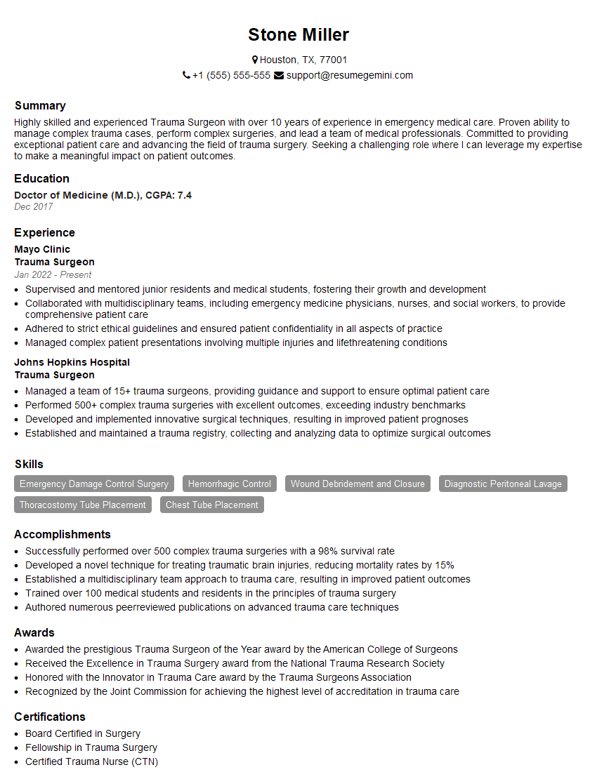
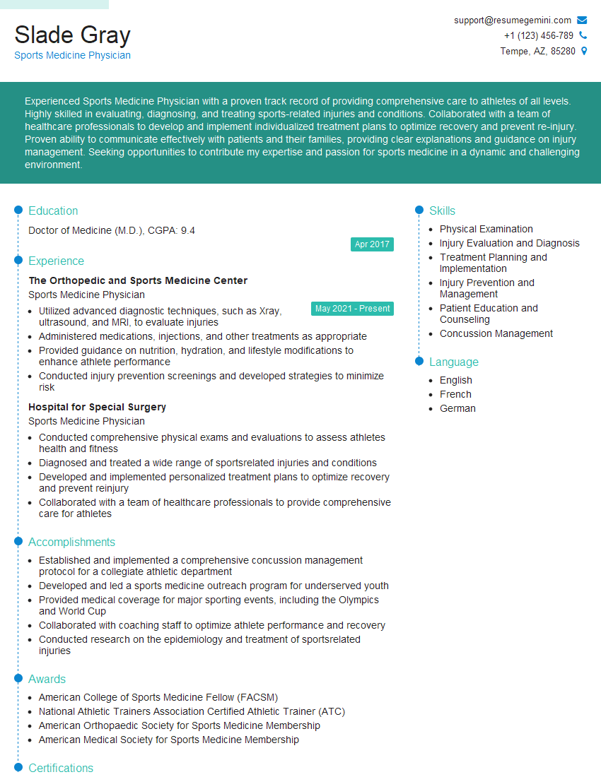
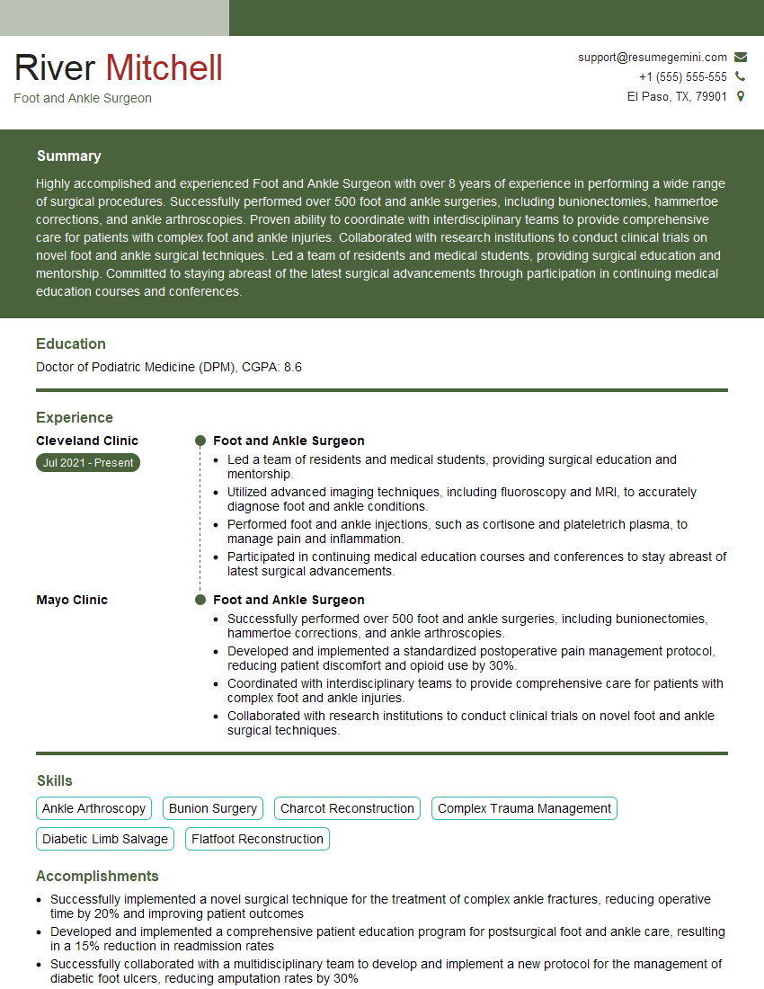
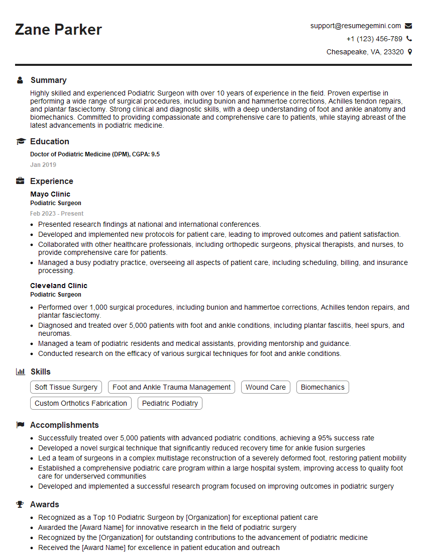
Mastering Lisfranc Fracture Repair significantly enhances your expertise in foot and ankle surgery, opening doors to specialized positions and advanced research opportunities. A strong resume is crucial to showcase your skills and experience to potential employers. Create an ATS-friendly resume that highlights your specific knowledge and accomplishments in this area. Use ResumeGemini to build a professional and impactful resume that will help you stand out from the competition. Examples of resumes tailored to Lisfranc Fracture Repair expertise are available to guide you. Take the next step towards your career goals today!
Explore more articles
Users Rating of Our Blogs
Share Your Experience
We value your feedback! Please rate our content and share your thoughts (optional).
What Readers Say About Our Blog
This was kind of a unique content I found around the specialized skills. Very helpful questions and good detailed answers.
Very Helpful blog, thank you Interviewgemini team.
