Preparation is the key to success in any interview. In this post, we’ll explore crucial Metatarsalgia Surgery interview questions and equip you with strategies to craft impactful answers. Whether you’re a beginner or a pro, these tips will elevate your preparation.
Questions Asked in Metatarsalgia Surgery Interview
Q 1. Describe the different types of metatarsalgia.
Metatarsalgia is a general term encompassing pain in the ball of the foot, specifically the metatarsal bones. It’s not a single condition but a symptom with several underlying causes. Different types of metatarsalgia are often classified based on the affected metatarsal head(s) and the underlying etiology.
- Intermetatarsal Bursitis: Inflammation of the bursa (fluid-filled sac) between the metatarsal heads. This is commonly seen between the second and third metatarsals, causing localized pain and sometimes a visible lump.
- Morton’s Neuroma: A thickening of the nerve tissue between the metatarsal heads, usually between the third and fourth toes. This causes burning, tingling, or numbness in the toes, radiating pain into the ball of the foot, and can mimic bursitis.
- Stress Fracture: A small crack in one of the metatarsal bones, typically caused by overuse or repetitive impact. This presents as localized pain that worsens with activity and may be accompanied by swelling.
- Sesamoiditis: Inflammation of the sesamoid bones (small bones embedded in the tendons under the big toe). This often leads to pain under the big toe, especially when flexing the foot.
- Metatarsalgia of inflammatory origin: Conditions such as rheumatoid arthritis can lead to inflammatory changes in the metatarsal joints, causing pain and stiffness.
- Other causes of metatarsal pain: Plantar fasciitis, tight calf muscles, improper footwear, and biomechanical abnormalities can contribute to pain in the metatarsal area, even if not strictly defined as metatarsalgia.
Q 2. Explain the diagnostic process for metatarsalgia, including imaging techniques.
Diagnosing metatarsalgia begins with a thorough history and physical examination. The doctor will assess the location and nature of the pain, as well as your activity levels, footwear, and medical history. A physical examination typically includes palpating the metatarsal heads and surrounding soft tissues to check for tenderness, swelling, or masses.
Imaging techniques play a crucial role in differentiating between various causes of metatarsal pain.
- X-rays: Are the primary imaging modality and help to rule out fractures, arthritis, or other bony abnormalities. They can also show the morphology of the metatarsal heads and sesamoids, providing clues to the cause of the problem.
- Ultrasound: May be used to visualize soft tissues, allowing for better assessment of bursitis or Morton’s neuroma. It can show fluid collection in bursae and nerve thickening.
- MRI: Offers more detailed images of soft tissues and bone and may be useful in complicated cases or when other imaging studies are inconclusive. MRI is particularly helpful in evaluating Morton’s neuroma and assessing for stress fractures more accurately.
Ultimately, the diagnosis is made based on a combination of history, physical examination, and imaging findings.
Q 3. What are the conservative treatment options for metatarsalgia?
Conservative treatment is the first-line approach for most cases of metatarsalgia. The goal is to alleviate pain, reduce inflammation, and restore normal foot biomechanics.
- Rest and Ice: Avoiding activities that aggravate the pain and applying ice packs to the affected area can significantly reduce inflammation and pain.
- Footwear Modification: Switching to low-heeled, supportive shoes with ample room in the toe box is crucial. Custom orthotics can provide additional arch support and cushioning, redistributing pressure away from the metatarsal heads.
- Medications: Over-the-counter pain relievers like ibuprofen or naproxen can reduce pain and inflammation. In some cases, your doctor may prescribe stronger pain relievers or corticosteroids for more severe inflammation.
- Physical Therapy: A physical therapist can teach exercises to strengthen the foot muscles, improve flexibility, and correct any biomechanical issues contributing to the problem. This often involves stretching exercises for the calf muscles and specific strengthening exercises for the intrinsic muscles of the foot.
- Injections: Corticosteroid injections may provide temporary relief from pain and inflammation, especially in cases of bursitis or Morton’s neuroma. However, repeated injections are generally avoided due to potential side effects.
Q 4. When is surgical intervention indicated for metatarsalgia?
Surgical intervention for metatarsalgia is considered when conservative treatments have failed to provide adequate relief for an extended period (typically 6-12 months). Surgical intervention is also indicated in cases of significant deformity, severe nerve compression (Morton’s neuroma), or intractable pain.
Examples include persistent pain despite appropriate orthotics and physical therapy, significant functional limitations impacting daily activities, and cases where conservative management poses a risk to the patient’s overall health and well-being. The decision to proceed with surgery should be made on a case-by-case basis with shared decision-making between the surgeon and patient.
Q 5. Describe the various surgical techniques used to treat metatarsalgia.
Several surgical techniques are employed to address the underlying causes of metatarsalgia, depending on the specific diagnosis.
- Morton’s Neuroma Excision: Surgical removal of the thickened nerve tissue responsible for Morton’s neuroma. This is often a minimally invasive procedure with a high success rate.
- Metatarsal Head Resection: A partial or complete removal of the metatarsal head, typically performed for severe degenerative changes or in cases of persistent pain despite other treatments. This procedure is more invasive.
- Osteotomy: A bone realignment procedure to correct deformities or abnormal alignment of the metatarsal bones. This might involve shortening or lengthening the metatarsal bone to improve biomechanics.
- Arthrodesis: This procedure involves the surgical fusion of a metatarsophalangeal (MTP) joint, creating a rigid connection. While effective for pain relief, it results in loss of motion at the affected joint.
- Distal Metatarsal Osteotomy: Involves cutting and repositioning of the metatarsal bone to improve alignment or to relieve pressure on the metatarsal head. This procedure is often performed in conjunction with soft tissue procedures.
The choice of surgical technique is determined by the underlying cause of metatarsalgia, the extent of the damage, and the patient’s overall condition and goals.
Q 6. What are the potential complications of metatarsalgia surgery?
As with any surgical procedure, metatarsalgia surgery carries potential complications, although they are relatively infrequent with experienced surgeons. These can include:
- Infection: A risk with any surgery, requiring prompt treatment with antibiotics.
- Nerve Damage: Potential damage to the nerves supplying sensation to the toes can lead to numbness, tingling, or altered sensation.
- Nonunion or Malunion: Failure of the bone to heal properly after osteotomy, requiring revision surgery.
- Painful Scar Tissue: Formation of excessive scar tissue can lead to persistent pain and stiffness.
- Recurrence of Symptoms: While surgery aims to address the underlying cause, symptoms may occasionally recur, possibly due to biomechanical factors or inadequate correction of the deformity.
- Persistent Swelling: Swelling can linger for several weeks even months post-operatively, but should eventually subside.
- Joint Stiffness: Limited joint mobility may occur following surgery, particularly after arthrodesis.
The surgeon will discuss these potential complications preoperatively and take steps to minimize the risks.
Q 7. How do you manage postoperative pain and swelling in metatarsalgia patients?
Postoperative management is crucial for a successful outcome after metatarsalgia surgery. Careful attention is paid to pain control and minimizing swelling to ensure optimal healing and patient comfort.
- Pain Management: Pain medications, ranging from over-the-counter analgesics to stronger prescription medications, are used to control post-surgical pain. Regular pain assessments are conducted, and medications are adjusted as needed.
- Elevation and Immobilization: Keeping the foot elevated helps reduce swelling. A cast, splint, or protective boot may be used to immobilize the foot, promoting proper healing and reducing pain.
- Ice Therapy: Regular application of ice packs helps reduce inflammation and pain.
- Physical Therapy: Once the incision has healed, physical therapy plays a key role in restoring range of motion, strengthening muscles, and improving overall foot function. A gradual progression of exercises is crucial to avoid re-injury.
- Weight-bearing Restrictions: The surgeon will provide guidance on weight-bearing restrictions following the procedure, starting with partial weight-bearing and gradually increasing as healing progresses.
- Wound Care: Meticulous wound care is essential to prevent infection and ensure proper healing. The patient will be instructed on proper cleaning and dressing techniques.
Close monitoring and follow-up appointments with the surgeon and physical therapist are essential to ensure optimal recovery and address any potential complications that may arise.
Q 8. What are the common post-operative rehabilitation protocols for metatarsalgia surgery?
Post-operative rehabilitation after metatarsalgia surgery is crucial for a successful outcome. The specific protocol varies depending on the surgical procedure performed and the individual patient’s needs, but generally involves a structured progression through several phases.
Immediate Post-operative Phase (0-2 weeks): This involves pain management (often with ice, elevation, and medication), non-weight-bearing on the affected foot, and range-of-motion exercises for the ankle and toes to prevent stiffness. The foot is typically protected with a surgical shoe or cast.
Early Weight-Bearing Phase (2-6 weeks): Gradual weight-bearing is introduced under the guidance of a physical therapist. This phase focuses on building strength and improving range of motion. Patients might use crutches or a walking boot. The use of custom orthotics may begin.
Late Weight-Bearing Phase (6-12 weeks): Increased weight-bearing and more advanced exercises are incorporated, targeting improved balance, coordination, and functional mobility. The goal is to return to normal activity levels. The patient might transition to regular footwear.
Return to Activity Phase (12+ weeks): This phase focuses on a gradual return to pre-operative activity levels, which varies significantly depending on the patient’s activity level and the type of surgery. It’s important to ensure proper mechanics to prevent reinjury.
Throughout the entire rehabilitation process, regular follow-up appointments with the surgeon and physical therapist are essential to monitor progress and adjust the plan as needed. For example, a patient who experiences unexpected pain might need to temporarily reduce weight-bearing or modify exercises.
Q 9. Explain the differences between proximal and distal metatarsalgia.
Proximal and distal metatarsalgia refer to the location of the metatarsal pain. They differ significantly in their underlying causes and the surgical approaches used to treat them.
Proximal Metatarsalgia: This refers to pain originating near the base of the metatarsals, often associated with problems in the Lisfranc joint (the articulation between the midfoot and forefoot). Conditions like plantar plate injury, stress fractures, or arthritis in this region can cause proximal metatarsalgia. Surgery may involve joint fusion or other procedures aimed at restoring joint stability.
Distal Metatarsalgia: This involves pain at the heads of the metatarsals, closer to the toes. Common causes include metatarsalgia, Morton’s neuroma, and stress fractures in the metatarsal heads. Surgical options for distal metatarsalgia often target the metatarsal heads themselves, like Chevron or Akin osteotomies.
The distinction is crucial for accurate diagnosis and appropriate surgical planning. Imaging studies such as X-rays are often used to pinpoint the exact location of the pain and identify any underlying bony abnormalities.
Q 10. Describe your experience with specific surgical procedures for metatarsalgia (e.g., Chevron osteotomy, Akin osteotomy).
I have extensive experience with various surgical procedures for metatarsalgia, including Chevron and Akin osteotomies. These are frequently used to address distal metatarsalgia caused by metatarsus primus varus (MPV) or other forefoot deformities.
Chevron Osteotomy: This procedure involves a V-shaped cut at the base of the first metatarsal, which is then repositioned to correct the deformity. This helps to relieve pressure and improve weight distribution. I often use this for mild to moderate MPV.
Akin Osteotomy: This involves a smaller, more localized cut on the first metatarsal head to correct the angle of the first metatarsal phalangeal (MTP) joint. This is particularly useful for patients with hallux valgus (bunion) deformity contributing to their metatarsalgia. I might choose this technique for patients with a more significant bunion deformity or where a Chevron osteotomy alone may not fully address the underlying problem.
Other procedures, like resection arthroplasty or bone fusion, are also in my repertoire depending on the patient’s specific anatomy and the severity of their condition. Pre-operative planning includes detailed analysis of radiographs and thorough patient assessment to select the optimal approach for each case. A recent case I managed involved a patient with severe MPV and a history of failed conservative management. A Chevron osteotomy and MTP joint fusion was successful.
Q 11. How do you counsel patients on the risks and benefits of metatarsalgia surgery?
Counseling patients about metatarsalgia surgery involves a thorough discussion of the risks and benefits, emphasizing shared decision-making. I start by explaining the condition clearly and highlighting the limitations of conservative treatment when it’s failed.
Benefits: I focus on the potential for pain relief, improved function, and return to desired activities. I discuss realistic expectations based on the individual’s condition and the selected surgical technique.
Risks: I openly discuss potential complications, including infection, nerve damage, nonunion (failure of the bone to heal), malunion (healing in an incorrect position), and persistent pain. I always address the possibility of a less than ideal outcome and the need for further procedures.
I encourage patients to ask questions, and I provide them with written information to review at their own pace. This open and honest communication helps patients make informed decisions aligned with their values and expectations. For instance, a highly active patient might be more willing to accept the higher risk of a complex procedure for the chance of a full return to their prior activity level.
Q 12. How do you select the appropriate surgical technique for each individual patient?
Selecting the appropriate surgical technique involves a multi-faceted approach tailored to the individual patient. It’s not a ‘one-size-fits-all’ scenario.
Patient Assessment: A comprehensive history, physical examination, and imaging studies (X-rays, CT scans if needed) provide a clear picture of the patient’s condition, including the severity and location of the deformity, presence of arthritis, and the overall foot biomechanics.
Surgical Goals: I collaboratively discuss the patient’s expectations and functional goals to align surgical intervention with their lifestyle and activity levels. For instance, a marathon runner will require a different surgical approach and rehabilitation protocol compared to someone with a sedentary lifestyle.
Technique Selection: The surgical plan considers the deformity’s specific characteristics, including the severity of the metatarsalgia and the presence of associated conditions such as hallux valgus or other foot deformities. The optimal approach balances the potential benefits and risks based on the patient’s individual factors.
For example, a patient with mild MPV might benefit from a simple Chevron osteotomy, whereas a patient with significant deformity and associated hallux valgus might require a more extensive procedure combining a Chevron or Akin osteotomy with a bunion correction.
Q 13. What are the criteria for determining successful outcome after metatarsalgia surgery?
Determining a successful outcome after metatarsalgia surgery is multifaceted, going beyond simple pain relief. It involves a combination of factors assessed over time.
Pain Relief: A significant reduction in pain is paramount. We often use standardized pain scales (like the visual analog scale) to quantify this improvement.
Functional Improvement: Patients should demonstrate improved ability to walk, stand, and participate in daily activities without pain limitations. This can be assessed through functional tests or patient-reported outcome measures (PROMs).
Radiographic Healing: X-rays are essential to confirm proper bone healing and the absence of malunion or nonunion.
Patient Satisfaction: Ultimately, the patient’s perception of success is critical. Follow-up assessments involve gathering information about their overall satisfaction with the surgical outcome and quality of life.
A successful outcome is characterized by lasting pain relief, restoration of function, and a high level of patient satisfaction, ideally assessed several months after surgery.
Q 14. How do you manage patients with complications after metatarsalgia surgery?
Managing complications after metatarsalgia surgery requires a prompt and proactive approach. The specific management strategy depends entirely on the nature of the complication.
Infection: This is managed with antibiotics, wound debridement (removal of infected tissue), or even surgical drainage depending on severity. Close monitoring is crucial.
Nonunion/Malunion: Nonunion (failure of the bone to heal) may require secondary surgery, such as bone grafting or internal fixation, to encourage healing. Malunion (healing in an incorrect position) might necessitate corrective osteotomy.
Nerve Damage: This can manifest as numbness, tingling, or pain. Conservative management, including physical therapy and pain medication, is initially attempted. Surgical exploration and repair may be needed in cases of significant nerve injury.
Persistent Pain: If post-operative pain persists despite appropriate management, further investigations might include imaging studies and nerve conduction studies to identify the cause and implement targeted treatment.
A multidisciplinary approach involving surgeons, physical therapists, and pain specialists often improves the management of complications. Open communication with the patient is key to ensuring optimal care and managing expectations.
Q 15. What are the latest advancements in surgical techniques for metatarsalgia?
Advancements in metatarsalgia surgery are focused on minimally invasive techniques and improved implant designs. One significant advance is the increased use of percutaneous procedures, which involve smaller incisions and less tissue trauma. This leads to faster recovery times and reduced scarring. Another area of advancement is the development of biocompatible and resorbable implants, minimizing the need for a second surgery for implant removal. We’re also seeing refined techniques in osteotomy (bone cutting), such as less invasive approaches like closing wedge osteotomies, designed to precisely correct metatarsal alignment with less bone removal. Finally, advanced imaging techniques like 3D CT scans are allowing for better pre-operative planning and more accurate surgical execution.
Career Expert Tips:
- Ace those interviews! Prepare effectively by reviewing the Top 50 Most Common Interview Questions on ResumeGemini.
- Navigate your job search with confidence! Explore a wide range of Career Tips on ResumeGemini. Learn about common challenges and recommendations to overcome them.
- Craft the perfect resume! Master the Art of Resume Writing with ResumeGemini’s guide. Showcase your unique qualifications and achievements effectively.
- Don’t miss out on holiday savings! Build your dream resume with ResumeGemini’s ATS optimized templates.
Q 16. Describe your experience with minimally invasive surgery for metatarsalgia.
My experience with minimally invasive surgery for metatarsalgia has been overwhelmingly positive. I’ve found that percutaneous procedures, using small incisions and specialized instruments, lead to significantly reduced pain and swelling post-surgery. Patients often report being able to bear weight sooner and return to their normal activities more quickly compared to traditional open surgeries. For example, I recently treated a patient with a Morton’s neuroma using a minimally invasive approach. They were walking with minimal discomfort within a week and returned to their active lifestyle within a month. The smaller incision resulted in much less scarring than a traditional open procedure would have caused.
Q 17. How do you assess patient suitability for different surgical techniques?
Assessing patient suitability for different surgical techniques involves a thorough evaluation of several factors. We start with a detailed history, including the patient’s symptoms, activity level, and past medical history. A comprehensive physical exam, including assessing the foot’s biomechanics, is crucial. Imaging studies, such as X-rays and sometimes MRI or CT scans, help to visualize the underlying bone structure and soft tissues. Based on this information, I determine the severity of the metatarsalgia, the presence of any associated conditions like arthritis, and the overall health of the patient. For instance, a patient with severe arthritis might be a better candidate for an arthrodesis (joint fusion), while a younger, more active patient with a less severe case might be suitable for a minimally invasive osteotomy. The final decision is always made in collaboration with the patient, ensuring they understand the risks and benefits of each procedure.
Q 18. What are the common causes of metatarsalgia?
Metatarsalgia, or pain in the ball of the foot, has several common causes. One frequent cause is Morton’s neuroma, a benign nerve tumor that develops between the toes. Another common cause is stress fractures, often occurring in runners or individuals with high-impact activities. High-heeled shoes can also contribute to metatarsalgia by putting excessive pressure on the forefoot. Additionally, underlying biomechanical issues, such as pes planus (flat feet) or metatarsus primus varus (a condition where the big toe is angled inward), can lead to uneven weight distribution, causing pain in the metatarsal bones. Finally, underlying systemic diseases like rheumatoid arthritis can also cause inflammation and pain in the metatarsals.
Q 19. What role do biomechanics play in the etiology and treatment of metatarsalgia?
Biomechanics play a crucial role in both the etiology and treatment of metatarsalgia. Abnormal foot mechanics, such as overpronation (the foot rolling inward excessively), can lead to increased stress on certain metatarsal heads, resulting in pain and inflammation. Conversely, the treatment often involves addressing these biomechanical issues. Custom orthotics, designed to support the arches and correct abnormal foot alignment, are frequently used to reduce stress on the metatarsals. In surgical cases, the goal is often to restore proper alignment and weight distribution. For example, correcting a bunion (hallux valgus) can alleviate pressure on the adjacent metatarsal heads, thereby improving metatarsalgia symptoms. Therefore, a thorough biomechanical evaluation is paramount for both diagnosis and treatment planning.
Q 20. Describe your experience with different types of implants used in metatarsalgia surgery.
My experience encompasses a range of implants used in metatarsalgia surgery. For osteotomy procedures, I often use small, biocompatible screws to stabilize the bone fragments after the osteotomy is performed. These screws are designed to be strong enough to provide sufficient fixation while being minimally invasive. In cases requiring arthrodesis (joint fusion), I may use plates and screws for robust fixation of the joint. The choice of implant depends heavily on the specific surgical technique and the patient’s individual needs. For instance, smaller screws and less invasive techniques are preferred in younger, more active patients to minimize potential long-term complications. Recently, there’s increasing use of resorbable implants that dissolve over time, eliminating the need for a second procedure to remove the implant material.
Q 21. How do you prevent complications such as infection or nerve damage during metatarsalgia surgery?
Preventing complications like infection and nerve damage during metatarsalgia surgery is a top priority. Strict adherence to sterile surgical techniques is paramount. This includes using appropriate sterilization methods for all instruments and ensuring a sterile surgical field. Meticulous dissection during the surgery to avoid inadvertent nerve injury is crucial. Intraoperative nerve monitoring is sometimes utilized, particularly in complex cases, to provide real-time feedback on the status of the nerves. Careful wound closure techniques are employed to minimize the risk of infection. Post-operatively, patients receive detailed instructions on wound care and are monitored closely for any signs of infection or other complications. Prompt identification and management of any potential complications are critical in ensuring a favorable outcome.
Q 22. What is your approach to managing patients with recurrent metatarsalgia?
Recurrent metatarsalgia, unfortunately, is a significant challenge. My approach focuses on a thorough reassessment to identify any overlooked factors contributing to the persistent pain. This begins with a detailed history, physical exam, and imaging review (X-rays, sometimes MRI or CT) to rule out any new pathology, like stress fractures, arthritis progression, or nerve entrapment. We meticulously analyze the initial surgery – what procedure was performed, was it successful initially, and were there any postoperative complications?
Often, recurrent pain stems from inadequate initial treatment, incomplete correction of the underlying biomechanical issue, or the development of secondary complications such as scar tissue formation or adjacent joint involvement. Conservative measures are often revisited first: customized orthotics, injections (corticosteroids or, occasionally, PRP), physical therapy focusing on strengthening and flexibility, and activity modification. If conservative measures fail, surgical revision may be necessary. The type of revision depends on the original surgery and the cause of recurrence. It might involve a different surgical approach altogether, addressing a previously overlooked problem. For example, if the original surgery addressed a plantar plate tear, but failed to address a significant metatarsalgia associated with Morton’s neuroma, a secondary neuroma resection might be indicated.
Q 23. Describe your experience with the use of platelet-rich plasma (PRP) or other regenerative therapies in metatarsalgia.
Platelet-rich plasma (PRP) is a regenerative therapy I use selectively in the management of metatarsalgia. It’s not a standalone solution, but rather an adjunct to other conservative treatments. I typically reserve PRP for patients who have not responded well to standard conservative measures such as orthotics and physical therapy, and who are not yet candidates for surgery. The procedure involves drawing the patient’s blood, processing it to concentrate the platelets, and then injecting this concentrate into the affected area. The platelets release growth factors that stimulate tissue healing and reduce inflammation.
My experience has shown that PRP can provide pain relief in some patients, but the results are not always predictable. Response varies depending on the underlying cause of the metatarsalgia, the severity of the condition, and the patient’s overall health. It is crucial to manage patient expectations as PRP is not a guaranteed fix. Other regenerative therapies, such as bone marrow aspirate concentrate (BMAC), are less commonly used but are a potential option in select cases. We always clearly discuss the likelihood of success and alternative options with the patient before proceeding with any regenerative therapy.
Q 24. How do you manage patients with co-morbidities affecting their surgical candidacy for metatarsalgia?
Managing patients with comorbidities significantly impacts surgical decision-making. Conditions like diabetes, peripheral artery disease (PAD), and obesity increase the risk of complications like infection, delayed wound healing, and poor surgical outcomes. A thorough preoperative evaluation is essential, which includes assessing the patient’s overall health, reviewing their medical history, and conducting relevant investigations such as blood tests and vascular assessments. For example, a patient with poorly controlled diabetes might require more intensive pre-operative and postoperative care, including glycemic control and meticulous wound management. Patients with significant PAD might require vascular consultation to evaluate perfusion to the foot, and may be unsuitable for surgery if perfusion is severely compromised.
In some cases, these comorbidities may make surgery too high-risk. When the risks outweigh the benefits, we prioritize optimizing conservative management. Multidisciplinary collaboration with other specialists (endocrinologists, cardiologists, vascular surgeons) is crucial to create a tailored management plan that addresses both the metatarsalgia and the co-morbidities. Sometimes, surgical timing needs to be deferred until comorbidities are better managed.
Q 25. What are the long-term outcomes of different surgical techniques for metatarsalgia?
Long-term outcomes of metatarsalgia surgeries vary considerably based on the specific procedure, the patient’s individual factors (age, activity level, compliance with post-operative instructions), and the underlying cause of the metatarsalgia. Studies on procedures like metatarsal osteotomy, plantar plate repair, and neuroma excision demonstrate varying success rates for pain reduction and functional improvement. In general, patients who undergo less invasive procedures often experience faster recovery times but may not always achieve the same long-term outcomes as more complex procedures. Furthermore, the presence of pre-existing conditions like arthritis can impact long-term results.
For instance, metatarsal osteotomy, while effective in addressing metatarsalgia caused by malalignment, may have a higher rate of recurrence in patients with significant arthritis. Longitudinal studies often show that pain relief and functional improvement are generally maintained over several years, with a gradual decline possible in some cases. Postoperative compliance with physical therapy, footwear adjustments, and activity modification significantly influences long-term outcomes. It’s important to counsel patients on the realistic expectations and potential long-term limitations based on their specific case.
Q 26. How do you assess the effectiveness of treatment for metatarsalgia?
Assessing treatment effectiveness in metatarsalgia involves a multifaceted approach. It is not solely reliant on pain scores. We use validated questionnaires like the Visual Analog Scale (VAS) for pain, the Foot and Ankle Outcome Score (FAOS), and the American Orthopaedic Foot and Ankle Society (AOFAS) score to objectively measure functional improvement and quality of life. The patient’s subjective assessment of their pain and function is equally crucial. We also assess changes in gait, range of motion, and any improvements in activities of daily living. Imaging studies, such as X-rays, can help monitor the healing process and detect any postoperative complications. For instance, if a patient reports a 70% reduction in pain as per VAS, a significant improvement in AOFAS scores indicating improved function, and a normalized gait pattern, we can conclude a positive outcome.
Furthermore, the duration of pain relief is significant. Transient relief is not as meaningful as sustained, long-term improvement. Regular follow-up appointments are essential to monitor progress, address any concerns, and make adjustments to the treatment plan as needed. The ultimate goal is to improve the patient’s quality of life by alleviating pain and restoring normal foot function.
Q 27. How do you use evidence-based medicine to guide your surgical decisions in metatarsalgia?
Evidence-based medicine (EBM) forms the cornerstone of my surgical decisions. I utilize a systematic approach that involves critically appraising the existing literature, considering the level of evidence supporting different surgical techniques and the potential benefits and risks of each. High-quality randomized controlled trials, meta-analyses, and systematic reviews guide my choices, prioritizing procedures with proven efficacy and safety. However, it’s important to acknowledge that EBM is not a rigid set of rules. Individual patient factors (age, activity level, comorbidities) must be integrated with the best available evidence to formulate a personalized treatment plan.
For example, when deciding between different osteotomy techniques, I consult the latest research on their comparative outcomes, complication rates, and patient satisfaction. I avoid using procedures lacking substantial evidence of effectiveness or with high complication rates. Continual professional development and staying abreast of the latest research are critical for maintaining an evidence-based approach. I actively participate in professional organizations, attend conferences, and regularly review relevant medical journals to ensure that my practice is consistently updated with the most current and reliable evidence.
Q 28. Describe a challenging case of metatarsalgia you successfully managed and the lessons learned.
One particularly challenging case involved a 55-year-old female marathon runner with severe, recurrent metatarsalgia after multiple failed conservative treatments and a previous failed metatarsal osteotomy. Her pain was debilitating and severely impacted her ability to run. Initial imaging revealed significant degenerative changes in the metatarsophalangeal joints, particularly the second and third. Simply repeating the osteotomy was not a viable option due to the advanced arthritis. This required a comprehensive approach.
We decided on a combination of procedures: a modified chevron osteotomy to address the metatarsalgia, and arthrodesis (joint fusion) of the most severely affected metatarsophalangeal joints. The osteotomy addressed the biomechanical issues, while the arthrodesis stabilized the arthritic joints. Postoperatively, she required extensive physiotherapy and a gradual return to running. The outcome was ultimately successful. She regained a pain-free functional range of motion, and she was able to return to running, albeit at a modified pace. The key lesson from this case was the importance of considering the entire foot and not just focusing on the immediate source of pain. A holistic approach, taking into account all the contributing factors, is essential for optimal results, especially in complex and recurrent cases.
Key Topics to Learn for Metatarsalgia Surgery Interview
- Anatomy and Biomechanics of the Forefoot: Understanding the intricate structure of the metatarsals, phalanges, and surrounding soft tissues is fundamental. Consider the biomechanical forces involved in weight-bearing and gait analysis.
- Etiology and Diagnosis of Metatarsalgia: Explore the various causes of metatarsalgia, including Morton’s neuroma, stress fractures, and inflammatory conditions. Master the diagnostic techniques used to identify the underlying pathology, from physical examination to imaging studies.
- Surgical Techniques for Metatarsalgia: Familiarize yourself with different surgical approaches, such as osteotomy, resection arthroplasty, and nerve decompression. Understand the indications and contraindications for each procedure.
- Pre- and Post-operative Management: Master the principles of patient care before and after surgery. This includes pre-operative planning, pain management, rehabilitation protocols, and potential complications.
- Implant Selection and Fixation: Learn about the various implants used in metatarsalgia surgery and their appropriate application. Understand the principles of fracture fixation and joint stability.
- Complications and Management: Prepare to discuss potential complications such as infection, non-union, malunion, and nerve damage. Know the management strategies for each complication.
- Evidence-Based Practice and Research: Demonstrate your understanding of current research and evidence-based practices in the field of metatarsalgia surgery. Be prepared to discuss recent advancements and their clinical implications.
Next Steps
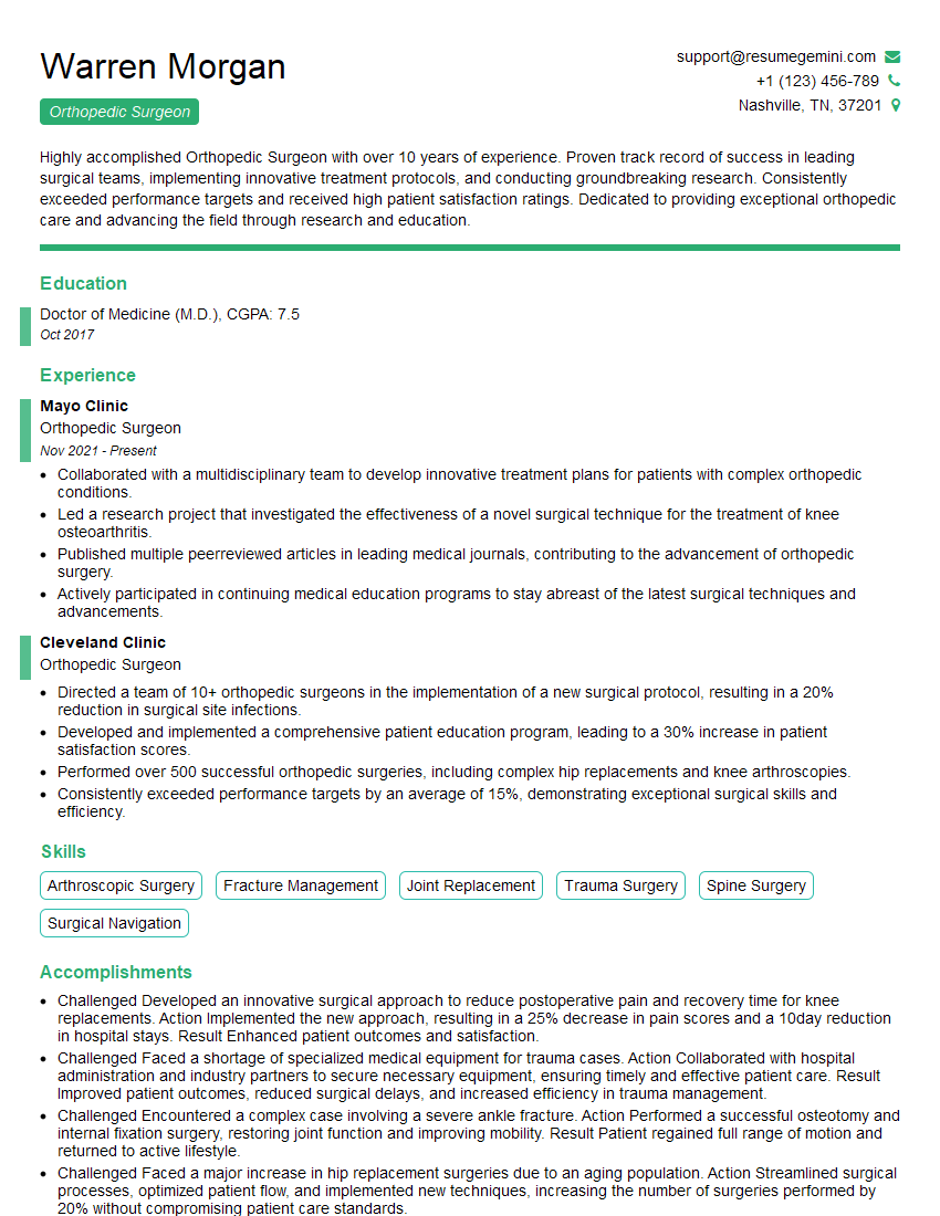
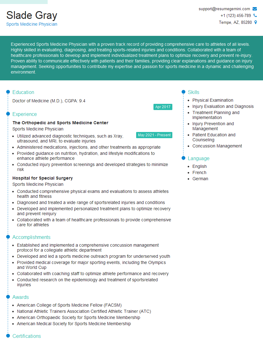
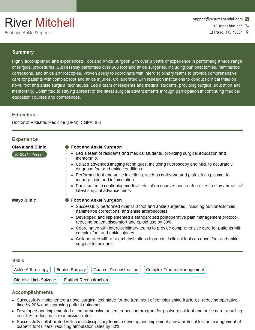
Mastering Metatarsalgia Surgery significantly enhances your career prospects in orthopedics and podiatric surgery. A strong understanding of these topics demonstrates expertise and commitment to patient care. To maximize your job search success, create an ATS-friendly resume that showcases your skills and experience effectively. ResumeGemini is a trusted resource that can help you build a professional and impactful resume. Take advantage of their tools and resources, including examples of resumes tailored to Metatarsalgia Surgery, to create a document that stands out to recruiters.
Explore more articles
Users Rating of Our Blogs
Share Your Experience
We value your feedback! Please rate our content and share your thoughts (optional).
What Readers Say About Our Blog
This was kind of a unique content I found around the specialized skills. Very helpful questions and good detailed answers.
Very Helpful blog, thank you Interviewgemini team.


