Unlock your full potential by mastering the most common Inhalation Therapy interview questions. This blog offers a deep dive into the critical topics, ensuring you’re not only prepared to answer but to excel. With these insights, you’ll approach your interview with clarity and confidence.
Questions Asked in Inhalation Therapy Interview
Q 1. Describe the different types of inhalers and their mechanisms of action.
Inhalers are devices used to deliver medication directly to the lungs, offering a targeted approach to treat respiratory conditions. There are several types, each with a unique mechanism of action:
Metered-Dose Inhalers (MDIs): These are pressurized canisters that release a measured dose of medication as a fine mist upon actuation. They often require a spacer device to enhance drug delivery to the lungs, especially for children or those with poor coordination. The mechanism relies on propellant to aerosolize the medication.
Dry Powder Inhalers (DPIs): DPIs contain medication in a dry powder form. Inhalation through the device causes the powder to be dispersed into the airways. These are often preferred for patients who have difficulty coordinating breathing and actuation, as the medication release is triggered by inhalation. They don’t require a propellant.
Soft Mist Inhalers (SMIs): SMIs deliver a fine mist of medication without the use of propellants. They offer a gentler and quieter delivery system compared to MDIs and are often easier to use for some patients. The mechanism relies on a pump to generate the mist.
Nebulizers (discussed in more detail in the next question): While not strictly an inhaler in the same sense as the others, nebulizers also deliver medication to the lungs as an aerosol, but through a different mechanism.
Choosing the right inhaler type depends on the patient’s individual needs, coordination abilities, and the specific medication being delivered. For example, a patient with asthma might use an MDI with a spacer, while a patient with COPD might use a DPI.
Q 2. Explain the process of administering medication via a nebulizer.
Nebulizers transform liquid medication into a fine mist that is easily inhaled. The process involves several steps:
Medication Preparation: The prescribed medication is added to the nebulizer cup according to the physician’s orders. The correct dose and dilution are crucial for efficacy and safety.
Nebulizer Setup: The nebulizer cup is securely attached to the nebulizer device, and the tubing is connected to an air compressor or oxygen source. The flow rate should be appropriately set for optimal aerosol generation.
Aerosolization: The air compressor or oxygen flow forces air through a small nozzle within the nebulizer, breaking the liquid medication into tiny droplets. This mist is then delivered to the patient’s airway.
Inhalation: The patient inhales the medication mist through a mouthpiece or face mask. Deep, slow breaths are encouraged for maximum medication delivery. Proper breathing techniques should be taught to patients.
Monitoring: Throughout the treatment, the respiratory therapist monitors the patient’s respiratory status, noting any adverse reactions or changes in their condition.
Cleaning and Maintenance: After each use, the nebulizer should be thoroughly cleaned and dried to prevent bacterial contamination. This is paramount to ensure patient safety and prevent future infections.
Nebulizers are particularly useful for patients who struggle to use inhalers effectively, or those who require a large volume of medication. For instance, a patient experiencing an acute asthma exacerbation might benefit from a nebulizer treatment to rapidly deliver bronchodilators.
Q 3. What are the indications and contraindications for oxygen therapy?
Oxygen therapy involves administering supplemental oxygen to increase the oxygen levels in a patient’s blood. Indications include conditions where oxygen levels are low or the body’s ability to utilize oxygen is compromised. Contraindications are less frequent but are critical to consider.
Indications: Hypoxia (low blood oxygen levels), respiratory distress, heart failure, chronic obstructive pulmonary disease (COPD) exacerbation, pneumonia, and post-surgical recovery are common indications. Essentially, any condition that impairs oxygen uptake or delivery.
Contraindications: The primary contraindication is hyperoxia (excessive oxygen levels), which can be toxic, especially in patients with certain lung diseases. Additionally, extreme caution is required for patients with specific conditions where increased oxygen may worsen underlying issues.
The decision to initiate oxygen therapy, the flow rate, and the delivery method are determined by the patient’s specific condition and oxygen saturation levels. Close monitoring is vital to prevent complications.
Q 4. How do you monitor a patient’s response to oxygen therapy?
Monitoring a patient’s response to oxygen therapy is crucial to ensure efficacy and safety. This involves several key measurements:
Pulse Oximetry: This non-invasive method measures the oxygen saturation (SpO2) in the blood. We aim for a target SpO2, which varies based on the individual and their underlying conditions. Continuous monitoring is key.
Arterial Blood Gas (ABG) Analysis: ABG provides a more comprehensive assessment of blood oxygen levels (PaO2), carbon dioxide levels (PaCO2), and pH. This test is usually ordered if pulse oximetry readings are concerning or if a more detailed analysis is required.
Respiratory Rate and Depth: Assessing the patient’s breathing pattern is vital. Improved respiratory rate and depth often indicate improved oxygenation.
Heart Rate and Rhythm: Changes in heart rate can be a sign of the patient’s response to oxygen therapy, and rhythm irregularities may indicate a problem.
Clinical Assessment: This involves observing the patient for improvements in symptoms such as shortness of breath, chest pain, and level of consciousness. A significant improvement in their mental state often points to successful oxygenation.
Based on these assessments, adjustments to the oxygen therapy may be necessary to optimize the patient’s oxygen levels while minimizing potential risks.
Q 5. What are the common complications of mechanical ventilation and how are they managed?
Mechanical ventilation, while life-saving, can lead to several complications. These complications require vigilant monitoring and prompt management:
Ventilator-Associated Pneumonia (VAP): A common infection of the lungs often due to the presence of an endotracheal tube. Prevention involves meticulous hand hygiene, aseptic techniques during suctioning, and elevation of the head of the bed.
Barotrauma and Volutrauma: These refer to lung injury caused by high pressures or volumes during ventilation, respectively. Careful adjustment of ventilator settings is critical to minimize this risk.
Oxygen Toxicity: Prolonged exposure to high oxygen concentrations can damage the lungs. We closely monitor oxygen levels and strive for the lowest effective FiO2 (fraction of inspired oxygen).
Atelectasis: Collapse of lung tissue, often due to reduced lung volume. Strategies include deep breathing exercises, incentive spirometry, and appropriate ventilator settings.
Infection: The invasive nature of mechanical ventilation increases the risk of infections beyond VAP, necessitating meticulous infection control protocols.
Management strategies often involve adjusting ventilator settings, administering appropriate medications (e.g., antibiotics for infection), performing chest physiotherapy, and providing supportive care to address any complications.
Q 6. Describe the different modes of mechanical ventilation and their applications.
Mechanical ventilation offers various modes, each suited to different patient needs:
Volume-Controlled Ventilation (VCV): Delivers a preset tidal volume with a variable respiratory rate. It’s often used for patients with weak respiratory muscles who need a consistent tidal volume.
Pressure-Controlled Ventilation (PCV): Delivers a preset airway pressure for a set inspiratory time. It’s beneficial for patients with lung disease who may need lower airway pressures to prevent barotrauma.
Pressure Support Ventilation (PSV): Provides pressure support to each breath initiated by the patient, augmenting their spontaneous breathing efforts. It’s often used during weaning from mechanical ventilation.
Synchronized Intermittent Mandatory Ventilation (SIMV): Combines controlled breaths with patient-initiated breaths, allowing for gradual weaning. It balances ventilator support with the patient’s respiratory efforts.
High-Frequency Ventilation (HFV): Uses very high respiratory rates with small tidal volumes. This mode is often used for patients with severe lung injury or acute respiratory distress syndrome (ARDS).
The selection of a ventilation mode is based on the patient’s respiratory status, disease severity, and response to therapy. Careful monitoring and adjustments are necessary to optimize ventilation and prevent complications.
Q 7. Explain the principles of airway clearance techniques.
Airway clearance techniques aim to remove mucus and secretions from the airways, improving lung function and reducing the risk of infection. Several techniques are employed:
Huff Coughing: A controlled technique that uses short, forceful exhales to loosen secretions without the strain of a full cough.
Chest Physiotherapy: Includes manual techniques like percussion, vibration, and postural drainage to mobilize secretions. These are often used in conjunction with other techniques to maximize effectiveness.
Active Cycle of Breathing (ACB): A series of breathing exercises that combine controlled breathing, huffing, and coughing to clear airways.
Autogenic Drainage (AD): A technique that encourages patients to use controlled breathing to move secretions from the periphery of the lungs towards the larger airways, where they can be coughed out.
High-Frequency Chest Wall Oscillation (HFCWO): Uses a vest that delivers high-frequency vibrations to loosen mucus. This is often used for patients with chronic conditions.
The choice of technique depends on the patient’s age, condition, and ability to cooperate. Education and training are essential for patients to effectively perform these techniques, ensuring optimal results and minimizing discomfort.
Q 8. How do you assess a patient’s respiratory status?
Assessing a patient’s respiratory status involves a comprehensive approach, combining observation, auscultation, and measurement. We begin with a thorough history, noting any pre-existing conditions like asthma or COPD. Then, we move to the physical assessment.
Inspection: Observing the patient’s breathing pattern – rate, depth, rhythm, and effort. For example, rapid, shallow breaths (tachypnea) may indicate distress, while labored breathing (dyspnea) suggests airway obstruction or lung disease. We also look for cyanosis (bluish discoloration of the skin and mucous membranes), a sign of low blood oxygen.
Auscultation: Listening to the lungs with a stethoscope to identify abnormal breath sounds like wheezes (high-pitched whistling sounds), crackles (wet, popping sounds), or rhonchi (low-pitched, rumbling sounds), each indicating different underlying issues.
Palpation: Assessing chest expansion to ensure both lungs are inflating equally. Unequal expansion suggests a possible collapse or obstruction in one lung.
Measurement: Measuring vital signs, including respiratory rate, heart rate, blood pressure, and oxygen saturation (SpO2) using a pulse oximeter. Low SpO2 indicates hypoxemia (low blood oxygen levels), a serious condition.
Finally, we consider the patient’s subjective experience; how breathless they feel and how this impacts their daily activities. This holistic approach gives us a complete picture of their respiratory health.
Q 9. What are the signs and symptoms of respiratory distress?
Respiratory distress manifests through a range of signs and symptoms, varying in severity depending on the underlying cause. Early signs can be subtle, progressing to life-threatening emergencies. Some key indicators include:
Increased respiratory rate (tachypnea): Rapid, shallow breathing, often exceeding 20 breaths per minute in adults.
Use of accessory muscles: Patients may use their neck and chest muscles to aid breathing, indicating increased effort.
Nasal flaring: Widening of the nostrils during inhalation.
Retractions: Indrawing of the skin between the ribs or above the clavicles during inhalation, indicating increased effort to draw in air.
Grunting: A sound made during exhalation, an attempt to keep the alveoli open.
Cyanosis: A bluish discoloration of the skin and mucous membranes due to low blood oxygen.
Altered mental status: Confusion, lethargy, or drowsiness due to hypoxia (lack of oxygen to the brain).
Wheezing, crackles, or rhonchi: Abnormal breath sounds heard on auscultation.
It’s crucial to remember that the combination and severity of these symptoms will vary widely depending on the cause of the respiratory distress, which could range from asthma to pneumonia to pneumothorax (collapsed lung).
Q 10. Describe the process of arterial blood gas interpretation.
Interpreting arterial blood gas (ABG) results requires understanding the interplay of several key variables: pH, PaO2, PaCO2, and HCO3-. Let’s break it down.
pH: Measures the acidity or alkalinity of the blood. A normal pH is 7.35-7.45. Acidosis (pH < 7.35) indicates an excess of acid, while alkalosis (pH > 7.45) indicates an excess of base.
PaO2 (Partial pressure of oxygen): Represents the amount of oxygen dissolved in the arterial blood. Normal values are typically 80-100 mmHg. Low PaO2 (hypoxemia) indicates inadequate oxygenation.
PaCO2 (Partial pressure of carbon dioxide): Reflects the amount of carbon dioxide in the arterial blood. Normal values are usually 35-45 mmHg. High PaCO2 (hypercapnia) suggests impaired ventilation, while low PaCO2 (hypocapnia) suggests hyperventilation.
HCO3- (Bicarbonate): A major buffer in the blood, helping to regulate pH. Normal values are generally 22-26 mEq/L.
For example, a patient with respiratory acidosis might have a low pH, high PaCO2, and a normal or slightly elevated HCO3-. This suggests that the lungs are not effectively removing carbon dioxide. In contrast, metabolic acidosis shows a low pH, low HCO3- and a normal or slightly low PaCO2. Careful analysis of all four parameters, along with the patient’s clinical picture, is essential for accurate interpretation and appropriate treatment.
Q 11. What are the different types of respiratory infections and their treatment?
Respiratory infections are broadly categorized into upper and lower respiratory tract infections. Treatment strategies vary greatly depending on the specific infection and the patient’s overall health.
Upper Respiratory Infections (URIs): These typically involve the nose, sinuses, pharynx, and larynx. Common examples include the common cold (rhinovirus), influenza (flu virus), and sinusitis (bacterial or viral infection of the sinuses). Treatment often focuses on supportive care, such as rest, fluids, and over-the-counter medications for symptom relief (pain relievers, decongestants).
Lower Respiratory Infections (LRIs): These affect the trachea, bronchi, and lungs. Examples include:
Pneumonia: Infection of the lung parenchyma (lung tissue). Treatment may involve antibiotics (bacterial pneumonia), antiviral medications (viral pneumonia), and supportive care.
Bronchitis: Inflammation of the bronchi. Treatment typically includes rest, fluids, and bronchodilators to help open the airways. Antibiotics are usually not indicated unless a bacterial infection is suspected.
Tuberculosis (TB): A bacterial infection that primarily affects the lungs. Treatment involves a course of multiple antibiotics for several months.
It is crucial to obtain a proper diagnosis through medical evaluation before initiating treatment, as the specific pathogen and patient factors will greatly influence the most appropriate treatment approach.
Q 12. How do you manage a patient with acute respiratory failure?
Managing acute respiratory failure is a critical situation requiring immediate intervention. This life-threatening condition necessitates a multi-faceted approach:
Ensure a patent airway: This may involve suctioning secretions, inserting an endotracheal tube (intubation) for mechanical ventilation, or performing a tracheostomy if necessary.
Provide supplemental oxygen: Oxygen therapy is crucial to improve oxygenation. The delivery method (nasal cannula, face mask, high-flow oxygen) will depend on the severity of the hypoxemia.
Mechanical ventilation: For patients who are unable to maintain adequate ventilation on their own, mechanical ventilation is essential to support breathing. Various modes of ventilation exist, tailored to the individual patient’s needs.
Address underlying cause: Treatment of the underlying condition (e.g., pneumonia, pulmonary edema, COPD exacerbation) is crucial. This might involve antibiotics, diuretics, bronchodilators, or other medications.
Monitor vital signs and ABGs: Close monitoring of respiratory rate, heart rate, blood pressure, oxygen saturation, and ABGs is essential to assess the effectiveness of treatment and make necessary adjustments.
Provide supportive care: This may involve fluid management, nutritional support, and management of other organ systems affected by the respiratory failure.
The management of acute respiratory failure is a dynamic process requiring a team approach involving physicians, nurses, and respiratory therapists working together to optimize patient outcomes. This often takes place in an intensive care setting.
Q 13. Explain the role of peak flow meters in asthma management.
Peak flow meters are handheld devices used to measure the speed of air exhaled forcefully from the lungs. They are particularly valuable in asthma management for several reasons:
Monitoring lung function: Daily peak flow measurements help track changes in lung function, providing early warning signs of worsening asthma.
Identifying triggers: By comparing peak flow readings, patients can identify potential triggers (e.g., allergens, pollutants, exercise) that worsen their asthma.
Guiding treatment: Peak flow readings help patients and their healthcare providers adjust medication, such as increasing inhaler use when peak flow decreases, preventing severe asthma attacks.
Personal best: Establishing a personal best peak flow helps determine the patient’s baseline lung function, providing a benchmark for comparison.
Action plan: Many patients with asthma have a personalized action plan guided by their peak flow measurements, advising them on medication changes based on different peak flow zones (green, yellow, red zones).
Think of a peak flow meter as a personal early warning system for asthma. Regular use empowers patients to actively manage their condition and prevent severe exacerbations.
Q 14. What are the safety precautions associated with oxygen therapy?
Oxygen therapy, while life-saving, carries inherent risks if not administered safely. Precautions must be taken to minimize these risks:
Fire hazard: Oxygen supports combustion, making it crucial to eliminate potential ignition sources (e.g., smoking, open flames, electrical equipment). ‘No smoking’ signs should be prominently displayed. All electrical equipment should be properly grounded.
Oxygen toxicity: High concentrations of oxygen for prolonged periods can damage lung tissue. Oxygen should be administered at the lowest effective concentration.
Carbon dioxide retention: In patients with chronic lung disease, excessive oxygen administration can suppress the respiratory drive, leading to carbon dioxide retention. Careful monitoring is crucial.
Skin irritation: High-flow oxygen delivery systems can cause skin irritation or breakdown. Padding and proper placement of equipment are essential.
Drying of mucous membranes: Oxygen can dry out mucous membranes. Humidification may be necessary, especially with high-flow systems.
Proper equipment: Using well-maintained and appropriate equipment is crucial for safe and effective oxygen delivery. Regular checks on oxygen tanks, tubing, and delivery devices are essential.
Safe oxygen therapy requires a thorough understanding of oxygen’s properties, risks, and appropriate administration techniques, stressing the importance of patient education and monitoring.
Q 15. How do you select the appropriate size and type of endotracheal tube?
Selecting the right endotracheal tube (ETT) is crucial for effective ventilation and airway management. It’s a balance of ensuring adequate airway patency while minimizing trauma. The size is determined primarily by the patient’s age, weight, and the predicted internal diameter of the trachea. We use size charts as a guide, but clinical judgment is key. For example, a smaller-than-expected ETT might lead to insufficient ventilation, while a larger one may cause tracheal damage.
The type of ETT depends on the patient’s specific needs and the duration of intubation. Standard ETTs are for short-term ventilation, while reinforced ETTs are better for long-term use to prevent kinking. Cuffed ETTs, which have an inflatable cuff at the distal end, are commonly used to seal the airway and prevent air leakage. Uncuffed ETTs are generally used for pediatric patients or short-term use where the risk of tracheal damage is higher.
In practice, we always consider factors like the patient’s anatomy (e.g., a short, thick neck might require a curved ETT), any pre-existing airway conditions (e.g., previous tracheostomy), and the anticipated duration of intubation. It’s not just about numbers; it’s about a comprehensive assessment to ensure patient safety and comfort. I always double-check the size and type with the physician before insertion.
Career Expert Tips:
- Ace those interviews! Prepare effectively by reviewing the Top 50 Most Common Interview Questions on ResumeGemini.
- Navigate your job search with confidence! Explore a wide range of Career Tips on ResumeGemini. Learn about common challenges and recommendations to overcome them.
- Craft the perfect resume! Master the Art of Resume Writing with ResumeGemini’s guide. Showcase your unique qualifications and achievements effectively.
- Don’t miss out on holiday savings! Build your dream resume with ResumeGemini’s ATS optimized templates.
Q 16. Describe the steps involved in performing suctioning.
Suctioning clears secretions from the airway, maintaining a patent airway. It’s a crucial skill, but it must be performed carefully to minimize patient discomfort and avoid complications. Before starting, we always ensure we have the right equipment – a sterile suction catheter (size chosen according to patient’s size and needs), a suction machine (with appropriate pressure settings – usually between 80-120 mmHg), sterile gloves, and appropriate personal protective equipment (PPE).
- Pre-oxygenation: We pre-oxygenate the patient with 100% oxygen for several minutes to prevent hypoxemia during the procedure.
- Catheter insertion: The catheter is gently inserted into the airway, usually through the ETT port if the patient is intubated. We only suction during withdrawal, and avoid applying too much pressure. Rotating the catheter may help to remove the secretions more effectively.
- Suctioning: Suction is applied intermittently during withdrawal, typically for no more than 10-15 seconds at a time, to avoid hypoxia and trauma.
- Post-suctioning: After suctioning, we provide supplemental oxygen to the patient to help compensate for any oxygen loss during the procedure. We monitor the patient’s heart rate, respiratory rate, oxygen saturation, and overall clinical status.
The whole process is meticulously documented in the patient’s chart, noting the amount and nature of secretions removed, the patient’s response, and any complications encountered.
Q 17. What are the complications associated with suctioning?
While essential, suctioning carries potential risks. The most significant complications include:
- Hypoxemia: Reduced oxygen levels in the blood due to prolonged suctioning or inadequate pre-oxygenation. This is why we carefully time and monitor oxygen saturation levels.
- Trauma to the airway: Excessive force or prolonged suctioning can damage the tracheal mucosa, leading to bleeding, infection, or even tracheal perforation. Careful technique is absolutely critical.
- Infection: Introduction of pathogens into the airway via a contaminated catheter or improper technique. Strict adherence to sterile procedures is paramount.
- Cardiac arrhythmias: Stimulation of the vagus nerve during suctioning can trigger irregular heart rhythms, especially in patients with underlying cardiac conditions. We monitor the heart rate throughout the process.
- Bronchospasm: Stimulation of the airways can cause bronchospasm, particularly in patients with asthma or COPD. We might administer bronchodilators to prevent or treat bronchospasm.
We mitigate these risks through proper training, adherence to sterile technique, appropriate suctioning time and pressure, and careful patient monitoring.
Q 18. How do you assess the effectiveness of respiratory treatments?
Assessing the effectiveness of respiratory treatments requires a multi-faceted approach. It’s not just about how the patient *feels* but about objective measurements and clinical observations. We utilize various methods to gauge improvement:
- Clinical Examination: Observing breathing patterns, respiratory rate and effort, auscultation of lung sounds (to detect wheezes, crackles, etc.), and overall patient comfort and level of consciousness.
- Pulse Oximetry: Monitoring blood oxygen saturation (SpO2) to assess oxygenation status. An improvement in SpO2 following treatment is a positive indicator.
- Arterial Blood Gas (ABG) Analysis: Provides a comprehensive assessment of blood oxygen and carbon dioxide levels, pH, and other parameters. ABGs are particularly useful in critical care settings.
- Peak Expiratory Flow (PEF): Used to monitor airflow in patients with asthma or other obstructive lung diseases. An increase in PEF after treatment indicates improvement.
- Spirometry: Measures lung volumes and airflow, giving detailed insight into respiratory function. It’s an invaluable tool to assess the effectiveness of bronchodilators and other treatments.
We always compare pre- and post-treatment values to determine the effectiveness of the intervention. For example, a significant improvement in SpO2 and a decrease in respiratory rate after bronchodilator administration would indicate the treatment’s efficacy.
Q 19. What are the ethical considerations involved in respiratory care?
Ethical considerations in respiratory care are central to our practice. We always prioritize patient autonomy, beneficence, non-maleficence, and justice. This means:
- Informed Consent: Patients or their surrogates must be fully informed about the risks and benefits of any respiratory treatment before it’s administered. We use clear, understandable language, avoiding technical jargon.
- Confidentiality: Protecting patient information is paramount. We adhere strictly to HIPAA regulations and maintain the confidentiality of all patient data.
- Resource Allocation: In situations of limited resources, we must make ethical decisions about how to allocate those resources fairly and equitably, considering the needs of all patients.
- End-of-life care: We need to provide compassionate and ethical care to patients at the end of life, ensuring comfort and dignity, while respecting their wishes and those of their families.
- Truth-telling and honesty: Always being honest with patients and their families about their condition and prognosis, even when it’s difficult. This promotes trust and allows patients to make informed decisions about their care.
Navigating these ethical dilemmas often requires collaboration with the physician, nursing staff, and the patient’s family, ensuring a comprehensive and ethical approach to care.
Q 20. Explain the importance of patient education in respiratory therapy.
Patient education is crucial for successful respiratory care. Empowered patients are better able to manage their conditions and improve their outcomes. We teach patients about their disease process, the rationale behind their treatments, how to use their medications correctly, and how to recognize and respond to potential complications.
For example, a patient with asthma needs to understand their triggers, how to use their inhalers properly, and when to seek medical attention. Similarly, a patient with COPD needs to learn effective breathing techniques, the importance of pulmonary rehabilitation, and how to manage their exacerbations effectively. We use various methods, including verbal instruction, written materials, videos, and demonstrations, tailoring our approach to each patient’s learning style and needs. We also encourage active participation and answer questions openly and honestly. Effective patient education fosters a partnership between the patient and the healthcare team.
I always strive to make the education process simple, relatable, and empowering. I believe this improves adherence to treatment plans, reduces hospital readmissions, and improves overall quality of life for my patients.
Q 21. Describe your experience with various respiratory monitoring devices.
My experience encompasses a wide range of respiratory monitoring devices. I’m proficient in using and interpreting data from:
- Pulse oximeters: Essential for continuous monitoring of SpO2 and heart rate. I understand the limitations and potential artifacts that can affect accuracy.
- Capnographs: Used to monitor end-tidal carbon dioxide (ETCO2), providing valuable information about ventilation and circulation. I can interpret capnograms and recognize abnormal waveforms indicative of respiratory issues.
- Mechanical ventilators: I have extensive experience with various ventilator modes and settings, including volume-controlled, pressure-controlled, and other advanced modes. I’m adept at adjusting ventilator settings based on patient responses and clinical parameters.
- Spirometers: I routinely use spirometry for pre- and post-bronchodilator assessments, as well as for evaluating lung volumes and airflow. I’m familiar with different spirometry techniques and can interpret the results accurately.
- Peak flow meters: These are frequently used in patients with asthma to self-monitor their lung function and manage their treatment appropriately. I’m proficient in educating patients on peak flow meter use.
I also have experience with more advanced monitoring tools like hemodynamic monitors, blood gas analyzers, and bedside echocardiography, which are crucial in managing critically ill patients with respiratory compromise. Proficiency in using these devices and interpreting their data is vital in making timely and effective clinical decisions.
Q 22. How do you handle difficult airway situations?
Managing difficult airways requires a systematic approach prioritizing patient safety and a calm, efficient demeanor. My approach begins with a thorough assessment, including reviewing the patient’s history, current medications, and any potential anatomical challenges. This assessment informs the choice of airway management technique.
For example, if a patient is experiencing upper airway obstruction, I might initially attempt maneuvers like chin lift, jaw thrust, or even oral or nasal suctioning to clear secretions. If these are unsuccessful, I would then progress to more advanced techniques, such as insertion of an oral or nasal airway, followed by laryngeal mask airway (LMA) or endotracheal intubation if necessary. Endotracheal intubation requires skilled use of laryngoscopes and a thorough understanding of airway anatomy to safely pass the tube into the trachea. If I encounter unforeseen difficulties, I wouldn’t hesitate to seek assistance from another experienced respiratory therapist or physician, ensuring the patient’s safety remains paramount.
Cricothyroidotomy, a surgical airway procedure, is a last resort, reserved for situations where other methods fail and immediate airway access is life-threatening. Throughout the process, continuous monitoring of oxygen saturation, heart rate, and respiratory rate is crucial to ensure the patient’s stability and to guide further intervention. Regular reassessment is vital, adapting the approach based on the patient’s response and the ongoing clinical picture.
Q 23. What is your experience with CPAP and BiPAP ventilation?
Continuous Positive Airway Pressure (CPAP) and Bilevel Positive Airway Pressure (BiPAP) are non-invasive ventilation modes I use frequently. CPAP delivers a constant positive pressure throughout the respiratory cycle, helping to keep the alveoli open and improving oxygenation, particularly beneficial in conditions like sleep apnea or acute respiratory distress syndrome (ARDS) in early stages. BiPAP, on the other hand, provides two different pressure levels: an inspiratory positive airway pressure (IPAP) and an expiratory positive airway pressure (EPAP). The higher IPAP assists inspiration, making it easier for the patient to breathe, while the lower EPAP helps prevent alveolar collapse during exhalation.
I have extensive experience in applying and managing both CPAP and BiPAP, including selecting appropriate pressure settings, troubleshooting equipment malfunctions, and monitoring patient response. For example, I’ve successfully used BiPAP to support patients with chronic obstructive pulmonary disease (COPD) exacerbations, providing respiratory support while minimizing the need for invasive mechanical ventilation. I am adept at recognizing signs of patient intolerance, such as respiratory distress, excessive air leakage, or discomfort, and adjusting the settings or transitioning to alternative strategies as needed. I am also skilled in patient and family education regarding the use and maintenance of these modalities at home.
Q 24. Describe your experience with non-invasive ventilation techniques.
Non-invasive ventilation (NIV) encompasses a range of techniques that deliver respiratory support without the need for endotracheal intubation. Beyond CPAP and BiPAP, I have experience with high-flow nasal cannula (HFNC) therapy, which delivers heated, humidified oxygen at high flow rates, improving oxygenation and reducing work of breathing. I’ve used this effectively in patients with hypoxemic respiratory failure, particularly those with acute respiratory distress syndrome. Furthermore, I am familiar with various NIV interfaces, such as nasal masks, full-face masks, and helmet systems, and I can select the most appropriate option based on the patient’s specific needs and comfort.
In my practice, a key element of successful NIV is close monitoring of the patient’s response to therapy, which includes evaluating their respiratory rate, heart rate, oxygen saturation, and level of comfort. I adjust NIV settings as needed and recognize when escalation to invasive ventilation is necessary. A good example is a case where I used NIV to successfully manage a patient with an acute exacerbation of COPD. By carefully titrating the BiPAP settings and closely observing the patient, I was able to avoid intubation, allowing for a faster recovery.
Q 25. What are the different types of pulmonary function tests and their interpretations?
Pulmonary function tests (PFTs) provide quantitative measures of respiratory function. Common tests include spirometry, measuring lung volumes and flow rates; diffusion capacity (DLCO), assessing the ability of the lungs to transfer gases; and lung volumes, determining the total volume of air the lungs can hold.
Spirometry helps diagnose obstructive lung diseases like asthma and COPD by assessing FEV1 (forced expiratory volume in 1 second) and FVC (forced vital capacity). A low FEV1/FVC ratio is indicative of obstruction. DLCO measures the lungs’ ability to transfer carbon monoxide from the alveoli into the bloodstream, helping diagnose interstitial lung diseases, such as pulmonary fibrosis. Lung volume measurements help identify restrictive lung diseases, like sarcoidosis or interstitial lung diseases, where lung expansion is limited. The interpretation of PFTs requires careful consideration of the patient’s age, gender, height, and clinical presentation, and often needs correlation with other clinical information.
For example, a patient with a significantly reduced FEV1 and a low FEV1/FVC ratio on spirometry, along with a history of cough and dyspnea, would strongly suggest an obstructive lung disease like COPD. This information would guide further diagnostic testing and treatment strategies.
Q 26. How do you manage a patient with a tracheostomy?
Managing a patient with a tracheostomy involves several key aspects, focusing on airway maintenance, secretion management, and preventing complications. Regular suctioning is crucial to remove secretions that accumulate around the tracheostomy tube, preventing airway obstruction and infection. The type and frequency of suctioning depend on the patient’s individual needs and the amount of secretions produced.
Proper tracheostomy tube care, including cleaning the inner cannula and changing the dressing, is also essential to minimize infection risk. I’m experienced in providing tracheostomy care, including securing the tracheostomy tube properly, monitoring for signs of infection (such as increased secretions, fever, or redness around the stoma), and educating patients and caregivers on proper home tracheostomy care. Furthermore, monitoring the patient’s respiratory status, including oxygen saturation and respiratory rate, is paramount. I’m proficient in assessing the need for additional respiratory support, like humidification or mechanical ventilation, as needed. For example, a patient with a tracheostomy who develops increased secretions might require more frequent suctioning, and if their respiratory status deteriorates, I would assess the need for mechanical ventilation.
Q 27. What is your experience with weaning patients from mechanical ventilation?
Weaning patients from mechanical ventilation is a gradual process that requires careful monitoring and assessment. The goal is to safely transition the patient from mechanical support to spontaneous breathing while minimizing complications. My approach involves a thorough assessment of the patient’s respiratory status, including their respiratory rate, tidal volume, oxygen saturation, and level of consciousness.
I start by gradually reducing ventilator support, usually beginning with a decrease in the fraction of inspired oxygen (FiO2) and then reducing the ventilator’s respiratory rate and tidal volume. I closely monitor the patient’s response to these changes, looking for signs of respiratory distress or intolerance. I utilize various weaning modes, like pressure support ventilation or synchronized intermittent mandatory ventilation (SIMV), to help the patient gradually assume more responsibility for their breathing. If the patient demonstrates readiness for weaning, they may undergo a spontaneous breathing trial (SBT), allowing them to breathe without ventilator assistance for a specified period. Throughout this process, I monitor the patient for signs of fatigue, increased respiratory rate, or decreased oxygen saturation, which may indicate the need to adjust the weaning strategy or provide additional support. Success in weaning involves meticulous observation, careful titration of ventilator support, and close collaboration with the medical team.
Q 28. Describe your experience with managing patients with cystic fibrosis.
Managing patients with cystic fibrosis (CF) involves a multifaceted approach focusing on airway clearance, infection control, and nutritional support. Airway clearance techniques such as chest physiotherapy, positive expiratory pressure (PEP) therapy, and high-frequency chest wall oscillation (HFCWO) are essential to remove thick, tenacious mucus from the airways, reducing the risk of lung infections.
I am experienced in instructing patients on appropriate techniques and devices for airway clearance. Aggressive management of pulmonary exacerbations using antibiotics and bronchodilators is crucial. Regular monitoring of pulmonary function tests (PFTs) is essential to track disease progression and the effectiveness of treatment. Nutrition is a critical aspect, as malabsorption can lead to poor growth and overall health. Close collaboration with other specialists, including pulmonologists, dieticians, and infectious disease specialists, is necessary for optimal patient care. I’ve worked with numerous CF patients, adapting my approach to their individual needs and preferences. A successful example includes a young adult patient who, through diligent airway clearance, nutritional support, and regular medical follow-up, has maintained stable lung function and a good quality of life despite their condition.
Key Topics to Learn for Your Inhalation Therapy Interview
- Respiratory Physiology: Understand the mechanics of breathing, gas exchange, and the impact of various lung diseases on these processes. Prepare to discuss ventilation, perfusion, and diffusion in detail.
- Medication Delivery Systems: Master the principles and operation of different inhalation devices, including metered-dose inhalers (MDIs), dry powder inhalers (DPIs), nebulizers, and high-flow oxygen delivery systems. Be ready to explain their advantages, disadvantages, and appropriate patient populations.
- Assessment and Monitoring: Familiarize yourself with techniques for assessing respiratory status, including auscultation, pulse oximetry, arterial blood gas interpretation, and spirometry. Practice explaining how you would monitor a patient’s response to therapy and identify potential complications.
- Pathophysiology of Respiratory Diseases: Develop a strong understanding of common respiratory illnesses like asthma, COPD, cystic fibrosis, and pneumonia. Focus on the disease mechanisms, clinical presentations, and how inhalation therapy is used in their management.
- Patient Education and Communication: Practice explaining complex medical concepts in simple, understandable terms. Be prepared to discuss effective strategies for patient education and adherence to treatment plans.
- Emergency Situations: Review your knowledge of managing respiratory emergencies, including acute respiratory distress, respiratory failure, and cardiac arrest. Be ready to explain your approach to these critical situations.
- Ethical Considerations and Legal Aspects: Understand the ethical implications of patient care and the legal frameworks governing respiratory therapy practice. This includes patient confidentiality and informed consent.
Next Steps: Launch Your Inhalation Therapy Career
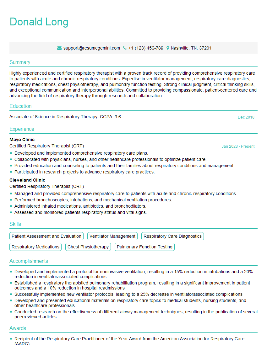
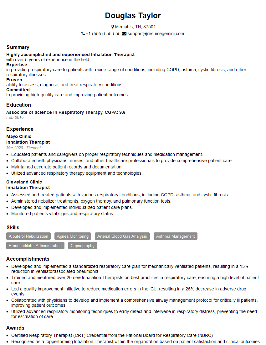
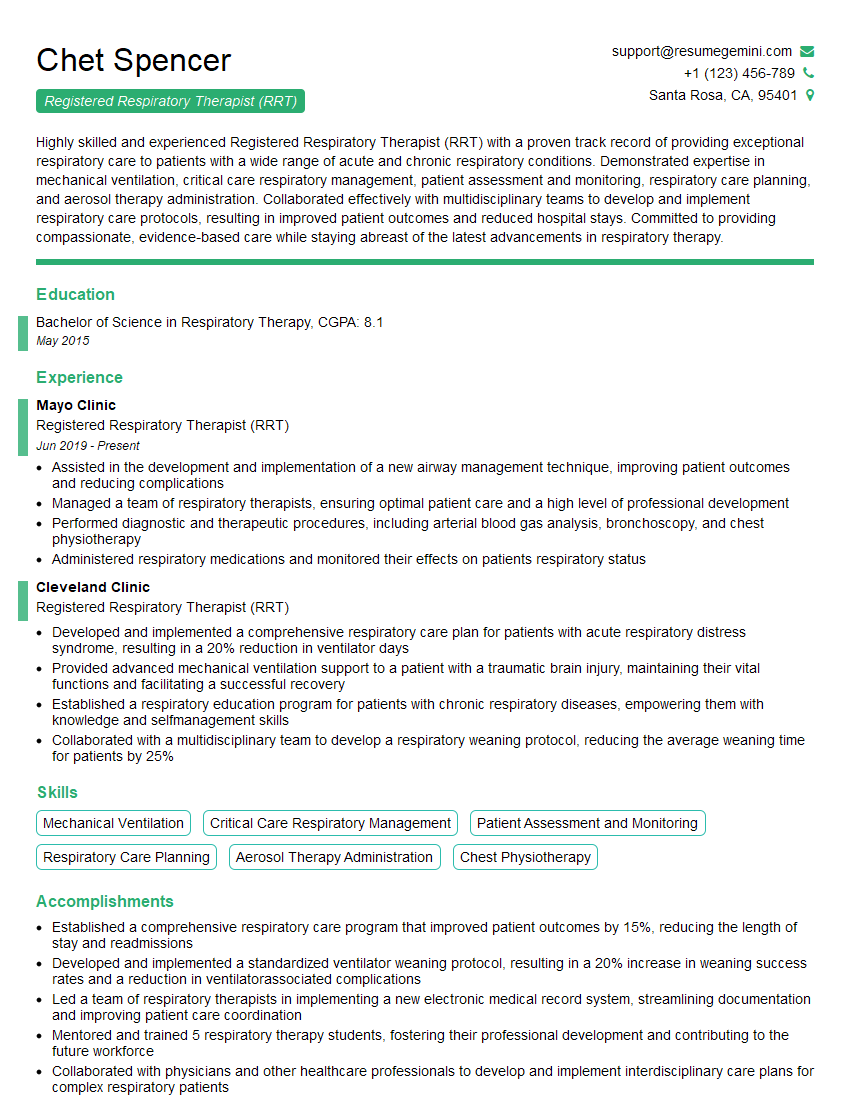
Mastering Inhalation Therapy opens doors to a rewarding career with diverse opportunities for growth and specialization. To maximize your job prospects, it’s crucial to present yourself effectively. Creating a strong, ATS-friendly resume is your first step. ResumeGemini is a trusted resource to help you build a professional resume that highlights your skills and experience. They offer examples of resumes tailored specifically to Inhalation Therapy, giving you a head start in crafting a compelling application. Invest the time to create a resume that truly showcases your potential – it’s an investment in your future.
Explore more articles
Users Rating of Our Blogs
Share Your Experience
We value your feedback! Please rate our content and share your thoughts (optional).
What Readers Say About Our Blog
This was kind of a unique content I found around the specialized skills. Very helpful questions and good detailed answers.
Very Helpful blog, thank you Interviewgemini team.


