The thought of an interview can be nerve-wracking, but the right preparation can make all the difference. Explore this comprehensive guide to Echocardiography Assisting interview questions and gain the confidence you need to showcase your abilities and secure the role.
Questions Asked in Echocardiography Assisting Interview
Q 1. Describe your experience with different echocardiography modalities (transthoracic, transesophageal, stress echo).
My experience encompasses all three major echocardiography modalities: transthoracic echocardiography (TTE), transesophageal echocardiography (TEE), and stress echocardiography. TTE, the most common, uses a transducer placed on the chest to image the heart. I’m proficient in acquiring various views, optimizing image quality, and identifying common cardiac pathologies. TEE provides higher-resolution images by placing a transducer into the esophagus, offering a unique perspective, particularly valuable in assessing valvular disease or detecting thrombi. I have extensive experience in performing and interpreting TEE studies, including those during cardiac surgery. Stress echocardiography combines exercise or pharmacological stress with echocardiography to assess the heart’s response to increased workload, aiding in the diagnosis of coronary artery disease. I’m skilled in managing patient stress levels, interpreting stress-induced wall motion abnormalities, and identifying ischemic areas.
Q 2. Explain the process of obtaining a quality echocardiogram image.
Obtaining a high-quality echocardiogram is a multi-step process requiring precision and attention to detail. It begins with proper patient preparation – ensuring appropriate fasting (if necessary) and a comfortable position. Next, I meticulously select the appropriate transducer and apply the coupling gel evenly to enhance sound transmission. Image optimization involves adjusting gain, depth, and focus to achieve clear visualization of cardiac structures. The angle of the transducer is critical; subtle adjustments can greatly improve image clarity. I carefully assess the image for artifacts (discussed in the next question) and adjust accordingly. Finally, consistent and careful transducer movement is essential to acquire comprehensive views of the heart from different angles. Think of it like sculpting; the transducer is the tool, and the heart is the clay. It takes practice and skill to ‘shape’ the images into a comprehensive assessment.
Q 3. How do you identify and troubleshoot common artifacts in echocardiography?
Identifying and troubleshooting artifacts is crucial for accurate interpretation. Common artifacts include reverberation (multiple echoes from a single reflection), shadowing (sound attenuation behind a highly reflective structure), and acoustic enhancement (increased brightness behind a fluid-filled structure). For example, reverberation might appear as multiple parallel lines behind the sternum. Shadowing can obscure the visualization of structures behind a calcified valve. Acoustic enhancement could be seen behind a pericardial effusion. Troubleshooting involves systematically reviewing the image acquisition techniques. Changing transducer position, adjusting the gain settings, or altering the frequency can often eliminate or reduce these artifacts. Understanding the underlying cause is key: reverberation is often due to air, shadowing from dense structures, and acoustic enhancement from fluid.
Q 4. What are the key anatomical structures you identify during a standard echocardiogram?
A standard echocardiogram focuses on identifying key anatomical structures, including the four chambers of the heart (right and left atria and ventricles), the heart valves (tricuspid, mitral, aortic, pulmonic), the major vessels (aorta, vena cava, pulmonary artery), and the pericardium. We also assess the thickness of the heart walls (septum and left ventricular wall), the size of the chambers, and the overall function of the heart. For example, we meticulously measure the ejection fraction (EF) – the percentage of blood ejected from the left ventricle with each beat – a vital indicator of heart function. We also evaluate the presence and nature of any masses, thrombi, or effusions. This comprehensive assessment allows for the diagnosis and monitoring of a wide array of cardiac conditions.
Q 5. Describe your experience with echocardiography equipment and its maintenance.
I’m experienced with various echocardiography machines from leading manufacturers, including their operating systems and software. This includes proficiency in using different transducers (phase array, sector, linear), adjusting settings, and optimizing image quality. Equipment maintenance is critical. This includes daily checks of machine functionality, transducer cleaning and sterilization, and timely reporting of any malfunctions to biomedical engineering. I understand the importance of following manufacturer’s guidelines for calibration and preventative maintenance to ensure the accuracy and reliability of the equipment. Regular quality control procedures, including phantom studies, ensure optimal machine performance and accurate diagnostic results.
Q 6. How do you ensure patient comfort and safety during an echocardiogram?
Patient comfort and safety are paramount. Before the procedure, I explain the process clearly, answer any questions, and obtain informed consent. During the examination, I maintain a calm and reassuring demeanor, explaining each step. I adjust the patient’s position to maximize comfort and minimize discomfort. The use of appropriate gel temperature is vital for patient comfort. Monitoring the patient’s vital signs during the procedure and being aware of any potential adverse reactions (e.g., dizziness, nausea) is crucial. In cases of TEE, meticulous attention is paid to ensure proper sedation and continuous monitoring by a qualified nurse or physician.
Q 7. Explain your understanding of different echocardiographic views (e.g., apical, parasternal).
Echocardiography employs multiple views to comprehensively visualize the heart. The apical views (acquired with the transducer placed at the apex of the heart) are useful for assessing overall cardiac function and valvular function. The parasternal views (obtained with the transducer placed along the sternum) are crucial for assessing the left ventricle and its function. Other views include subcostal and suprasternal, each providing a unique perspective. Think of it as taking a picture of a 3D object – each view shows a different aspect, contributing to a complete picture. Different views offer optimized visualization of specific structures and functions of the heart. The appropriate selection and execution of these views are essential for a thorough echocardiographic assessment.
Q 8. How do you interpret basic echocardiographic measurements (e.g., ejection fraction, wall thickness)?
Interpreting basic echocardiographic measurements is fundamental to assessing cardiac function. Let’s break down two key measurements: ejection fraction (EF) and wall thickness.
Ejection Fraction (EF): This represents the percentage of blood ejected from the left ventricle (the heart’s main pumping chamber) with each contraction. A normal EF is generally considered to be between 55% and 70%. An EF below 55% suggests reduced pumping ability, potentially indicating heart failure. For instance, an EF of 30% signifies significantly impaired ventricular function, requiring close monitoring and potentially medical intervention. We look at the size of the ventricle before and after contraction to calculate this percentage. A smaller end-systolic volume (the volume after contraction) compared to a larger end-diastolic volume (volume before contraction) points towards a healthy EF. Conversely, a larger end-systolic volume suggests a lower EF.
Wall Thickness: This refers to the thickness of the heart muscle (myocardium). Increased wall thickness, or hypertrophy, can be a sign of underlying conditions like hypertension (high blood pressure). We measure wall thickness in different segments of the heart, comparing them to established norms. For example, a significantly thickened left ventricular wall could indicate long-standing hypertension, leading to the heart working harder to pump blood against increased resistance. We must consider the patient’s age and other clinical factors when interpreting wall thickness as normal ranges vary.
In summary, interpreting these measurements requires considering them in the context of the entire echocardiogram, the patient’s medical history, and other clinical findings. It’s not simply about the numbers; it’s about understanding what those numbers *mean* for the patient’s overall cardiac health.
Q 9. Describe your experience with patient preparation and education for echocardiography procedures.
Patient preparation and education are critical for a successful echocardiogram. My approach begins with a thorough explanation of the procedure. I use clear, simple language, avoiding medical jargon whenever possible. I describe what will happen during the test, how long it will take (typically 30-45 minutes), and what sensations they might experience (usually none, but some slight pressure from the transducer).
For transthoracic echocardiograms (TTEs), the most common type, I explain that they will need to lie on their left side, and that I will apply a gel to their chest to allow the ultrasound waves to pass through the skin. I emphasize the importance of remaining still during the scan to obtain clear images. For transesophageal echocardiograms (TEEs), which involve a probe going down the esophagus, I explain the process in detail, emphasizing the importance of good cooperation to improve image quality and reduce any potential discomfort.
I address any anxieties or concerns the patient might have. Many patients are nervous about medical procedures, and addressing their concerns builds trust and ensures a more comfortable experience. After the procedure, I provide a brief explanation of what we saw and emphasize that a full report will be available shortly from the cardiologist. Clear and empathetic communication is key.
Q 10. How do you handle emergency situations during an echocardiography procedure?
Emergency situations during an echocardiogram are rare but must be handled swiftly and efficiently. The most common emergencies involve patient discomfort or changes in vital signs.
For instance, if a patient experiences significant chest pain or shortness of breath, I immediately stop the procedure, notify the physician immediately, and assess their vital signs. Depending on the situation, this may involve administering oxygen, contacting emergency medical services, or following protocols established by the hospital. Knowing and following these protocols is vital.
I’m trained to recognize signs of hypotension, arrhythmias, and other life-threatening events. My primary responsibility during an emergency is to ensure patient safety and provide the attending physician with the necessary information to facilitate appropriate treatment. Regular training on emergency protocols, including CPR and basic life support, is crucial to my role.
Q 11. What is your experience with documenting echocardiogram results?
Accurate and comprehensive documentation is vital. My echocardiogram documentation includes all relevant patient information: name, date of birth, medical record number, and referring physician. The report details the type of echocardiogram performed (TTE or TEE), the equipment used, and a description of the images obtained.
I meticulously document all measurements, including ejection fraction, wall thickness, valve function, and chamber dimensions. Any abnormalities or observations are noted, along with the method of measurement. For example, I might note “Mild mitral regurgitation, estimated by color Doppler to be
Finally, the report summarizes the findings and their clinical significance, noting potential diagnoses and recommendations for further investigation or treatment. This information is essential for the cardiologist to make an informed decision regarding the patient’s care.
Q 12. How do you maintain patient confidentiality?
Maintaining patient confidentiality is paramount. I adhere strictly to HIPAA regulations and hospital policies. All patient information, including echocardiogram images and reports, is handled with the utmost care. Access to these records is restricted to authorized personnel only.
I never discuss patient information with unauthorized individuals. Even in casual conversations, I avoid disclosing identifying details. All digital records are password-protected and stored securely. Physical records are kept in locked cabinets. In summary, I understand that preserving patient confidentiality is not only an ethical obligation but a legal requirement.
Q 13. Describe your knowledge of cardiac anatomy and physiology.
A strong understanding of cardiac anatomy and physiology is essential for interpreting echocardiograms accurately. I have a thorough knowledge of the heart’s chambers (atria and ventricles), valves (mitral, aortic, tricuspid, and pulmonary), and major blood vessels (aorta, vena cava, pulmonary arteries and veins). I understand the intricate relationship between the electrical conduction system and the mechanical function of the heart, as this is reflected in the images produced by echocardiography.
My knowledge extends to the physiology of cardiac function, including blood flow dynamics, cardiac output, and pressure gradients. I can identify abnormal flow patterns and structural abnormalities, such as valve stenosis, regurgitation, and septal defects. For instance, I can visualize and measure the pressure gradient across the aortic valve to assess the severity of aortic stenosis. This detailed understanding enables me to interpret the echocardiogram’s findings accurately and provide valuable input to the cardiologist.
Q 14. What is your understanding of the role of echocardiography in diagnosing cardiac conditions?
Echocardiography plays a crucial role in diagnosing a wide range of cardiac conditions. It provides a non-invasive method of visualizing the heart’s structure and function in real-time. This allows for the detection of abnormalities that might otherwise go unnoticed.
Echocardiography is used to diagnose conditions such as:
- Valve diseases (stenosis and regurgitation)
- Congenital heart defects
- Heart failure
- Cardiomyopathies
- Pericardial effusion
- Myocardial infarction (heart attack)
- Arrhythmias
The information obtained from an echocardiogram helps clinicians assess the severity of the condition, guide treatment decisions, and monitor the effectiveness of therapy. In many cases, echocardiography can provide essential information without the need for more invasive procedures.
Q 15. Explain your knowledge of different cardiac pathologies and their echocardiographic presentations.
Echocardiography allows us to visualize the heart’s structure and function, revealing various pathologies. Understanding these presentations is crucial for accurate diagnosis and management. For example, in Mitral Regurgitation, we’d see echocardiographic evidence of regurgitant flow across the mitral valve during systole, often quantified by measuring the regurgitant jet area and its velocity. This can lead to chamber enlargement (left atrium and ventricle) which we can measure directly. Similarly, in Hypertrophic Cardiomyopathy, we’d observe thickened ventricular septum and walls, often with dynamic outflow tract obstruction that can be visualized during the procedure. In Aortic Stenosis, we’d see a diminished or delayed aortic valve opening, leading to decreased aortic flow velocity, resulting in a smaller aortic valve opening during systole (the opening is also less round and smooth than normal) and increased left ventricular afterload (pressure). We’d also see left ventricular hypertrophy. Each pathology has unique echocardiographic features, allowing for differential diagnosis.
- Ischemic Heart Disease: Echocardiography can reveal wall motion abnormalities, reflecting areas of reduced blood flow.
- Dilated Cardiomyopathy: We see enlarged chambers, particularly the left ventricle, and reduced ejection fraction.
- Pericardial Effusion: This shows as fluid accumulation around the heart, compressing the heart and affecting its function.
The combination of qualitative (visual assessment) and quantitative (measurements) information obtained allows for a thorough assessment of the cardiac condition.
Career Expert Tips:
- Ace those interviews! Prepare effectively by reviewing the Top 50 Most Common Interview Questions on ResumeGemini.
- Navigate your job search with confidence! Explore a wide range of Career Tips on ResumeGemini. Learn about common challenges and recommendations to overcome them.
- Craft the perfect resume! Master the Art of Resume Writing with ResumeGemini’s guide. Showcase your unique qualifications and achievements effectively.
- Don’t miss out on holiday savings! Build your dream resume with ResumeGemini’s ATS optimized templates.
Q 16. How do you manage challenging patients during echocardiography?
Managing challenging patients requires adaptability and a patient-centered approach. For example, patients with severe anxiety or claustrophobia may require sedation or a more gradual approach to the exam. We can start with a brief explanation of the procedure, focusing on what they can expect. Sometimes, adjusting the positioning of the patient or using a smaller transducer can make a difference. For patients with respiratory issues or limited mobility, we adjust the examination to minimize stress, using shorter examination times and frequent breaks. In cases of arrhythmias, careful synchronization with the ECG signals is essential for accurate image acquisition. Ultimately, effective communication and empathy are key to ensuring patient comfort and obtaining high-quality images. Using appropriate transducer frequencies for the patient’s size and body habitus is also important for image acquisition.
Q 17. Describe your experience with quality assurance and quality control measures in echocardiography.
Quality assurance and quality control are paramount in echocardiography to ensure accurate and reliable results. We follow rigorous protocols to maintain equipment calibration and image quality, using phantoms to evaluate the systems periodically. Regular testing of the machines ensures accuracy of measurements, image resolution, and overall functionality. Our team regularly reviews image quality and measurement accuracy, comparing results with established standards. We also participate in inter-institutional quality control programs to compare our results against other echocardiography laboratories; this ensures our consistency in interpreting images, measurements, and reporting. Finally, we maintain meticulous records of all equipment maintenance and quality control procedures.
Q 18. How do you stay updated on advancements in echocardiography technology and techniques?
Staying current in echocardiography requires a multifaceted approach. I actively participate in continuing medical education (CME) courses and workshops focused on new technologies and techniques. I regularly review peer-reviewed journals and attend national and international conferences in the field. Online resources, such as professional society websites, offer access to the latest research and guidelines. Furthermore, collaborating with experienced colleagues and engaging in case discussions help to broaden my knowledge and refine my skills.
Q 19. Explain your proficiency in using echocardiography software and reporting systems.
I am proficient in various echocardiography software and reporting systems. My expertise includes using these systems for image acquisition, processing, measurements, and report generation. I am familiar with different software packages such as GE Vivid, Philips Epiq, and Siemens Acuson, and I can efficiently utilize their features for various echocardiographic modalities including 2D, M-mode, Doppler, and tissue Doppler imaging. My experience includes the generation of detailed reports that conform to accepted standards, including the use of standardized nomenclature and terminology. I am adept at generating high-quality images for use in diagnosis and follow-up patient care.
Q 20. How do you collaborate with other members of the healthcare team?
Collaboration is integral to providing optimal patient care. I work closely with cardiologists, nurses, and other healthcare professionals to ensure seamless information flow and coordinated care. I participate in multidisciplinary team meetings, providing echocardiographic insights to aid in diagnosis and treatment planning. Clear and concise communication of findings to the cardiologist, often including images or clips, is essential. I also assist in educating patients and their families about the results of their echocardiogram in understandable terms.
Q 21. Describe a time you had to solve a technical problem during an echocardiography procedure.
During a recent transesophageal echocardiogram (TEE), we encountered a problem with the image quality due to air bubbles in the esophagus. The images were significantly degraded and the view of the heart was obstructed. After several attempts to reposition the probe, I recognized the issue was caused by the patient inadvertently swallowing air during the procedure. We asked the patient to hold their breath and then calmly repositioned the TEE probe. We then proceeded to slowly withdraw the probe with careful attention to the position of the patient to help remove the air from the esophagus. We repeated this method until the air bubbles were eliminated from the esophagus, yielding excellent images and enabling completion of the exam without having to repeat it.
Q 22. How do you handle conflicting priorities or a high workload in the echocardiography lab?
In the fast-paced environment of an echocardiography lab, conflicting priorities and high workloads are common. My approach is multifaceted. First, I prioritize tasks based on urgency and clinical significance. For example, a patient requiring immediate echocardiography for a suspected acute event takes precedence over a routine study. Second, I leverage effective time management techniques, such as creating detailed schedules and breaking down large tasks into smaller, manageable chunks. This allows me to stay organized and focused. Third, I communicate proactively with the cardiologist and other team members. If I foresee a potential delay or conflict, I immediately communicate the situation and explore potential solutions collaboratively. Finally, I’m adept at multitasking and efficiently utilizing available resources, like delegating appropriate tasks when possible, to ensure all patients receive timely and high-quality care.
For example, during a particularly busy afternoon with multiple urgent cases, I prioritized patients with chest pain and shortness of breath, while simultaneously preparing equipment for the next patient and maintaining communication with the cardiology team. By utilizing effective workflow strategies and strong team communication, we managed to complete all studies without compromising the quality of care.
Q 23. Describe your experience with performing stress echocardiography procedures.
I have extensive experience in assisting with stress echocardiography procedures, both pharmacological (using dobutamine or adenosine) and exercise stress tests. My responsibilities include preparing the patient, ensuring proper equipment setup, monitoring vital signs closely throughout the procedure, and assisting the cardiologist with image acquisition and interpretation. This involves understanding the nuances of different stress protocols and recognizing potential complications like arrhythmias or hypotension. I’m proficient in using the echocardiography machine to optimize image quality during periods of stress and in identifying hemodynamic changes reflected in the echocardiogram. I also assist in post-procedure documentation and report generation.
A particularly memorable experience involved a patient with a history of hypertension who exhibited significant ST-segment changes during exercise stress testing. My careful monitoring of vital signs and prompt communication with the cardiologist enabled us to recognize the potential for an acute coronary event and quickly implement appropriate interventions, resulting in the patient receiving immediate treatment.
Q 24. What are your strengths and weaknesses related to echocardiography assisting?
My strengths lie in my meticulous attention to detail, my ability to work effectively under pressure, and my excellent communication skills. I am highly proficient in operating various echocardiography machines and possess a thorough understanding of echocardiographic principles. I am also a quick learner and adapt easily to new technologies and techniques. A key strength is my ability to anticipate the needs of the cardiologist and other team members, ensuring a smooth and efficient workflow.
One area I am actively working on is further developing my proficiency with advanced echocardiographic techniques, such as 3D echocardiography. While I possess foundational knowledge, I am eager to enhance my expertise in this area through continued learning and training.
Q 25. Why are you interested in this specific Echocardiography position?
I am highly interested in this specific Echocardiography position because of [Hospital/Clinic Name]’s reputation for excellence in cardiovascular care and its commitment to innovation. The opportunity to work alongside a team of experienced professionals, using state-of-the-art equipment, is particularly appealing. The description of the role highlights a collaborative environment which aligns perfectly with my own working style. Moreover, the emphasis on [mention specific aspects of the job description, e.g., patient care, teamwork, professional development] resonates with my professional goals and values.
Q 26. What are your salary expectations?
My salary expectations are in line with the market rate for an experienced echocardiography assistant with my skills and experience in this region. I am open to discussing a competitive compensation package based on the specifics of the position and benefits offered.
Q 27. What are your long-term career goals in echocardiography?
My long-term career goals include continuing to advance my expertise in echocardiography. I am interested in pursuing further certifications and potentially specializing in a specific area like transesophageal echocardiography (TEE) or stress echocardiography. Ultimately, I aspire to contribute significantly to the field through high-quality patient care, mentorship of other echocardiography technicians, and possibly through research or educational opportunities within the cardiovascular field.
Q 28. Do you have any questions for me?
Yes, I have a few questions. First, could you elaborate on the opportunities for professional development and continuing education provided by the hospital? Second, what is the typical workflow and team structure within the echocardiography lab? And finally, what are some of the key performance indicators or goals that the echocardiography team is currently focused on?
Key Topics to Learn for Echocardiography Assisting Interview
- Echocardiography Equipment and Operation: Understanding the functionality of various echocardiography machines, transducers, and their appropriate applications. This includes troubleshooting common technical issues.
- Image Acquisition Techniques: Mastering the skills necessary to obtain high-quality images, including proper patient positioning, transducer manipulation, and gain/depth adjustments. Practical experience in optimizing image quality for different clinical scenarios is crucial.
- Cardiac Anatomy and Physiology: A strong grasp of the heart’s structure and function is essential for accurately interpreting echocardiograms. This includes understanding normal and abnormal cardiac cycles, valve function, and chamber dimensions.
- Echocardiographic Measurements and Calculations: Proficiency in performing accurate measurements (e.g., ejection fraction, chamber volumes) and calculations related to cardiac function is vital. Understanding the clinical significance of these measurements is equally important.
- Patient Care and Safety: Demonstrate knowledge of proper patient preparation, infection control protocols, and emergency response procedures in the echocardiography setting. This includes understanding patient comfort and anxiety management.
- Basic Hemodynamics and Cardiovascular Pathology: Familiarity with common cardiovascular diseases and their echocardiographic manifestations is necessary. This enables you to anticipate potential challenges and contribute effectively to the team.
- Communication and Teamwork: Highlight your ability to communicate effectively with cardiologists, other healthcare professionals, and patients. Explain your teamwork skills and how you contribute to a positive and productive work environment.
Next Steps
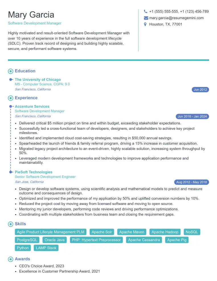
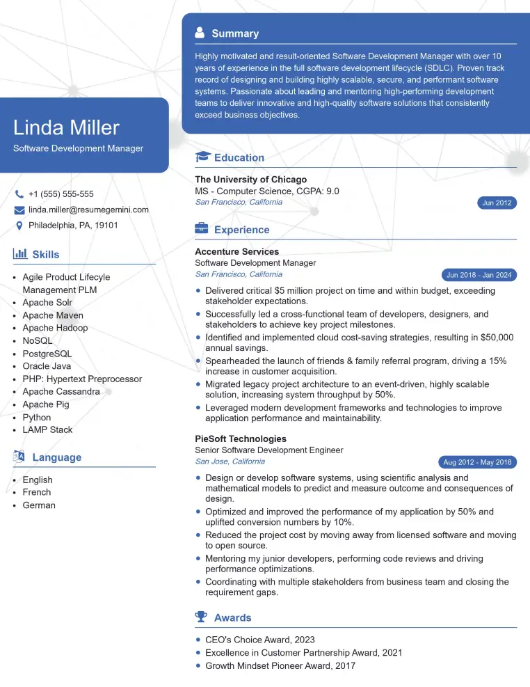
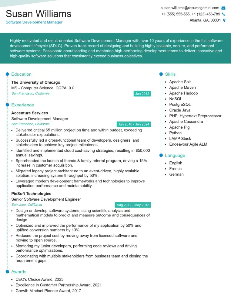
Mastering echocardiography assisting opens doors to a rewarding career with excellent growth potential within the healthcare sector. You’ll be contributing directly to patient care and working alongside experienced medical professionals. To maximize your job prospects, creating a strong, ATS-friendly resume is key. ResumeGemini is a trusted resource that can help you build a professional and impactful resume that highlights your skills and experience effectively. Examples of resumes tailored to Echocardiography Assisting are available to guide you through the process. Invest the time to craft a compelling resume – it’s your first impression to potential employers.
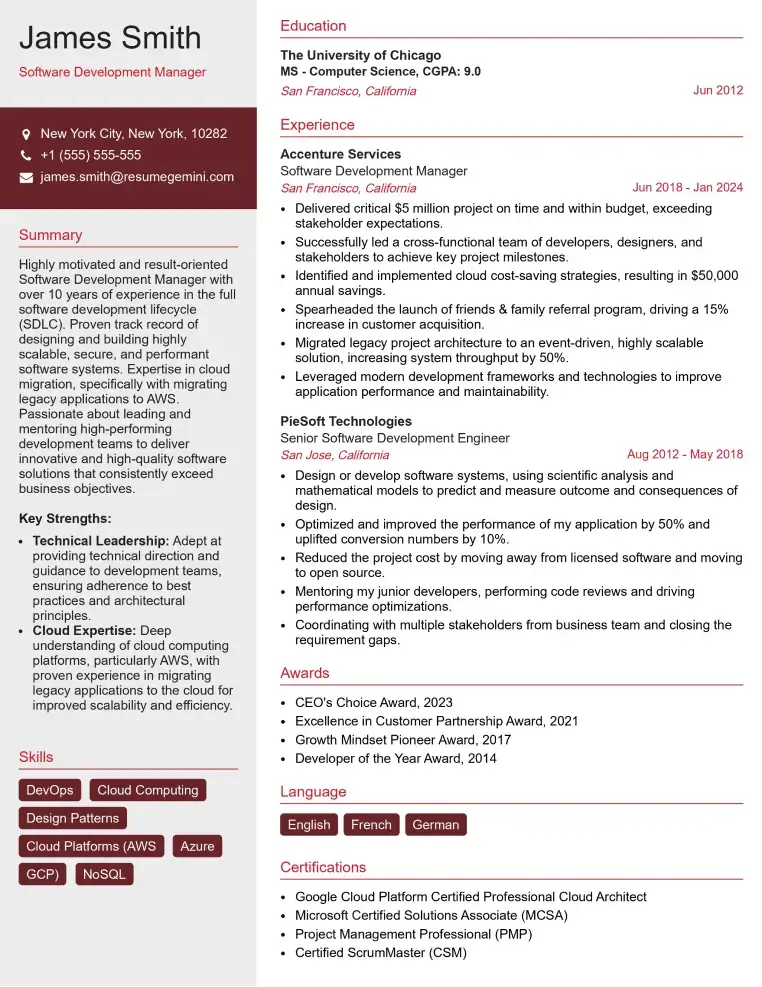
Check Out Resume Samples at ResumeGemini
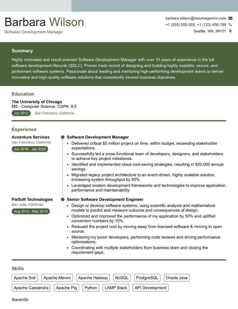
Check Out Resume Samples at ResumeGemini
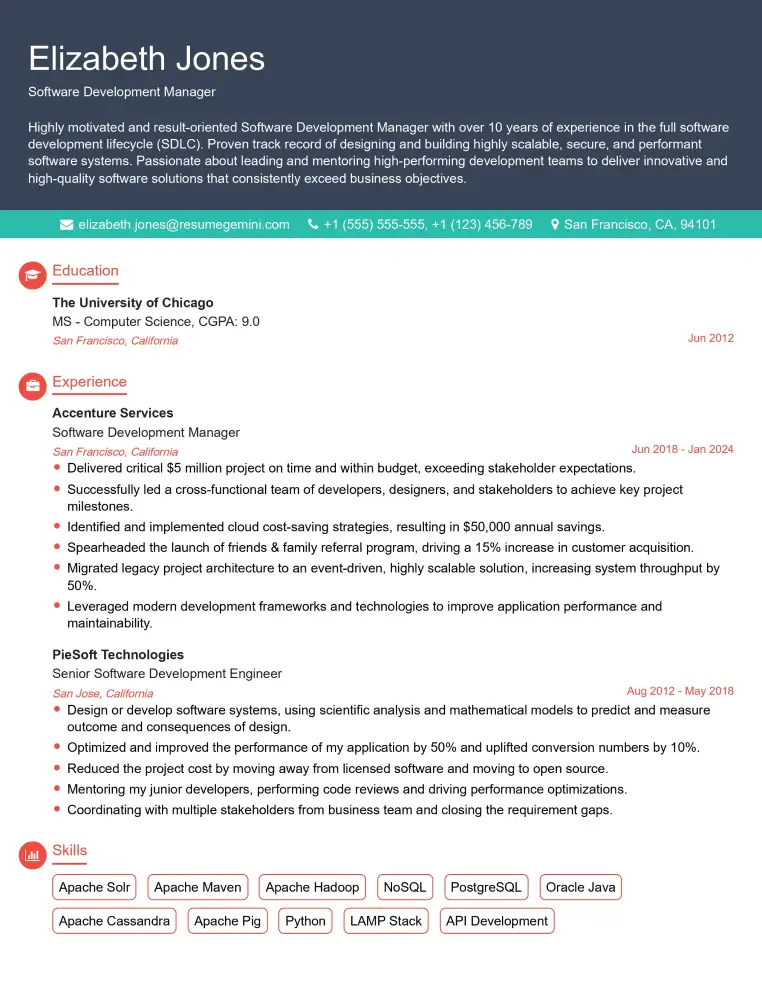
Check Out Resume Samples at ResumeGemini
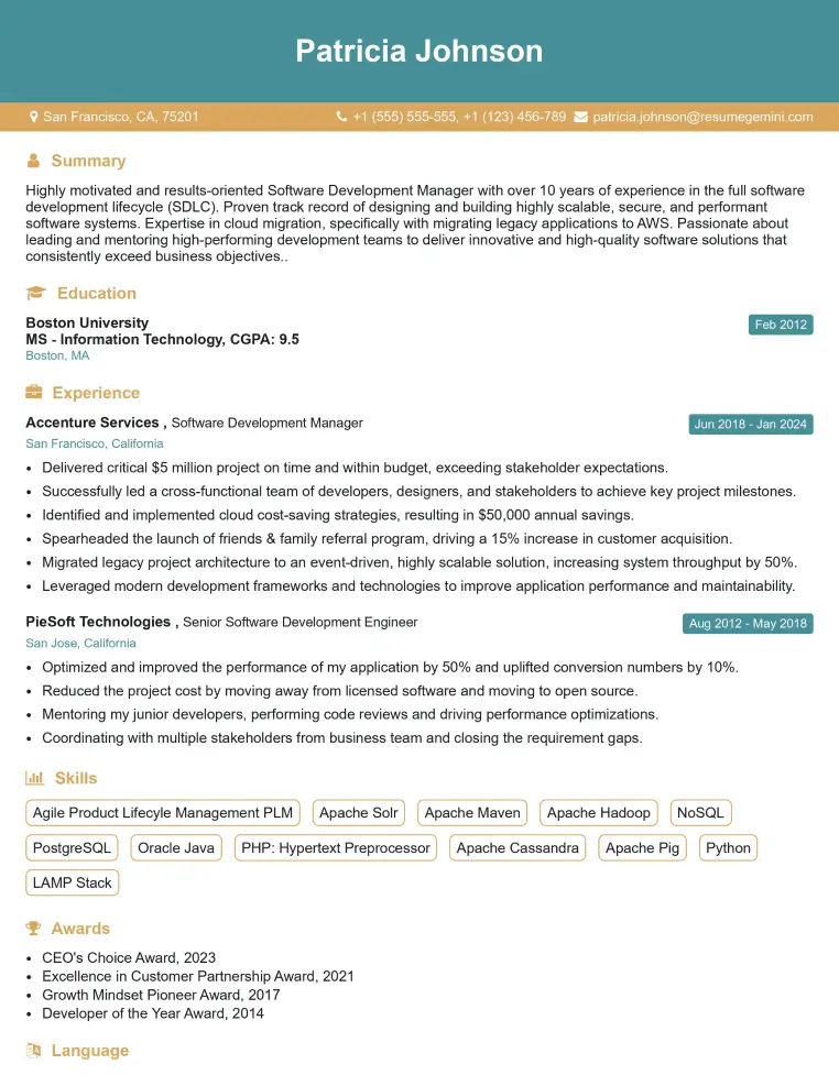
Check Out Resume Samples at ResumeGemini
Check Out Resume Samples at ResumeGemini
Check Out Resume Samples at ResumeGemini
Check Out Resume Samples at ResumeGemini
Explore more articles
Users Rating of Our Blogs
Share Your Experience
We value your feedback! Please rate our content and share your thoughts (optional).
What Readers Say About Our Blog
To the interviewgemini.com Webmaster.
Very helpful and content specific questions to help prepare me for my interview!
Thank you
To the interviewgemini.com Webmaster.
This was kind of a unique content I found around the specialized skills. Very helpful questions and good detailed answers.
Very Helpful blog, thank you Interviewgemini team.