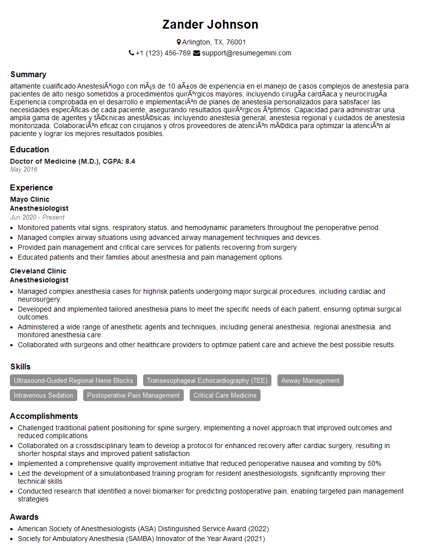
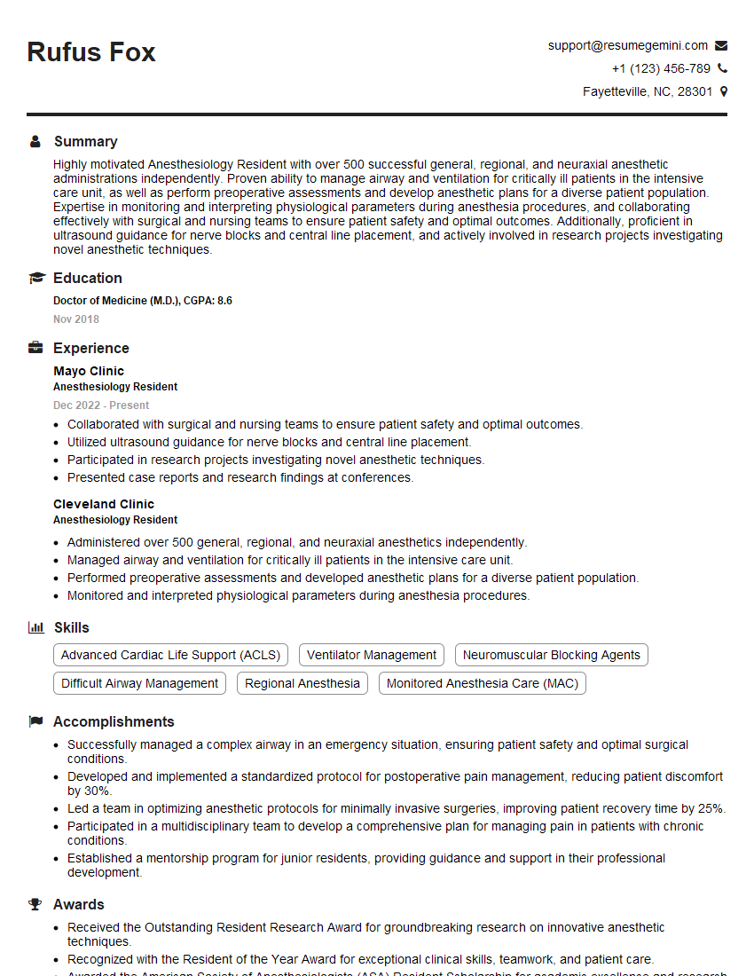
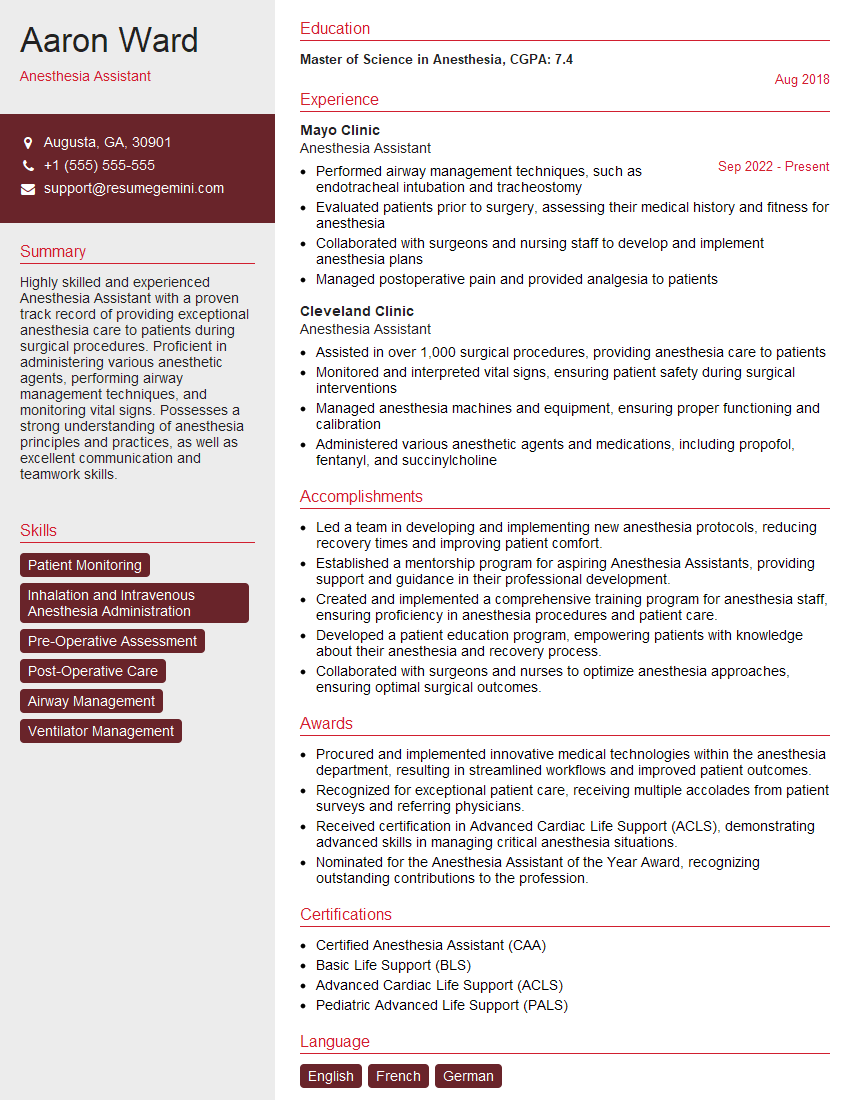
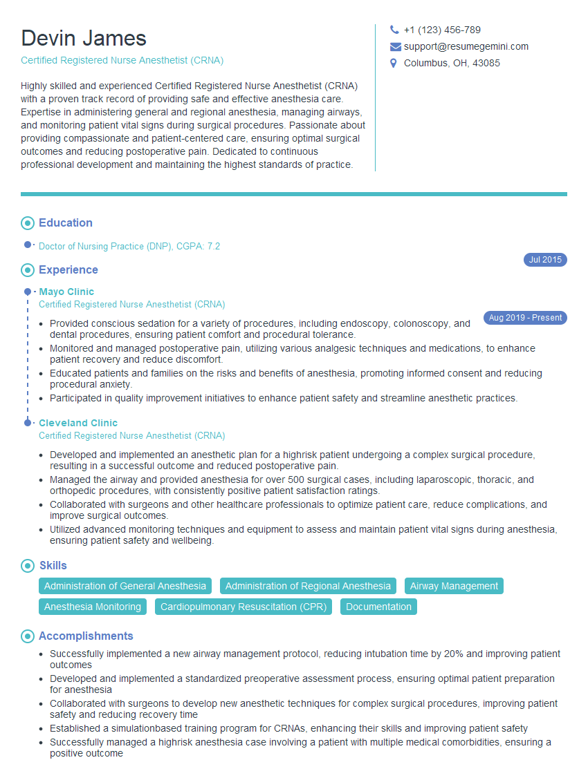
Interviews are more than just a Q&A session—they’re a chance to prove your worth. This blog dives into essential Anesthesia induction interview questions and expert tips to help you align your answers with what hiring managers are looking for. Start preparing to shine!
Questions Asked in Anesthesia induction Interview
Q 1. Describe the steps involved in a rapid sequence induction.
Rapid Sequence Induction (RSI) is a technique used to rapidly induce general anesthesia and secure the airway in situations where a patient is at high risk of aspiration. It’s crucial for patients with a full stomach, who might vomit and aspirate during induction. The speed is essential to minimize this risk.
- Preoxygenation: We begin by giving the patient 100% oxygen for several minutes to increase the oxygen reserve in their lungs. Think of it like filling a gas tank before a long drive.
- Cricoid Pressure: Next, we apply cricoid pressure, gently pressing on the cricoid cartilage in the neck. This helps to occlude the esophagus, preventing stomach contents from entering the airway. This is a vital step, often practiced with team-based drills.
- Induction Agent Administration: A rapid-acting intravenous anesthetic, such as propofol or etomidate, is then given quickly. This renders the patient unconscious.
- Neuromuscular Blocker Administration: Simultaneously or immediately after the induction agent, a neuromuscular blocking agent (like succinylcholine) is administered to paralyze the muscles, including those involved in breathing. This allows for easy intubation without resistance.
- Endotracheal Intubation: Once the patient is unconscious and paralyzed, a breathing tube (endotracheal tube) is quickly inserted into the trachea, securing the airway. This ensures controlled ventilation and prevents aspiration.
- Confirmation of Tube Placement: We then verify the tube’s proper placement using techniques such as auscultation (listening to breath sounds) and capnography (measuring carbon dioxide in the exhaled breath).
The entire process needs to be coordinated, swift, and precisely executed to minimize the risk of aspiration. It’s a team effort involving anesthesiologist, a nurse anesthetist, and often other surgical personnel.
Q 2. Explain the difference between inhalational and intravenous induction agents.
Inhalational and intravenous induction agents both achieve general anesthesia but differ significantly in their method of administration and onset of action.
- Intravenous induction agents are administered directly into a vein. They act quickly, producing unconsciousness within seconds. Examples include propofol, etomidate, ketamine, and barbiturates. The rapid onset is advantageous in emergency situations.
- Inhalational induction agents are gases or volatile liquids that are inhaled. Their onset is slower, and induction time can be affected by factors like the patient’s respiratory rate and depth of breathing. Sevoflurane and desflurane are common examples. They are often used for maintenance of anesthesia rather than the initial induction.
The choice between intravenous and inhalational induction depends on various factors, including patient characteristics, the urgency of the procedure, and the anesthesiologist’s preference. For instance, rapid sequence intubation would typically favor intravenous agents due to their rapid onset.
Q 3. What are the advantages and disadvantages of using propofol for induction?
Propofol is a widely used intravenous anesthetic induction agent. It’s known for its rapid onset and short duration of action, making it ideal for many procedures.
- Advantages: Relatively smooth induction and emergence from anesthesia, rapid onset, short context-sensitive half-time (meaning it clears the body quickly), and antiemetic properties (it reduces nausea and vomiting).
- Disadvantages: Potential for hypotension (low blood pressure), apnea (cessation of breathing), pain on injection, and rarely, propofol infusion syndrome (a severe metabolic disorder associated with prolonged infusions of high doses).
We always carefully monitor vital signs, particularly blood pressure and heart rate, during and after propofol administration. The potential for hypotension is managed through careful fluid management and the use of vasoconstrictors if needed. The pain on injection can be mitigated by using a rapid injection technique and co-administering a local anesthetic. Propofol infusion syndrome is rare and is mostly associated with very prolonged high-dose infusions.
Q 4. How do you manage a difficult airway during anesthesia induction?
Managing a difficult airway is a critical skill in anesthesia. Difficult airways can be predicted based on patient factors like obesity, facial deformities, or a history of difficult intubations. But sometimes, difficulties arise unexpectedly.
- Preoperative Assessment: A thorough airway assessment before induction is crucial. This includes evaluating the patient’s mouth opening, neck mobility, and Mallampati score (which assesses the visibility of the posterior pharynx).
- Alternative Airways: If intubation proves difficult, we have several strategies. Laryngeal mask airways (LMA) provide a less invasive alternative, offering a seal to the airway without requiring full endotracheal intubation. Fiberoptic intubation allows visualization of the airway using a flexible scope, enabling intubation even in difficult cases.
- Surgical Airway Techniques: In extreme cases, a surgical airway may be necessary. This involves creating a surgical opening into the trachea to secure the airway. This is a last resort, but it’s a critical skill to have and utilize properly.
- Teamwork and Communication: Effective communication and teamwork are essential in managing difficult airways. Having a skilled team experienced in airway management is crucial for a positive outcome. An experienced anesthesiologist will not hesitate to call for backup.
Difficult airway management requires practice and proficiency in various techniques. Regular simulations and advanced training courses are essential to staying updated on the latest airway management techniques. The priority is always to secure the patient’s airway safely and efficiently, even if that means adapting our strategy as needed.
Q 5. What are the signs of malignant hyperthermia, and how would you manage it?
Malignant hyperthermia (MH) is a rare but potentially fatal genetic disorder that can be triggered by certain anesthetic agents, such as volatile anesthetics (like sevoflurane and desflurane) and succinylcholine. It’s characterized by a rapid rise in body temperature and muscle rigidity.
- Signs and Symptoms: Early signs can be subtle and include muscle rigidity (especially masseter muscle rigidity), tachycardia (rapid heart rate), tachypnea (rapid breathing), and unexplained increase in end-tidal carbon dioxide (ETCO2).
- Management: Prompt recognition and management are crucial. The mainstay of treatment is dantrolene, a muscle relaxant that helps to reduce muscle rigidity and prevent further heat production. Cooling measures (e.g., ice packs, cooling blankets) are also essential to lower body temperature. Supportive care, including managing electrolyte imbalances and cardiac arrhythmias, is also critical.
Suspecting MH is a true medical emergency. We have established protocols in place to address this immediately. If MH is suspected, we promptly stop the triggering agents, administer dantrolene, and initiate cooling measures while simultaneously contacting the surgical team and hospital’s MH task force.
Q 6. Discuss your experience with regional anesthesia techniques used in conjunction with general anesthesia.
Regional anesthesia techniques, such as spinal, epidural, and peripheral nerve blocks, are frequently used in conjunction with general anesthesia. This multimodal approach can offer significant advantages to the patient.
- Reduced Opioid Requirements: Regional anesthesia can reduce the need for postoperative opioids, minimizing the risk of opioid-related side effects, such as nausea, vomiting, constipation, and respiratory depression. This is becoming increasingly important due to the opioid crisis.
- Improved Postoperative Analgesia: Regional blocks provide excellent postoperative pain control, allowing patients to be more comfortable and mobile postoperatively. This faster recovery time reduces the chances of complications.
- Enhanced Recovery After Surgery (ERAS) Protocols: Combined regional and general anesthesia is often integrated into ERAS protocols, aiming to minimize surgical stress and accelerate postoperative recovery.
For example, a patient undergoing a lower extremity surgery might receive a spinal anesthetic for operative analgesia, combined with general anesthesia to provide unconsciousness. This combination allows for adequate analgesia without requiring high doses of systemic opioids. In my experience, the combination frequently leads to a better patient experience and a more efficient recovery.
Q 7. Explain the principles of airway assessment before induction.
Airway assessment before induction is critical for identifying potential difficulties and planning a safe and effective anesthetic course. This assessment involves a combination of visual inspection, palpation, and sometimes additional diagnostic tests.
- Visual Inspection: We assess the patient’s mouth opening (inter-incisor distance), the size and shape of their teeth, the presence of any abnormalities in the jaw or neck, and the position of the tongue. The Mallampati classification helps to determine the visibility of the posterior pharynx.
- Palpation: We check for neck mobility, evaluating the ability to extend and flex the neck. Limited neck mobility can impede intubation.
- Thyromental Distance: Measuring the distance between the thyroid cartilage and the mentum (chin) helps assess the space available for intubation.
- Additional Assessments: Depending on the patient’s risk profile, we may use other tools, such as a flexible fiberoptic laryngoscope, to better visualize the airway. This is particularly valuable for patients with anticipated difficult airways.
A thorough airway assessment allows us to anticipate potential challenges and select the appropriate induction and airway management strategies. This proactive approach ensures a safer and more efficient procedure for the patient. Careful evaluation and documentation of the airway assessment are essential for good medical practice.
Q 8. Describe your preferred method for securing an airway.
My preferred method for securing an airway involves a tiered approach, prioritizing safety and patient comfort. The first step is always a thorough assessment of the patient’s airway, considering factors such as Mallampati score (assessing mouth opening and tongue size), thyromental distance (distance between thyroid cartilage and chin), and neck mobility. This assessment helps predict potential difficulties.
If the airway assessment is favorable, I’ll typically use a laryngeal mask airway (LMA) for ease of insertion and patient comfort, especially for shorter procedures. For longer procedures or those requiring controlled ventilation, I’ll opt for endotracheal intubation. I always have backup plans, including a difficult airway cart readily available, containing alternative airway devices like a bougie, gum elastic bougie, and various sized endotracheal tubes. The choice of technique is patient-specific and guided by the anticipated difficulty of intubation and the planned surgical procedure. For example, a patient with a known difficult airway might necessitate fiberoptic intubation, guided by a skilled anesthesiologist. I emphasize a gentle, step-wise approach minimizing patient discomfort and trauma.
Q 9. How do you monitor vital signs during anesthesia induction?
Monitoring vital signs during anesthesia induction is crucial for patient safety and is done continuously. This involves using a multi-modal approach. We use pulse oximetry to monitor oxygen saturation (SpO2), aiming for values above 95%. Non-invasive blood pressure monitoring (NIBP) provides continuous readings of systolic, diastolic, and mean arterial pressure (MAP), allowing for early detection of hypotension or hypertension. Electrocardiography (ECG) monitors the heart rhythm, providing immediate alerts for arrhythmias. Capnography continuously measures the end-tidal carbon dioxide (EtCO2) confirming proper ventilation and tube placement. Respiratory rate and waveform capnography are essential for assessing the adequacy of ventilation and detecting hypoventilation. Finally, temperature monitoring helps prevent hypothermia, a common complication of anesthesia. The integration of these monitoring modalities allows for a comprehensive assessment of the patient’s physiological response throughout the induction process.
For example, if the SpO2 suddenly drops, it would immediately signal the need to assess the adequacy of oxygenation and ventilation, possibly requiring adjustments to the anesthetic gases or airway management.
Q 10. What are the potential complications of anesthesia induction, and how do you mitigate them?
Potential complications of anesthesia induction are numerous and range in severity. Some common ones include hypotension (low blood pressure), bradycardia (slow heart rate), tachycardia (fast heart rate), hypoxia (low blood oxygen), hypercapnia (high carbon dioxide levels), airway complications like difficult intubation or esophageal intubation, and malignant hyperthermia (a rare but life-threatening condition).
Mitigation strategies vary depending on the specific complication. Hypotension can be addressed with fluid resuscitation and vasopressors. Bradycardia might necessitate atropine administration. Hypoxia is tackled by optimizing ventilation and supplemental oxygen. Hypercapnia is treated by increasing ventilation. Airway complications require immediate intervention with alternative airway techniques. Malignant hyperthermia is a medical emergency requiring immediate cessation of triggering agents and administration of dantrolene. Careful patient assessment, proper medication selection and dosage, continuous monitoring, and rapid response to adverse events are key to minimizing the risk of complications. A thorough pre-operative assessment, including a detailed history and physical examination, plays a crucial role in identifying and managing potential risk factors.
Q 11. Explain your understanding of the different phases of anesthesia.
Anesthesia is typically divided into four phases: pre-induction, induction, maintenance, and emergence.
- Pre-induction: This phase involves the pre-operative evaluation, preparation of the patient (including NPO status), and the initial establishment of intravenous access. It’s a crucial period for establishing a baseline and identifying potential risks.
- Induction: This is the phase of administering anesthetic agents to transition the patient from consciousness to a state of general anesthesia. This phase includes airway management (intubation or LMA placement).
- Maintenance: Once anesthesia is established, the maintenance phase involves the continued administration of anesthetic agents and other medications to keep the patient unconscious, pain-free, and hemodynamically stable. This is often the longest phase and is adjusted throughout the surgery based on the patient’s response and the surgeon’s needs.
- Emergence: This involves the gradual discontinuation of anesthetic agents and reversal of neuromuscular blockade if used, allowing the patient to regain consciousness. It’s a crucial phase that requires careful monitoring as the patient may experience side effects such as nausea, vomiting, or confusion.
Understanding these phases is essential for safe and effective anesthetic management.
Q 12. How do you adjust your induction technique for patients with different medical conditions (e.g., hypertension, diabetes)?
Adjusting the induction technique for patients with different medical conditions is crucial for optimizing safety. For example, patients with hypertension might require a slower induction to prevent rapid changes in blood pressure. Pre-operative medication, such as beta-blockers, may be used to control hypertension and reduce the risk of complications during induction. Similarly, patients with diabetes may have increased susceptibility to hypoglycemia (low blood sugar) during anesthesia. These patients need careful monitoring of blood glucose levels throughout the procedure, and intravenous glucose may be required. Patients with asthma may need a modified induction strategy to minimize bronchospasm. Premedication with bronchodilators, avoidance of certain anesthetic agents, and close monitoring of respiratory function are important considerations. The specific adjustments made depend heavily on the individual patient’s overall health status and the severity of their condition. A collaborative approach involving consultation with other specialists, such as cardiologists or endocrinologists, is often beneficial.
Q 13. Describe your experience with managing patients with compromised cardiovascular function.
Managing patients with compromised cardiovascular function requires careful attention to detail and close monitoring. It starts with a thorough pre-operative assessment focusing on cardiac function, including an echocardiogram if necessary. Preoperative optimization might include optimizing blood pressure and heart rate with medications and making sure the patient is well hydrated. During induction, I aim to minimize hemodynamic instability by using anesthetic agents that have minimal cardiovascular effects. I utilize techniques like slow induction and titration of anesthetic drugs to avoid sudden shifts in blood pressure and heart rate. Close monitoring of vital signs is imperative. Intraoperative management often includes careful fluid management, inotropic support (medications that increase contractility), and close collaboration with the cardiac team if needed. In post-operative care, close observation for arrhythmias, myocardial ischemia, and fluid balance is crucial. Each case is unique and requires individual tailoring of the anesthetic plan.
Q 14. What are the considerations for administering anesthesia to elderly patients?
Elderly patients often present unique challenges during anesthesia due to age-related physiological changes. These changes can include decreased cardiac reserve, reduced renal and hepatic function, and increased sensitivity to anesthetic agents. The goal is to minimize these risks and provide safe and effective anesthesia. A thorough pre-operative assessment is critical, including a comprehensive review of the patient’s medical history, current medications, and functional status. I often use a modified induction technique with lower doses of anesthetic agents to reduce the risk of hypotension and respiratory depression. Careful monitoring is essential, with close attention paid to blood pressure, heart rate, oxygen saturation, and respiratory function. Post-operative care involves close observation for postoperative cognitive dysfunction and potential complications related to age-related organ compromise. Collaboration with the geriatrician or the patient’s primary care physician is often beneficial in planning and optimizing anesthetic management and post-operative care for elderly patients. For instance, using shorter acting anesthetic agents that are more rapidly cleared from the body can speed up recovery and reduce post-operative confusion.
Q 15. Explain your understanding of the pharmacokinetics and pharmacodynamics of commonly used induction agents.
Understanding the pharmacokinetics (what the body does to the drug) and pharmacodynamics (what the drug does to the body) of induction agents is crucial for safe and effective anesthesia. Let’s look at some common examples:
- Propofol: This is a widely used agent. Its pharmacokinetics involve rapid distribution into highly perfused tissues like the brain, followed by redistribution to less perfused tissues and ultimately metabolism in the liver. Its pharmacodynamics involve depressing the CNS, leading to unconsciousness. The rapid onset and short context-sensitive half-time make it ideal for short procedures.
- Etomidate: Etomidate also provides rapid onset and short duration of action, primarily through its action on GABA receptors in the brain. Its metabolism is primarily hepatic, but it’s known for its potential to cause adrenal suppression, a factor we carefully consider in patient selection.
- Ketamine: Unlike propofol and etomidate, ketamine is a dissociative anesthetic. It acts on NMDA receptors, resulting in a state of analgesia and amnesia, but also maintaining some airway reflexes. Its metabolism is primarily hepatic, and it can cause bronchodilation which can be beneficial in certain patients.
- Thiopental (less commonly used now): This barbiturate has a rapid onset but a longer duration of action compared to propofol. It’s highly lipid-soluble, allowing rapid brain penetration, but its metabolism and context-sensitive half-time need to be taken into account, especially in elderly or liver-compromised patients.
Understanding these differences in pharmacokinetics and pharmacodynamics allows us to tailor our induction technique to the individual patient’s needs and the specifics of the surgical procedure. For example, a patient with liver disease might require a lower dose of propofol or an alternative agent altogether.
Career Expert Tips:
- Ace those interviews! Prepare effectively by reviewing the Top 50 Most Common Interview Questions on ResumeGemini.
- Navigate your job search with confidence! Explore a wide range of Career Tips on ResumeGemini. Learn about common challenges and recommendations to overcome them.
- Craft the perfect resume! Master the Art of Resume Writing with ResumeGemini’s guide. Showcase your unique qualifications and achievements effectively.
- Don’t miss out on holiday savings! Build your dream resume with ResumeGemini’s ATS optimized templates.
Q 16. How do you calculate drug dosages for anesthesia induction?
Calculating drug dosages for anesthesia induction is not a simple formula; it’s a clinical judgment based on multiple factors. We don’t use a single equation but rather a clinical approach that considers:
- Patient factors: Age, weight, body mass index (BMI), overall health status (including cardiac, hepatic, and renal function), and pre-existing conditions (e.g., hypertension, diabetes).
- Procedure type: The duration and complexity of the surgical procedure influence the choice of induction agent and the desired depth of anesthesia.
- Co-administered medications: The effects of other drugs the patient is taking (e.g., beta-blockers, opioids) must be considered.
- Individual response: Patients react differently to medications. Previous anesthetic experiences can provide valuable insight.
While we may use weight-based dosing as a starting point (e.g., mg/kg), we often adjust the dose based on our clinical assessment and monitoring of vital signs like heart rate, blood pressure, and oxygen saturation. This is why experience and continuous monitoring are key to safe anesthetic practice. For example, a frail elderly patient might receive a much lower dose of propofol than a healthy young adult.
Q 17. What is your approach to managing a patient experiencing unexpected hemodynamic instability during induction?
Unexpected hemodynamic instability during induction is a serious event requiring immediate action. My approach involves:
- Immediate assessment: I quickly assess the patient’s vital signs (heart rate, blood pressure, oxygen saturation, ECG), looking for arrhythmias or signs of hypotension or hypertension.
- Identify the cause: This is critical. The cause could be related to the anesthetic drugs, underlying patient conditions, or a reaction to the surgical stimulation. For example, a rapid drop in blood pressure could be caused by a vagal response to intubation or a direct effect of the induction agent.
- Treatment: This is tailored to the identified cause. Hypotension might require intravenous fluids, vasopressors (e.g., epinephrine, norepinephrine), or the administration of atropine if bradycardia is present. Hypertension might require administering antihypertensive medications.
- Continuous monitoring: Vital signs, ECG, and oxygen saturation are continuously monitored post treatment.
- Communication: Open communication with the surgical team and other members of the anesthesia care team is crucial.
In summary, managing hemodynamic instability requires rapid assessment, identification of the underlying cause, and immediate intervention based on the specific problem. Each case is unique and calls for quick thinking and decisive action.
Q 18. Describe your experience with using neuromuscular blocking agents.
Neuromuscular blocking agents (NMBAs) are essential for facilitating tracheal intubation and surgical procedures by providing skeletal muscle relaxation. I have extensive experience with both depolarizing (e.g., succinylcholine) and non-depolarizing (e.g., rocuronium, vecuronium) agents.
- Succinylcholine: This depolarizing agent causes initial fasciculations (muscle twitches) followed by paralysis. Its rapid onset is advantageous for rapid-sequence intubation, but it can trigger hyperkalemia in susceptible patients (e.g., those with burns or severe muscle trauma), which I carefully screen for beforehand.
- Rocuronium and Vecuronium: These non-depolarizing agents offer a more predictable and longer-lasting paralysis without the risk of hyperkalemia. The choice between them often depends on the patient’s renal and hepatic function, as they are metabolized and excreted differently.
Monitoring the depth of neuromuscular blockade is vital to avoid prolonged paralysis and to ensure adequate relaxation for intubation and surgery. I routinely use peripheral nerve stimulators to assess the degree of neuromuscular block. This allows for titrating the dose of NMBAs and administering reversal agents (e.g., neostigmine, sugammadex) when appropriate. I always prioritize a smooth and controlled emergence from neuromuscular blockade to prevent post-operative muscle pain. For instance, Sugammadex is used to reverse Rocuronium blockade, shortening the post-operative recovery time.
Q 19. How do you monitor the depth of anesthesia?
Monitoring the depth of anesthesia is paramount for patient safety. We use a combination of methods to assess the depth of anesthesia including:
- Clinical signs: Observing the patient’s response to stimuli (e.g., verbal commands, surgical incision), heart rate, blood pressure, and respiratory rate.
- Electroencephalography (EEG): Bispectral Index (BIS) monitoring provides a numerical representation of the EEG, offering a quantitative assessment of the depth of anesthesia. This helps ensure the patient is adequately anesthetized, reducing the incidence of intraoperative awareness.
- Entropy monitoring: Similar to BIS, entropy monitoring uses processed EEG and other physiological signals to estimate the level of anesthesia, providing another quantitative measure.
- Autonomic nervous system signs: Heart rate variability and blood pressure fluctuations can indicate the depth of anesthesia.
The ideal level of anesthesia will vary depending on the patient’s specific condition and the surgical procedure. For example, a highly anxious patient might require a deeper level of anesthesia than a calm patient. The combination of clinical assessment and technological monitoring allows us to tailor anesthesia to optimize patient safety and comfort.
Q 20. What are the different types of laryngoscopes, and which one do you prefer and why?
Several types of laryngoscopes are available, each with its own advantages and disadvantages. The most common are:
- Miller blade: This straight blade lifts the epiglottis directly to expose the larynx. It is excellent for visualizing the glottic opening in infants and children. It requires a more anterior approach.
- Macintosh blade: This curved blade sits in the vallecula (the space between the base of the tongue and the epiglottis) to indirectly lift the epiglottis. It is often preferred for adults because it may result in a less traumatic intubation procedure.
- Other specialized blades: There are also other blades designed for specific anatomical challenges or patient populations (e.g., pediatric blades).
My personal preference is for the Macintosh blade in most adult patients because of its ease of use and its gentler approach to the airway. However, the choice of blade ultimately depends on the patient’s anatomy, the clinician’s experience, and the particular circumstances of the situation. Sometimes, a Miller blade may be preferable if intubation is particularly difficult.
Q 21. Explain your understanding of the different types of endotracheal tubes.
Endotracheal tubes come in various sizes, materials, and designs, tailored to specific patient needs. Key differences include:
- Size: Sized by internal diameter (e.g., 7.0 mm, 8.0 mm), the size is selected based on the patient’s age, sex, and anticipated airway size.
- Material: Most commonly made of polyvinyl chloride (PVC), but other materials like silicone are available; silicone tubes are more comfortable and less irritating, but are less durable.
- Cuff type: High-volume, low-pressure cuffs are commonly used to prevent air leaks while minimizing pressure on the tracheal wall. The cuff is essential to sealing the airway.
- Features: Some tubes have features like radiopaque markings for easy identification on x-ray, Murphy eyes (small side holes) to improve ventilation if the tube is slightly malpositioned, or reinforced cuffs for improved durability.
- Specialized tubes: Specialized tubes are available for particular situations, such as double-lumen tubes for lung isolation during surgery.
Choosing the right endotracheal tube is crucial for successful airway management. This requires a thorough understanding of the patient’s anatomy and potential complications. Proper size selection and an appropriate cuff pressure are vital for patient safety and to prevent airway injury. Pre-operative assessment plays a major role in making sure the right size is selected.
Q 22. How do you confirm proper endotracheal tube placement?
Confirming proper endotracheal tube (ETT) placement is paramount to patient safety. It’s a multi-step process relying on several methods, none of which should be considered in isolation. We utilize a combination of techniques to ensure accurate placement.
Visual Confirmation: Direct visualization of the ETT passing through the vocal cords during insertion is ideal. This is the gold standard, but it’s not always feasible.
Auscultation: Listening for bilateral breath sounds over the lungs confirms airflow into both lungs. However, auscultation alone is unreliable, as sounds can be misleading due to factors like a partially obstructed airway or misplaced tube in the esophagus.
Capnography: This is the most reliable method. A capnograph continuously monitors the concentration of carbon dioxide in exhaled air. The presence of a waveform indicating exhaled CO2 confirms ventilation of the lungs. Absence of a waveform strongly suggests esophageal intubation.
Chest X-Ray: A chest x-ray provides definitive confirmation of ETT position. It shows the tip of the tube in the appropriate location within the trachea (avoiding the right main bronchus, for example).
In summary, I always strive for direct visualization, followed by immediate capnography confirmation. If any doubt remains, a chest X-ray is obtained immediately. Relying on a single method is simply unacceptable.
Q 23. Describe your experience with managing post-induction complications like nausea and vomiting.
Post-induction nausea and vomiting (PONV) are common complications we actively try to prevent and manage. My approach is multifaceted and involves proactive strategies and effective treatment if PONV occurs. Prevention is key. This involves considering risk factors such as patient history, the type of surgery, and the anesthetic agents used. I frequently utilize antiemetic prophylaxis, often a combination of drugs like ondansetron and dexamethasone, tailored to individual patient risk profiles. For example, patients undergoing laparoscopic surgery have a higher risk and receive more robust prophylaxis.
If PONV develops despite prophylaxis, I employ a stepwise approach to treatment. This starts with non-pharmacological interventions like antiemetic medications tailored to the specific type and severity of PONV. This might include administering additional antiemetics or using other supportive care such as intravenous fluids to improve hydration.
Q 24. What are some strategies for minimizing the risk of post-operative cognitive dysfunction?
Post-operative cognitive dysfunction (POCD) is a serious concern, and minimizing its risk is a high priority. While the exact mechanisms aren’t fully understood, several strategies are employed to mitigate this risk. These include:
Minimizing anesthetic exposure: Using shorter anesthetic durations and less potent agents where possible is crucial.
Optimizing intraoperative physiological variables: Maintaining normothermia (normal body temperature), normoglycemia (normal blood sugar), and adequate blood pressure helps reduce cognitive impairment.
Careful fluid management: Preventing dehydration and fluid overload are important, as both can contribute to cognitive dysfunction.
Postoperative analgesia: Appropriate and effective pain management is essential, as poorly managed pain can lead to stress and cognitive impairment. We aim for multimodal analgesia, combining different pain relief strategies.
Patient education and support: Preoperative education about expectations and recovery helps patients cope and manage post-surgical stress.
Remember, POCD is a complex issue, and research is ongoing. Our aim is to use a multi-modal approach, targeting several risk factors simultaneously.
Q 25. How do you handle unexpected events during anesthesia induction?
Unexpected events during anesthesia induction are a reality, demanding rapid assessment and decisive action. My approach hinges on maintaining situational awareness, recognizing warning signs early, and having a structured response plan.
For example, if a patient experiences unexpected hypotension (low blood pressure), the first step involves confirming the reading and assessing the patient’s clinical status. Then, I’d address the cause, which could range from hypovolemia (low blood volume) to drug-induced effects. Treatment may include intravenous fluids, vasopressors (medication to raise blood pressure), or adjusting anesthetic medications. Another example is malignant hyperthermia, a rare but life-threatening complication. Recognition is crucial, and immediate treatment involves stopping the triggering anesthetic agent and administering dantrolene. Teamwork and communication are paramount; I’d always call for immediate assistance from other members of the anesthesia team.
My response is always guided by a structured approach: assess, diagnose, treat, and monitor, ensuring appropriate documentation throughout.
Q 26. What are your preferred methods for post-operative pain management?
My preferred methods for postoperative pain management are based on the principles of multimodal analgesia, aiming to provide effective pain relief with reduced side effects. This strategy typically involves a combination of techniques:
Regional anesthesia: Techniques like epidural or peripheral nerve blocks provide excellent localized pain control with reduced opioid requirements.
Opioid analgesics: While opioids can be effective, their use is carefully titrated to minimize side effects such as nausea, constipation, and respiratory depression. We emphasize patient-controlled analgesia (PCA) to allow patients more control over their pain relief.
Non-opioid analgesics: Drugs like NSAIDs (nonsteroidal anti-inflammatory drugs) or acetaminophen are often combined with opioids to enhance pain relief and reduce opioid needs.
Adjuvant analgesics: Medications targeting specific pain pathways, such as gabapentinoids for neuropathic pain, may be used.
The specific combination of methods is always tailored to the patient’s individual needs, surgical procedure, and anticipated pain level. Regular pain assessments and patient communication are vital to ensure effectiveness and safety.
Q 27. Describe a time you had to adapt your induction plan due to unforeseen circumstances.
I recall a case where a patient scheduled for a routine laparoscopic cholecystectomy (gallbladder removal) arrived with an unexpectedly low hemoglobin level (anemia). This significantly altered my induction plan, as it increased the risk of intraoperative blood loss and hypotension. My initial plan involved a general anesthetic with volatile agents. However, I adjusted the strategy to minimize anesthetic-induced hypotension and optimize oxygen-carrying capacity. This included ensuring adequate pre-operative hydration, a slower induction, and careful attention to hemodynamic parameters throughout the surgery. I also coordinated closely with the surgical team to prepare for potential blood transfusion if necessary. The surgery proceeded smoothly, and the patient recovered well. This experience highlighted the importance of flexibility and adapting the anesthetic plan based on a thorough assessment of individual patient factors.
Q 28. Explain your understanding of the ethical considerations in anesthesia practice.
Ethical considerations are fundamental to anesthesia practice. Our primary ethical obligation is patient safety and well-being. This includes:
Informed Consent: Patients must understand the procedure, risks, benefits, and alternatives. I ensure clear and comprehensive communication before anesthesia is administered.
Beneficence and Non-maleficence: We must act in the patient’s best interest and avoid harm. This involves careful medication selection, monitoring for complications, and providing appropriate pain relief.
Respect for Autonomy: Patients have the right to make informed decisions about their care, including refusing treatment. Their wishes must be respected.
Justice: We must provide equitable access to care and avoid discrimination. All patients deserve the same standard of quality anesthesia regardless of their background.
Confidentiality: Patient information must be kept confidential and protected.
Ethical dilemmas occasionally arise, requiring careful consideration and consultation with colleagues or ethical committees to ensure decisions align with professional standards and patient values. Maintaining a strong ethical compass is paramount to our profession.
Key Topics to Learn for Anesthesia Induction Interview
- Pharmacology of Induction Agents: Understanding the mechanism of action, pharmacokinetics, and pharmacodynamics of commonly used induction agents (e.g., propofol, etomidate, ketamine). Consider comparing and contrasting their properties and suitability for different patient populations.
- Monitoring During Induction: Mastering the use and interpretation of vital signs monitoring (ECG, pulse oximetry, capnography, blood pressure) during induction and the immediate post-induction period. Practice recognizing and responding to common complications.
- Airway Management Techniques: Proficiency in various airway management techniques, including rapid sequence induction (RSI), difficult airway management strategies, and the use of airway adjuncts. Develop a strong understanding of the indications and contraindications for each technique.
- Hemodynamic Considerations: Understanding the impact of induction agents on cardiovascular function and the ability to manage potential hemodynamic instability. This includes recognizing and treating hypotension, hypertension, and arrhythmias.
- Preoperative Assessment and Patient Selection: The importance of a thorough preoperative assessment to identify and manage potential risks associated with anesthesia induction. This includes understanding patient-specific factors and comorbidities that might influence the choice of induction agents and techniques.
- Complications and Management: A comprehensive understanding of potential complications during anesthesia induction (e.g., airway complications, hemodynamic instability, allergic reactions) and the appropriate management strategies. Develop your problem-solving skills in this area.
- Understanding the Anesthesia Machine: Familiarity with the components and function of the anesthesia machine, including ventilator settings and gas delivery systems. Practice troubleshooting common issues.
Next Steps
Mastering anesthesia induction is crucial for career advancement and opens doors to specialized roles and increased responsibilities within the field of anesthesiology. A strong resume is your first impression on potential employers. Creating an ATS-friendly resume significantly increases your chances of getting your application noticed. ResumeGemini is a trusted resource to help you build a professional and impactful resume. We provide examples of resumes tailored specifically to Anesthesia induction positions to give you a head start. Invest in your future – build a resume that reflects your skills and experience effectively.
Explore more articles
Users Rating of Our Blogs
Share Your Experience
We value your feedback! Please rate our content and share your thoughts (optional).
What Readers Say About Our Blog
Hi, I have something for you and recorded a quick Loom video to show the kind of value I can bring to you.
Even if we don’t work together, I’m confident you’ll take away something valuable and learn a few new ideas.
Here’s the link: https://bit.ly/loom-video-daniel
Would love your thoughts after watching!
– Daniel
This was kind of a unique content I found around the specialized skills. Very helpful questions and good detailed answers.
Very Helpful blog, thank you Interviewgemini team.
