The right preparation can turn an interview into an opportunity to showcase your expertise. This guide to Bone and Joint Injections interview questions is your ultimate resource, providing key insights and tips to help you ace your responses and stand out as a top candidate.
Questions Asked in Bone and Joint Injections Interview
Q 1. Describe the procedure for performing a cortisone injection into the knee joint.
A cortisone injection into the knee joint, also known as an intra-articular injection, aims to reduce pain and inflammation directly within the joint. The procedure begins with thorough skin preparation using antiseptic solution. The physician then uses either fluoroscopy (live X-ray imaging) or ultrasound guidance to visualize the joint and precisely locate the injection site, typically avoiding major blood vessels and nerves. A small-gauge needle is inserted into the joint space. Aspiration is often performed first to remove any pre-existing fluid, which can then be analyzed. After aspiration, the corticosteroid medication is injected slowly and deliberately into the joint. Following the injection, the needle is withdrawn, and a sterile dressing is applied. The patient is usually monitored briefly for any immediate reactions. The entire process is usually quite quick, taking only a few minutes.
For example, a patient with osteoarthritis experiencing significant knee pain might benefit greatly from this procedure. The cortisone helps to reduce the swelling and inflammation in the joint, offering temporary pain relief and improved mobility. Post-injection, the patient should avoid strenuous activity for a day or two and monitor the knee for any adverse effects.
Q 2. What are the contraindications for a facet joint injection?
Facet joint injections are used to diagnose and treat pain originating from the facet joints in the spine. However, several contraindications exist. These include:
- Active infection: Injecting into an infected area could spread the infection.
- Bleeding disorders or use of anticoagulants: There’s a higher risk of bleeding and hematoma formation at the injection site. Careful consideration of risk vs benefit is needed.
- Allergy to the medication: A history of allergy to corticosteroids or local anesthetics must be carefully checked.
- Pregnancy or breastfeeding: The effects of the injected medication on the fetus or infant are not fully understood, so caution is warranted.
- Patient refusal or lack of informed consent: The procedure should always be performed with the patient’s full understanding and agreement.
- Anatomical abnormalities: Conditions that alter the normal anatomy of the facet joint might make the injection technically difficult or risky.
For example, a patient on blood thinners would require careful evaluation before undergoing a facet joint injection. A thorough assessment of the bleeding risk versus the potential benefits of the injection is necessary.
Q 3. Explain the different types of viscosupplementation injections and their indications.
Viscosupplementation involves injecting hyaluronic acid (HA) into a joint to improve its lubrication and cushioning. Different types exist, varying in their molecular weight and concentration. These include:
- Hyaluronan: This is the most common type, with various preparations available. It works by supplementing the natural HA in the synovial fluid, thereby improving joint viscosity and reducing friction.
- High Molecular Weight Hyaluronan (HMW-HA): These have shown greater efficacy in pain relief compared to lower molecular weight counterparts.
Indications for viscosupplementation typically include osteoarthritis of the knee, hip, or other joints that are not responding adequately to conservative measures such as physical therapy or analgesics. This is usually for mild-to-moderate osteoarthritis. The aim is to alleviate pain and improve joint function, usually taking several injections over a series of weeks to show effectiveness.
For example, a patient with mild knee osteoarthritis who experiences significant pain and stiffness despite regular exercise and over-the-counter pain relievers might benefit from a course of viscosupplementation injections. The improvements are usually gradual and not immediately apparent.
Q 4. How do you manage a patient experiencing a post-injection flare?
A post-injection flare, characterized by increased pain and inflammation after a joint injection, can occur in a small percentage of patients. Management involves:
- Rest and Ice: Resting the affected joint and applying ice packs can help reduce inflammation.
- Over-the-counter pain relievers: Nonsteroidal anti-inflammatory drugs (NSAIDs), such as ibuprofen or naproxen, can provide pain relief.
- Elevation: Elevating the injected joint can minimize swelling.
- Physical therapy: Gentle range-of-motion exercises guided by a physical therapist can help restore joint function.
- Oral corticosteroids: In more severe cases, short courses of oral corticosteroids might be prescribed.
For instance, a patient experiencing a post-injection flare after a knee injection might benefit from resting the knee, icing it regularly, and taking ibuprofen as directed. If the pain is severe or doesn’t improve, they should contact their physician for further management.
Q 5. What imaging modalities are used to guide bone and joint injections, and why?
Various imaging modalities are used to guide bone and joint injections to ensure accuracy and safety. These include:
- Fluoroscopy: This uses live X-ray imaging to visualize the needle’s position relative to the target anatomical structure. It’s particularly useful for injections into the spine or deeper joints.
- Ultrasound: Ultrasound uses sound waves to create real-time images of soft tissues and bones. It’s advantageous for visualizing superficial structures and avoiding nerves and blood vessels during injections.
- Computed Tomography (CT): CT scans provide detailed cross-sectional images. While less frequently used for real-time guidance during the procedure itself, they can be helpful in pre-injection planning to identify anatomical landmarks.
- Magnetic Resonance Imaging (MRI): MRI is excellent for visualizing soft tissues with high resolution, but is usually not used for real-time guidance during the procedure due to its slower image acquisition.
The choice of imaging modality depends on the specific injection site and the physician’s preference. Fluoroscopy is often used for spine injections, while ultrasound is common for guiding injections into smaller joints or soft tissues. Accurate guidance minimizes the risk of complications and maximizes the effectiveness of the injection.
Q 6. Describe the anatomy relevant to a subacromial bursa injection.
A subacromial bursa injection targets the subacromial bursa, a fluid-filled sac located between the acromion process (part of the shoulder blade) and the rotator cuff tendons. Relevant anatomy includes:
- Acromion: The bony prominence that forms part of the shoulder joint.
- Subacromial bursa: The target of the injection, located beneath the acromion.
- Rotator cuff tendons: These tendons surround the shoulder joint and are susceptible to impingement, often causing inflammation in the bursa.
- Deltoid muscle: This large muscle covers the shoulder joint and can be used as a landmark for needle placement.
- Humeral head: The head of the upper arm bone.
Understanding the precise location of these structures is crucial for accurate needle placement during the injection. Incorrect placement can lead to ineffective treatment or complications.
Q 7. What are the potential complications of a trigger point injection?
Trigger point injections target hyperirritable spots in muscle tissue that cause pain in other areas. While generally safe, potential complications include:
- Infection: Proper sterilization techniques are crucial to avoid infection at the injection site. This is relatively rare.
- Bleeding or hematoma: Although uncommon, bleeding can occur, especially in patients on anticoagulants. Usually resolves spontaneously.
- Nerve damage: Accidental needle placement near a nerve could cause temporary or permanent nerve damage. This is highly dependent on the accuracy of the injection.
- Allergic reaction: Allergic reactions to the injected medication are rare but possible. Pre-injection assessment is important.
- Pain worsening: While the goal is pain reduction, some patients might experience a temporary increase in pain post-injection, usually resolves quickly.
For example, inaccurate needle placement during a trigger point injection in the neck could potentially damage a nearby nerve, leading to temporary or lasting pain or numbness. Careful technique and anatomical knowledge are paramount to minimize risks.
Q 8. How do you select the appropriate needle gauge and length for a specific injection?
Selecting the right needle gauge and length for a bone and joint injection is crucial for successful and safe procedure. The gauge refers to the needle’s diameter – smaller gauge numbers indicate thicker needles. Needle length is determined by the target depth. We consider several factors:
- Target Anatomy: A deeper joint like the hip requires a longer needle than a shallower joint like the knee. For example, a 22-gauge, 1.5-inch needle might suffice for a knee injection, while a 20-gauge, 3.5-inch needle may be needed for a hip injection.
- Patient Body Habitus: Overweight or obese patients may require longer needles to reach the target.
- Imaging Guidance: The chosen needle’s size and length should be compatible with the imaging modality used (fluoroscopy or ultrasound). A longer needle may be required to compensate for the angle in fluoroscopy.
- Injection Type: The viscosity of the injected material (e.g., corticosteroids are thicker than local anesthetic) influences needle choice. Thicker substances might require a larger gauge needle for easier flow.
Think of it like choosing the right tool for a job: a small screwdriver for tiny screws, a larger one for bigger ones. Similarly, the right needle ensures accurate placement and minimizes trauma.
Q 9. Explain the difference between fluoroscopy and ultrasound guidance in injections.
Both fluoroscopy and ultrasound are image-guidance techniques used to ensure accurate needle placement during bone and joint injections, minimizing the risk of complications. However, they differ significantly in their mechanisms and applications:
- Fluoroscopy: Uses X-rays to produce real-time images of the bones and joints. It’s excellent for visualizing bony landmarks and needle placement relative to the joint space, particularly useful for injections near critical structures (e.g., nerves). However, it exposes the patient and the clinician to ionizing radiation.
- Ultrasound: Uses high-frequency sound waves to generate real-time images of soft tissues, including tendons, ligaments, muscles, and the joint capsule. It provides excellent visualization of the joint fluid and soft tissue structures surrounding the joint. It’s radiation-free, making it safer for repeated use and for pregnant patients. It might be less ideal for deeply located joints.
The choice depends on the specific injection site, patient factors, and the practitioner’s expertise and available equipment. For example, fluoroscopy is often preferred for hip injections due to the deep location and proximity to critical structures, whereas ultrasound might be more suitable for knee injections where clear visualization of the joint fluid and surrounding soft tissues is crucial.
Q 10. How do you assess a patient’s suitability for a bone and joint injection?
Assessing patient suitability for a bone and joint injection involves a thorough evaluation, including a detailed history, physical examination, and review of relevant medical imaging. We check for several factors:
- Diagnosis: The patient must have a condition that may benefit from the injection, such as osteoarthritis, rheumatoid arthritis, tendinitis, or bursitis.
- Infection: Any sign of local or systemic infection (fever, redness, swelling, elevated white blood cell count) is a contraindication, as injection risks spreading the infection.
- Bleeding Disorders: Patients with bleeding disorders or those on anticoagulant medication require careful assessment of the risk of bleeding and hematoma formation. Sometimes, adjustments to medication regimens are necessary prior to the injection.
- Allergies: We need to check for allergies to the injected substance (e.g., corticosteroids) or to local anesthetics.
- Patient Expectations: It’s important to have a frank conversation about the expected benefits and limitations of the injection, managing unrealistic expectations.
For instance, a patient with a suspected septic joint would not be a candidate, and a patient on high-dose anticoagulants may require consultation with their hematologist prior to proceeding.
Q 11. What are the potential risks associated with intra-articular injections?
Intra-articular injections, while generally safe, carry potential risks. These include:
- Infection: The risk, although low, is always present. Strict aseptic techniques are essential to minimize this risk.
- Bleeding (Hematoma): Especially in patients on anticoagulants or with bleeding disorders.
- Nerve Damage: Accidental needle placement near nerves can result in pain, numbness, or weakness. This is less likely with appropriate imaging guidance.
- Joint Damage: Repeated injections may, in rare cases, contribute to cartilage damage, though this is typically less of a concern than the benefits.
- Crystallization of medication: Some substances can crystallize within the joint if injected improperly.
- Allergic Reactions: Rare but possible, particularly with corticosteroids.
It’s imperative to weigh these potential risks against the potential benefits of the injection for each individual patient.
Q 12. Describe your experience with managing post-injection infections.
Managing post-injection infections requires prompt recognition and treatment. If a patient develops signs of infection (e.g., increasing pain, swelling, redness, warmth, fever) following an injection, I would immediately:
- Assess the patient thoroughly: This includes taking a detailed history, performing a physical exam, and ordering relevant blood tests (complete blood count with differential, inflammatory markers).
- Obtain joint fluid analysis: Aspiration of the joint fluid is crucial to identify the causative organism and guide antibiotic therapy.
- Initiate appropriate antibiotic therapy: The choice of antibiotic is guided by the results of the joint fluid analysis, often starting with broad-spectrum coverage until culture results are available.
- Close monitoring: Patients require close monitoring for clinical improvement. If no improvement is seen, we may need to adjust the antibiotic regimen.
- Surgical intervention: In severe cases, surgical intervention (e.g., arthrotomy, arthroscopy) may be necessary to drain the infected joint and debride necrotic tissue.
Early recognition and prompt treatment are crucial for minimizing the severity and long-term consequences of a post-injection infection. One memorable case involved a patient who developed a significant knee infection after an injection. Prompt treatment with appropriate antibiotics and close monitoring led to a successful outcome, highlighting the importance of rapid intervention.
Q 13. How do you differentiate between septic arthritis and other causes of joint pain?
Differentiating septic arthritis from other causes of joint pain is critical. Septic arthritis is a serious bacterial infection of the joint, requiring urgent treatment. Several features help distinguish it:
- Rapid Onset: Septic arthritis typically presents with a very rapid onset of severe pain, swelling, and inflammation, unlike many other conditions.
- Systemic Symptoms: Fever, chills, and malaise are frequently associated with septic arthritis.
- Joint Findings: The affected joint is often erythematous, swollen, warm to the touch, and exquisitely tender to palpation. Range of motion is severely restricted.
- Joint Fluid Analysis: Synovial fluid analysis is crucial: Septic arthritis typically shows a high white blood cell count (predominantly neutrophils), cloudy or purulent fluid, and positive cultures for bacteria.
- Other Causes of Joint Pain: Osteoarthritis, rheumatoid arthritis, gout, and psoriatic arthritis have different clinical presentations and diagnostic findings (e.g., specific crystals in gout, characteristic radiological changes in osteoarthritis).
If septic arthritis is suspected, immediate joint aspiration and antibiotic therapy are essential to prevent permanent joint damage.
Q 14. What are the indications for platelet-rich plasma (PRP) injections?
Platelet-rich plasma (PRP) injections are a relatively new therapeutic option used to stimulate tissue regeneration and healing. The indications are expanding, but common uses include:
- Osteoarthritis: PRP injections aim to reduce pain and improve function in patients with osteoarthritis by stimulating cartilage repair and reducing inflammation.
- Tendinopathies: Conditions like rotator cuff tendinitis and lateral epicondylitis (tennis elbow) may benefit from PRP to promote tendon healing.
- Ligament injuries: PRP can be used in conjunction with other treatment modalities to accelerate ligament healing.
- Musculoskeletal Injuries: In some cases, PRP injections can be used to enhance the healing of other soft tissue injuries.
It’s important to note that the efficacy of PRP injections is still under investigation, and the results are not always consistent. Patient selection and appropriate injection technique are crucial for optimal outcomes. It’s usually reserved for patients who have not responded adequately to more conservative treatments.
Q 15. Discuss the role of local anesthetics in bone and joint injections.
Local anesthetics play a crucial role in bone and joint injections by providing pain relief during the procedure and immediately afterward. They work by temporarily blocking nerve impulses, preventing the sensation of pain. This is essential for patient comfort and allows for a more precise injection. Commonly used local anesthetics include lidocaine and bupivacaine, often combined with epinephrine to prolong the anesthetic effect and minimize bleeding. For example, in a knee injection, lidocaine is often injected first to numb the area before administering the corticosteroid. The concentration and volume of local anesthetic used are carefully chosen based on the specific injection site and the patient’s individual needs. This helps minimize discomfort and ensure a smooth procedure.
Career Expert Tips:
- Ace those interviews! Prepare effectively by reviewing the Top 50 Most Common Interview Questions on ResumeGemini.
- Navigate your job search with confidence! Explore a wide range of Career Tips on ResumeGemini. Learn about common challenges and recommendations to overcome them.
- Craft the perfect resume! Master the Art of Resume Writing with ResumeGemini’s guide. Showcase your unique qualifications and achievements effectively.
- Don’t miss out on holiday savings! Build your dream resume with ResumeGemini’s ATS optimized templates.
Q 16. Explain the process of obtaining informed consent for a bone and joint injection.
Obtaining informed consent is paramount in any medical procedure, and bone and joint injections are no exception. The process involves a detailed discussion with the patient about the procedure, including its purpose, benefits, risks, and potential alternatives. I explain the procedure in clear, simple terms, answering any questions the patient may have. This includes discussing potential complications, such as infection, bleeding, nerve damage, or failure to achieve the desired outcome. I ensure the patient understands the injection’s limitations and that it might not be a permanent solution. Once the patient fully comprehends the information, I provide them with a consent form to sign, ensuring they understand they’re voluntarily agreeing to the procedure. For example, if a patient is considering a steroid injection for osteoarthritis, I’ll discuss the potential for temporary pain relief but also the risks of infection and the possibility that the injection may not resolve their pain entirely. I document this conversation meticulously in their medical record.
Q 17. How do you manage patient expectations regarding injection outcomes?
Managing patient expectations is crucial for a positive outcome. I always emphasize that bone and joint injections are not a cure-all and explain that the degree and duration of pain relief vary from patient to patient. Realistic expectations are set by clearly outlining the potential benefits (e.g., reduced inflammation, pain relief) and potential limitations (e.g., temporary relief, need for repeat injections). I explain that while some patients experience immediate relief, others may see gradual improvement over several days or weeks. For instance, I might tell a patient receiving a shoulder injection that they can expect reduced pain and improved range of motion within a few days, but complete resolution might not occur, and the relief might only last for a few months. Open communication and realistic expectations help prevent disappointment and foster a trusting patient-physician relationship.
Q 18. Describe your experience with various types of injection needles and cannulas.
My experience encompasses a range of needles and cannulas, chosen according to the injection site, the depth of the target area, and patient-specific factors. For smaller joints like fingers or toes, I might use a very fine needle. For larger joints like knees or hips, longer needles or cannulas are necessary to reach the target area accurately. I frequently use 22-gauge to 25-gauge needles for steroid injections, and for larger volumes, I might use a cannula for better distribution of medication and reduced risk of tissue damage. Cannulas are also advantageous in situations where multiple injections are needed, minimizing the number of puncture wounds. The choice of needle or cannula is always carefully considered to ensure optimal patient safety and efficacy.
Q 19. How do you document the injection procedure and patient response?
Documentation of the injection procedure is essential for legal and medical reasons. My documentation includes the date, time, and location of the injection; the type and amount of medication administered; the needle or cannula used; patient positioning; any relevant clinical findings; and the patient’s response to the injection (both immediate and follow-up). I meticulously record any adverse events and the steps taken to manage them. This documentation is usually done electronically within the patient’s electronic health record (EHR) system. A clear, comprehensive record provides a valuable resource for the patient’s future care and allows for effective communication among healthcare professionals.
Q 20. What are the common adverse events associated with steroid injections?
While steroid injections are generally safe and effective, potential adverse events exist. Common side effects include temporary pain, swelling, or bruising at the injection site. Infection is a rare but serious complication. More rarely, we might see skin atrophy or depigmentation at the injection site. In some cases, patients may experience systemic effects such as increased blood sugar levels or a temporary increase in blood pressure. The risk of these adverse events is minimized by strict adherence to sterile technique and appropriate patient selection. These risks and potential side effects are thoroughly discussed with the patient before the procedure. For example, we discuss the potential for a temporary increase in blood sugar levels in patients with diabetes before administering a steroid injection.
Q 21. How do you address patient concerns or anxieties about injections?
Addressing patient concerns and anxieties is crucial. I take the time to listen actively, empathize with their feelings, and explain the procedure in simple, understandable terms. I often answer questions repeatedly to ensure complete understanding. If a patient expresses strong anxiety, I might offer relaxation techniques or consider using a small amount of additional local anesthetic to alleviate their discomfort. A calm, reassuring approach helps alleviate anxiety and build trust, ensuring the patient feels safe and comfortable throughout the procedure. For example, some patients are afraid of needles. I explain the process step-by-step, offering distractions and allowing them to ask questions at any time to ease their concerns.
Q 22. What are the key differences between injections guided by ultrasound vs. fluoroscopy?
Ultrasound and fluoroscopy are both imaging modalities used to guide bone and joint injections, but they differ significantly in their mechanisms and applications. Ultrasound uses high-frequency sound waves to create real-time images of soft tissues, making it ideal for visualizing tendons, ligaments, and muscles. Fluoroscopy, on the other hand, uses X-rays to produce real-time images of bones and joints, providing excellent visualization of bony landmarks.
- Ultrasound: Offers excellent soft tissue visualization, is portable, relatively inexpensive, and doesn’t involve ionizing radiation. It’s particularly useful for guiding injections into areas like the shoulder, hip bursitis, or tendon sheaths where soft tissue structures are crucial. For example, accurately placing a corticosteroid injection into a specific tendon in the rotator cuff is much easier with ultrasound guidance.
- Fluoroscopy: Provides superior bone visualization, allowing precise placement of needles into joints and around bone structures. It’s often preferred for injections into the spine, sacroiliac joints, or facet joints where accurate needle placement relative to the bone is critical. The risk of nerve or vessel injury is reduced when using fluoroscopy in these areas. However, it involves ionizing radiation, requires specialized equipment, and is more expensive.
The choice between ultrasound and fluoroscopy depends heavily on the target anatomy and the specific clinical scenario. Often, a combination of both techniques might be considered for complex procedures to provide a comprehensive view.
Q 23. Discuss your experience with managing patients with bleeding disorders who require injections.
Managing patients with bleeding disorders who require injections requires a meticulous and cautious approach. The primary concern is minimizing the risk of hematoma formation, which can be significant in these patients. We begin by obtaining a thorough history, including the specific bleeding disorder, current medication regimen (e.g., anticoagulants), and any recent bleeding episodes.
Pre-injection assessment is crucial. A detailed coagulation profile including PT/PTT and platelet count is vital. We often adjust the injection technique to minimize trauma. This might involve using smaller gauge needles, applying prolonged pressure at the injection site after the procedure, and in some cases, selecting an alternative treatment. For example, for a patient with severe hemophilia, we might opt for a conservative approach, such as physical therapy and pain management, before considering injections. If an injection is deemed necessary, we may consult with a hematologist to determine the optimal approach and minimize bleeding risk, possibly including prophylactic measures like Factor VIII or IX infusions. Post-injection monitoring is paramount, including close observation for any signs of bleeding or hematoma formation.
Q 24. Describe your experience with injecting different joints (shoulder, hip, knee, etc.)
My experience encompasses injections into various joints, each requiring a unique approach.
- Shoulder: Injections into the shoulder often target the subacromial-subdeltoid bursa (bursitis) or the rotator cuff tendons (tendinitis/tears). Ultrasound guidance is frequently employed to visualize these soft tissue structures precisely.
- Hip: Hip injections usually target the hip joint itself (arthritis) or the bursae around the hip (bursitis). Fluoroscopy or ultrasound can be employed. The technique necessitates careful consideration of anatomical structures including the sciatic nerve and the femoral artery.
- Knee: Knee injections can be intra-articular (into the joint itself for osteoarthritis or inflammatory conditions) or periarticular (into structures surrounding the joint such as the bursa). Intra-articular knee injections usually do not require imaging for experienced practitioners.
- Other Joints: I have experience injecting the ankle, wrist, elbow, and smaller joints, each with their specific anatomical considerations and optimal injection techniques. Ultrasound frequently provides superior soft-tissue visualization in these areas.
In each case, patient positioning, needle insertion angle, and depth are crucial factors. The use of imaging guidance (ultrasound or fluoroscopy) enhances the accuracy and safety of the procedure, particularly in complex cases or when dealing with patients with anatomical abnormalities.
Q 25. How do you determine the appropriate volume of injectate for a specific joint?
Determining the appropriate volume of injectate for a specific joint is crucial to maximize therapeutic benefit while minimizing adverse effects. Several factors are considered:
- Joint Size: Larger joints like the knee typically accommodate larger volumes than smaller joints like the wrist. The amount of injectate should be proportional to the joint’s capacity to distribute the fluid evenly.
- Patient Size and Weight: Larger patients may benefit from slightly increased volumes.
- Type of Injectate: Some medications, such as corticosteroids, may have specific recommendations regarding the maximum dosage per injection or per anatomical area. The viscosity of the injectate can affect the spread and distribution within the joint.
- Clinical Indication: The clinical condition being treated influences the needed volume. Severe osteoarthritis might require more injectate than mild bursitis.
Experience and familiarity with injecting different joints are essential to determining appropriate volumes. It’s important to avoid over-distension of the joint, which can cause pain and discomfort.
Q 26. What are the criteria for selecting patients for regenerative medicine injections (e.g., stem cells)?
Patient selection for regenerative medicine injections, such as stem cell therapy, is rigorous and involves careful consideration of several factors.
- Diagnosis: The patient should have a specific diagnosis that may potentially benefit from regenerative therapies such as osteoarthritis, tendon injuries, or cartilage damage. The condition must be refractory to conventional treatments.
- Disease Severity: The severity of the condition is a key factor, as regenerative therapies are more likely to be effective in early-stage disease.
- Patient Age and Overall Health: Age and general health are taken into account. Older patients or those with other significant health issues might not be suitable candidates due to higher risk of complications and potential interference with the therapeutic process.
- Realistic Expectations: Patients must have realistic expectations regarding the potential outcomes and limitations of the therapy. The procedure is not a miracle cure.
- Exclusion Criteria: Patients with active infections, certain autoimmune diseases, uncontrolled bleeding disorders, or cancer are usually excluded from receiving stem cell injections.
A thorough evaluation, including imaging studies, physical examination, and review of medical history, is necessary before considering a patient for regenerative medicine injections.
Q 27. How do you handle a situation where an injection is unsuccessful?
An unsuccessful injection can stem from several factors: incorrect needle placement, anatomical variations, or technical difficulties. The immediate response involves reassessing the situation calmly and systematically.
- Re-evaluate the Imaging: If imaging guidance (ultrasound or fluoroscopy) was used, we review the images carefully to identify any technical errors or anatomical features that might have hindered accurate needle placement.
- Adjust the Approach: Based on the reassessment, we may adjust the needle angle, depth, or insertion point. Sometimes, a different approach (e.g., switching from a lateral to a medial approach) may be considered.
- Consider Alternative Techniques: If the initial attempt remains unsuccessful, we may consider alternative injection techniques or even an alternative treatment modality.
- Patient Communication: Open communication with the patient is vital to discuss the challenges encountered and to manage their expectations. We thoroughly explain the reasons for the unsuccessful attempt and discuss potential alternative treatments.
- Documentation: Meticulous documentation of the procedure, including the challenges faced and the corrective steps taken, is critical for patient safety and medical record accuracy.
The goal is to ensure patient safety and, if possible, to achieve the therapeutic objective through appropriate adjustments or alternative strategies. In some cases, referring the patient to another specialist might be considered.
Q 28. Discuss your knowledge of various injection techniques (e.g., transarticular, periarticular).
Various injection techniques are employed depending on the target anatomical structure and clinical indication.
- Intra-articular Injections: These injections are delivered directly into the joint space, targeting the synovial fluid. They are commonly used for conditions such as osteoarthritis and rheumatoid arthritis. Accuracy is essential to ensure the medication reaches the target area.
- Periarticular Injections: These injections are administered into the tissues surrounding the joint, such as the bursae, tendons, or ligaments. This technique is used for conditions like bursitis or tendinitis. Ultrasound guidance often improves accuracy.
- Transarticular Injections: This involves injecting medication across the joint, typically from one compartment to another. It’s less commonly used and requires precise needle placement to avoid damage to intervening structures.
- Bursal Injections: These target the inflamed bursa, a fluid-filled sac that cushions joints. Ultrasound is extremely helpful in precise location.
- Epidural Injections: These injections deliver medication into the epidural space of the spine for pain management. This is usually done under fluoroscopic guidance due to the sensitive structures involved.
The selection of the appropriate technique is crucial to ensure the effectiveness and safety of the injection. The choice depends on the specific anatomical location, the target tissue, the type of medication administered, and the individual patient factors.
Key Topics to Learn for Bone and Joint Injections Interview
- Anatomy and Physiology: Thorough understanding of relevant bone and joint structures, including ligaments, tendons, and surrounding tissues. Consider the variations across different joints.
- Injection Techniques: Mastering various injection methods (e.g., intra-articular, periarticular, trigger point injections) including patient positioning, needle selection, and aspiration techniques. Practice describing your approach for different injection sites.
- Indications and Contraindications: Develop a strong understanding of when injections are appropriate and when they are not. This includes recognizing potential risks and complications.
- Pain Management Strategies: Familiarize yourself with different anesthetic and corticosteroid options, understanding their mechanisms of action, potential side effects, and appropriate dosages.
- Patient Assessment and Communication: Practice effectively assessing patient needs, explaining the procedure, managing expectations, and addressing patient concerns. Strong communication skills are crucial.
- Complications and Management: Be prepared to discuss potential complications (e.g., infection, bleeding, nerve damage) and how to prevent and manage them. Knowing how to respond to adverse events is vital.
- Post-Injection Care and Patient Education: Understand the importance of providing clear post-injection instructions to patients, including activity modifications and potential side effect monitoring.
- Legal and Ethical Considerations: Familiarize yourself with relevant legal and ethical guidelines related to patient consent, informed consent, and proper documentation.
- Advanced Techniques and Emerging Technologies: Research and understand any advanced techniques or technologies related to bone and joint injections that may be relevant to the field.
Next Steps
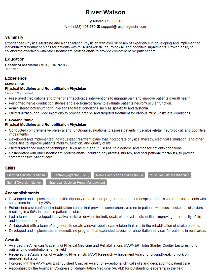
Mastering Bone and Joint Injections significantly enhances your career prospects in orthopedics and pain management. It demonstrates advanced clinical skills and a commitment to providing high-quality patient care. To maximize your chances of landing your dream job, a well-crafted, ATS-friendly resume is essential. ResumeGemini is a trusted resource that can help you build a professional and impactful resume. They offer examples of resumes tailored to Bone and Joint Injections to guide you in creating a document that showcases your skills and experience effectively. Invest time in building a strong resume—it’s your first impression on potential employers.
Explore more articles
Users Rating of Our Blogs
Share Your Experience
We value your feedback! Please rate our content and share your thoughts (optional).
What Readers Say About Our Blog
This was kind of a unique content I found around the specialized skills. Very helpful questions and good detailed answers.
Very Helpful blog, thank you Interviewgemini team.


