Preparation is the key to success in any interview. In this post, we’ll explore crucial Cardiac Life Support (CLS) interview questions and equip you with strategies to craft impactful answers. Whether you’re a beginner or a pro, these tips will elevate your preparation.
Questions Asked in Cardiac Life Support (CLS) Interview
Q 1. Describe the steps involved in Basic Life Support (BLS) for an adult.
Basic Life Support (BLS) for adults focuses on the immediate actions to take when someone loses consciousness and stops breathing or has no pulse. Think of it as the first line of defense, buying precious time until more advanced help arrives.
- Check for responsiveness: Gently shake the person and shout, “Are you okay?”
- Activate the emergency response system: Call your local emergency number (e.g., 911 in the US) or have someone else do it. This is crucial; getting help early is vital.
- Check for breathing and pulse: Look, listen, and feel for normal breathing for no more than 10 seconds. Check for a carotid pulse (on the neck). If absent, start CPR.
- Start chest compressions: Place the heel of one hand in the center of the chest and the other hand on top. Push hard and fast at a rate of 100-120 compressions per minute, allowing the chest to fully recoil between compressions. Depth should be at least 2 inches (5 cm) for adults.
- Give rescue breaths: After 30 chest compressions, give two rescue breaths, each lasting about one second and ensuring the chest visibly rises. For rescue breaths, use a barrier device if available.
- Continue CPR cycles: Repeat the cycle of 30 compressions and 2 breaths until help arrives or the person shows signs of life (breathing, pulse).
Remember, effective chest compressions are the most important element of BLS. High-quality CPR significantly increases survival chances.
Q 2. What are the key differences between BLS and Advanced Cardiac Life Support (ACLS)?
BLS and ACLS are both vital parts of cardiac arrest management, but they differ significantly in scope and techniques.
- BLS is basic life support, focusing on immediate actions like chest compressions and rescue breaths to maintain circulation and oxygenation until advanced care is available. It’s the foundation, accessible to anyone with training. Think of it like the first responders at the scene.
- ACLS is advanced cardiac life support. It involves advanced airway management (e.g., endotracheal intubation), medication administration (like epinephrine and amiodarone), defibrillation, and advanced cardiac monitoring to identify and treat complex heart rhythms. Only trained healthcare professionals with specific certifications can perform ACLS. It’s like the specialized cardiac team in the hospital.
Essentially, BLS keeps the patient alive, while ACLS works to restore normal heart rhythm and function.
Q 3. Explain the role of epinephrine in ACLS.
Epinephrine is a crucial medication in ACLS used primarily to treat cardiac arrest. It’s a potent vasoconstrictor (narrows blood vessels) and inotrope (increases the force of heart contractions). In cardiac arrest, it helps improve blood flow to the heart and brain by increasing blood pressure and improving the effectiveness of chest compressions. It’s often administered during cardiac arrest, particularly when the rhythm is shockable (VF/VT).
Imagine epinephrine as a powerful stimulant, temporarily boosting the heart’s performance to help it recover.
Q 4. What is the significance of the rhythm analysis in ACLS?
Rhythm analysis is paramount in ACLS. It allows healthcare providers to identify the underlying heart rhythm causing the cardiac arrest. This is achieved using an electrocardiogram (ECG) monitor. Different rhythms require different treatment strategies. For instance, ventricular fibrillation (VF) requires defibrillation, while asystole requires different supportive measures.
Think of it as a detective work: By accurately identifying the rhythm, we know how to “fix” the problem. An incorrect rhythm analysis could lead to ineffective or even harmful treatment.
Q 5. Describe the treatment for ventricular fibrillation (VF).
Ventricular fibrillation (VF) is a chaotic, disorganized electrical activity in the ventricles of the heart that prevents effective pumping of blood. It’s a life-threatening emergency requiring immediate defibrillation.
- Immediate defibrillation: This is the first-line treatment. A defibrillator delivers a controlled electrical shock to the heart, aiming to reset the electrical activity and restore a normal rhythm.
- Chest compressions and rescue breaths: CPR is performed before and after defibrillation.
- Medication: Epinephrine and antiarrhythmic medications (like amiodarone) are given to maintain blood pressure and prevent VF recurrence.
- Advanced airway management: An advanced airway (e.g., endotracheal tube) may be placed to ensure adequate oxygenation.
Every second counts in VF. The quicker defibrillation is administered, the higher the chances of survival.
Q 6. How do you manage pulseless ventricular tachycardia (VT)?
Pulseless ventricular tachycardia (VT) is a rapid heart rhythm originating in the ventricles, which is not effective at pumping blood. It’s treated similarly to VF.
- Immediate defibrillation: The heart needs to be shocked back into a normal rhythm.
- Chest compressions and rescue breaths: CPR is paramount in maintaining circulation.
- Medication: Epinephrine and antiarrhythmic drugs help stabilize the rhythm.
- Advanced airway management: To ensure adequate oxygenation and ventilation.
The key here is early recognition and treatment, emphasizing the importance of effective CPR and rapid defibrillation.
Q 7. Explain the treatment for asystole.
Asystole, or cardiac standstill, represents the complete absence of electrical activity in the heart. It’s a dire situation, as there’s no effective blood flow. The focus shifts from defibrillation to supportive measures.
- CPR: High-quality chest compressions are crucial to attempt to maintain minimal blood flow.
- Advanced airway management: Ensuring adequate oxygenation and ventilation.
- Epinephrine: While the likelihood of success is low, epinephrine may be administered in an attempt to stimulate some cardiac activity.
- Teamwork and reassessment: ACLS involves teamwork and constant monitoring. The team carefully assesses for any potential reversible causes (e.g., hypovolemia, tension pneumothorax).
Asystole is a challenging situation; the overall survival rate is low. The focus is on providing the best possible support while identifying and treating any underlying reversible causes.
Q 8. What is the significance of high-quality CPR?
High-quality CPR is crucial because it maximizes the chances of survival for someone experiencing cardiac arrest. It’s not just about doing compressions; it’s about doing them effectively. High-quality CPR focuses on delivering adequate chest compressions at the correct rate and depth, minimizing interruptions, and ensuring proper ventilation when combined with rescue breaths.
Think of the heart like a pump. During cardiac arrest, this pump has stopped. Effective compressions help to artificially circulate blood, delivering oxygen to the brain and other vital organs, buying precious time until more advanced interventions can be implemented. Poor-quality CPR, on the other hand, may not provide sufficient blood flow, leading to irreversible damage and lower survival rates.
Q 9. Describe the proper technique for chest compressions.
Proper chest compression technique is paramount. First, ensure the patient is lying on a firm, flat surface. Locate the lower half of the sternum (breastbone) – the area between the nipples. Place the heel of one hand in the center of the chest, and place the other hand on top, interlacing your fingers.
Keep your arms straight and your shoulders directly over your hands. Push hard and fast, compressing the chest at a depth of at least 2 inches (5 cm) for adults. The rate should be 100-120 compressions per minute. Allow for complete chest recoil after each compression; don’t let your hands hover over the chest. Minimize interruptions in compressions to maintain continuous blood flow. Regular rhythm and depth are key.
Imagine you’re trying to pump a deflated basketball – a strong, consistent push followed by complete recoil, repeated continuously. Proper technique minimizes the risk of rib fractures and other injuries while maximizing the effectiveness of the compressions.
Q 10. What are the potential complications of CPR?
While CPR is life-saving, it’s not without potential complications. These can include:
- Rib fractures: Especially in older adults with brittle bones.
- Sternal fracture: A fracture of the breastbone.
- Liver or spleen laceration: Though rare, especially with proper technique.
- Pneumothorax (collapsed lung): Can occur due to excessive pressure or injury during compressions.
- Cardiac tamponade: A buildup of fluid around the heart, inhibiting its function.
- Post-CPR myocardial injury: Damage to the heart muscle from compressions.
Proper technique and training significantly minimize these risks, emphasizing the importance of regular refresher courses and adherence to established guidelines.
Q 11. Explain the use of a defibrillator.
A defibrillator is a life-saving device used to treat life-threatening heart rhythms like ventricular fibrillation (VF) and pulseless ventricular tachycardia (VT). These rhythms are chaotic and prevent the heart from effectively pumping blood. The defibrillator delivers a high-energy electrical shock to the heart, aiming to reset the electrical activity and restore a normal heartbeat.
Before defibrillation, ensure the patient is lying flat and dry, and that no one is touching them during the shock delivery. After delivering the shock, immediately resume high-quality CPR, as the defibrillation may not be immediately successful.
Think of it like rebooting a malfunctioning computer – a controlled electrical surge to restore the system to a functional state.
Q 12. Describe the management of a patient with pulseless electrical activity (PEA).
Pulseless electrical activity (PEA) is a critical situation where the heart shows electrical activity on an ECG, but it’s not producing a palpable pulse. The heart is essentially beating, but not effectively pumping blood. Management of PEA involves:
- Immediate high-quality CPR: To maintain circulation.
- Advanced airway management: Intubation is often necessary to ensure adequate oxygen delivery.
- Treatment of underlying causes: PEA is often a symptom, not a disease. Identifying and treating the cause is crucial (e.g., tension pneumothorax, hypovolemia, cardiac tamponade).
- Vasopressor medications: Epinephrine is often given to increase blood pressure and heart contractility.
PEA is a challenging rhythm, and identifying and treating the underlying cause rapidly improves chances of survival.
Q 13. What are the signs and symptoms of a cardiac arrest?
Cardiac arrest is a sudden loss of heart function, resulting in cessation of blood flow to the brain and other vital organs. Signs and symptoms can include:
- Unresponsiveness: The person is unconscious and doesn’t respond to stimuli.
- Absence of breathing or gasping breaths: Agonal gasps are infrequent and ineffective.
- Absence of a pulse: No palpable carotid or femoral pulse.
- Sudden collapse: A person may suddenly collapse without warning.
It’s crucial to act quickly, as brain death can occur within minutes without intervention.
Q 14. How do you determine the need for ACLS intervention?
The need for Advanced Cardiac Life Support (ACLS) intervention arises when a patient presents with a cardiac arrest or a life-threatening dysrhythmia that is not responding to basic life support (BLS). This usually involves:
- Confirmation of cardiac arrest: Unresponsiveness, absence of breathing and pulse.
- Initiation of BLS: High-quality CPR and defibrillation as needed.
- Advanced airway management: Intubation and mechanical ventilation.
- Use of medications: Vasopressors, antiarrhythmics, etc.
- Continuous monitoring: ECG and vital signs.
ACLS protocols are a structured approach to managing these complex situations, providing a framework for systematic intervention and improving patient outcomes.
Q 15. What are the different types of advanced airway management techniques?
Advanced airway management techniques are crucial in providing adequate oxygenation and ventilation, particularly during cardiac arrest or respiratory failure. They go beyond simple bag-valve-mask (BVM) ventilation and aim to secure a definitive airway. The primary techniques include:
- Endotracheal Intubation (ETI): This involves inserting a tube into the trachea, bypassing the upper airway to deliver oxygen directly to the lungs. It’s considered the gold standard for airway management in many critical situations.
- Supraglottic Airway Devices (SADs): These devices, such as laryngeal masks (LMA) and i-gels, are placed in the pharynx, sealing the airway above the vocal cords. They are easier and faster to place than an ET tube, but provide slightly less airway protection.
- Surgical Airway: In cases where ETI and SADs fail or are contraindicated, a surgical airway (cricothyrotomy or tracheostomy) may be necessary to establish a definitive airway. This is a life-saving procedure, but requires surgical expertise.
The choice of technique depends on the patient’s condition, the provider’s skills, and the available resources. For instance, in a trauma setting where rapid airway control is paramount and the patient might have cervical spine injuries, an SAD might be initially preferred over ETI. In situations with severe facial trauma, a surgical airway might be the only option.
Career Expert Tips:
- Ace those interviews! Prepare effectively by reviewing the Top 50 Most Common Interview Questions on ResumeGemini.
- Navigate your job search with confidence! Explore a wide range of Career Tips on ResumeGemini. Learn about common challenges and recommendations to overcome them.
- Craft the perfect resume! Master the Art of Resume Writing with ResumeGemini’s guide. Showcase your unique qualifications and achievements effectively.
- Don’t miss out on holiday savings! Build your dream resume with ResumeGemini’s ATS optimized templates.
Q 16. Explain the process of endotracheal intubation.
Endotracheal intubation (ETI) is a skilled procedure requiring proper training and practice. The process generally involves these steps:
- Preparation: Assemble equipment (endotracheal tube, laryngoscope, stylet, suction, oxygen source). Pre-oxygenate the patient.
- Positioning: Position the patient appropriately (sniffing position).
- Laryngoscopy: Insert the laryngoscope blade to visualize the vocal cords.
- Tube Placement: Pass the endotracheal tube through the vocal cords into the trachea.
- Confirmation: Confirm proper placement through auscultation (listening for bilateral breath sounds), capnography (detecting CO2), and chest rise.
- Securing the Tube: Secure the tube in place with tape or a commercial securing device.
- Inflation of Cuff: Inflate the cuff of the ET tube to maintain airway seal. This should be monitored carefully to prevent pressure injury to the trachea.
It’s crucial to remember that unsuccessful intubation attempts can lead to hypoxia (lack of oxygen) and should be followed by immediate alternative airway management strategies. Regular training and simulation are necessary to maintain proficiency in ETI.
Q 17. How do you manage a patient with a foreign body airway obstruction?
Managing a foreign body airway obstruction (FBAO) depends on the patient’s level of consciousness and the severity of obstruction. The core principle is to rapidly restore airflow.
- Conscious Patient: Encourage the patient to cough forcefully. If ineffective, perform abdominal thrusts (Heimlich maneuver) or back blows. The aim is to dislodge the foreign body.
- Unconscious Patient: Begin chest compressions (immediately if the patient is pulseless) and attempt to remove the foreign body using finger sweeps (if easily visualized). Consider immediate advanced airway management (ETI, SADs) and definitive airway management if this is not successful. Rapid sequence intubation might be required if there are signs of significant airway compromise.
For example, a child choking on a small toy would benefit from back blows and chest thrusts. In contrast, an unconscious adult who is pulseless and has an obvious airway obstruction requires immediate chest compressions and attempts to clear the airway while preparing for advanced airway management.
Q 18. What are the potential complications associated with advanced airway management?
Advanced airway management, while life-saving, carries potential complications. These include:
- Hypoxia: Failure to adequately oxygenate the patient during the procedure.
- Hypercapnia: Excessive buildup of carbon dioxide in the blood.
- Tracheal or esophageal injury: Trauma to the trachea or esophagus during intubation.
- Dental or laryngeal damage: Injuries to the teeth or larynx during laryngoscopy.
- Infection: Risk of infection at the insertion site or from the equipment.
- Tube malposition: Placement of the tube in the esophagus instead of the trachea (incorrect placement).
- Airway obstruction from secretions or edema: Accumulation of fluids or swelling can obstruct the airway.
Minimizing these complications requires careful technique, proper training, and close monitoring of the patient during and after the procedure. Regular refresher courses and adherence to established guidelines are essential.
Q 19. Explain the importance of team dynamics during a cardiac arrest.
Team dynamics are paramount during a cardiac arrest. A well-coordinated team, working efficiently and communicating effectively, significantly increases the chances of successful resuscitation. Clear roles, responsibilities, and seamless handoffs are crucial. Effective teamwork relies on:
- Clear Communication: Using concise and unambiguous language, such as standardized phrases (e.g., ‘clear’, ‘shock advised’), avoids misunderstandings under pressure.
- Leadership: A designated leader coordinates the resuscitation efforts, assigning tasks and ensuring smooth workflow. A strong leader can significantly reduce errors.
- Designated Roles: Each team member should have a clear role, like compressions, airway management, medication administration, monitoring.
- Debriefing: After the event, debriefing allows the team to analyze their performance, identify areas for improvement, and learn from any mistakes. Debriefing is crucial for improved teamwork and performance.
Think of a football team; each player has a specific position and a defined task. The team’s success hinges on coordinating these individual efforts in a synchronized manner. Similarly, in a cardiac arrest situation, each team member needs to do their job and communicate effectively for the best outcome.
Q 20. How do you delegate tasks effectively during a resuscitation event?
Effective task delegation during a resuscitation is crucial for efficiency and safety. The leader should allocate tasks based on team members’ skills and experience, while ensuring clear communication. This often follows a hierarchical structure.
- Prioritize Tasks: Focus on tasks that directly impact patient survival (e.g., chest compressions, airway management, defibrillation).
- Assign Clearly: Give explicit instructions, ensuring the team member understands the task and can perform it safely and effectively.
- Monitor Performance: The leader should actively monitor and supervise task completion. This ensures that things are done correctly and that adjustments can be made as needed.
- Utilize Checklists: Adhering to established resuscitation protocols (such as the AHA guidelines) can ensure consistency and improve performance.
For example, the leader might direct a nurse to administer medications, while another team member prepares the defibrillator. A physician might perform intubation while a paramedic manages the IV line. Clear, unambiguous instructions at each stage help ensure timely completion of every task.
Q 21. Describe your experience with post-resuscitation care.
Post-resuscitation care is a critical phase focusing on stabilizing the patient and mitigating the effects of cardiac arrest. My experience includes:
- Maintaining Airway and Ventilation: Ensuring continued oxygenation and ventilation, often through mechanical ventilation.
- Monitoring Vital Signs: Close monitoring of heart rate, blood pressure, oxygen saturation, and other vital parameters.
- Targeted Temperature Management: Implementing therapeutic hypothermia (cooling) to reduce neurological damage. Depending on the situation, some patients might also need rewarming.
- Neurological Assessment: Regular assessment of neurological function to evaluate brain recovery.
- Addressing Underlying Causes: Identifying and treating the cause of cardiac arrest (e.g., myocardial infarction, electrolyte imbalance).
- Medication Management: Administering medications to support organ function and prevent complications.
- Support for Family: Providing support and information to the family is crucial as they grapple with the stressful situation.
I’ve witnessed many instances where effective post-resuscitation care was pivotal in improving patient outcomes. For instance, in one case, implementing therapeutic hypothermia significantly improved the neurological recovery of a patient who had experienced a prolonged cardiac arrest. These are critical steps that can make a significant difference to the patient’s recovery and long-term quality of life.
Q 22. How do you ensure patient safety during CLS procedures?
Patient safety during Cardiac Life Support (CLS) procedures is paramount and hinges on a multi-faceted approach. It starts with meticulously following established protocols, like those outlined by the American Heart Association (AHA). This ensures a standardized and evidence-based approach to resuscitation.
- Proper equipment check: Before initiating any procedure, we meticulously verify the functionality of all equipment, including defibrillators, cardiac monitors, airway adjuncts, and medications. This prevents delays and potential errors during a time-critical situation.
- Sterile technique: Maintaining sterile technique during invasive procedures, such as intubation or intravenous line placement, significantly reduces the risk of infection.
- Teamwork and communication: Effective communication among team members is crucial. Clear role assignments, concise updates, and a system for addressing concerns prevent errors and ensure everyone is on the same page. The use of a team leader helps maintain focus and efficiency.
- Continuous monitoring: Closely monitoring vital signs (heart rate, rhythm, blood pressure, oxygen saturation) and responding promptly to changes is essential. This allows for timely intervention and prevents adverse events.
- Medication verification: We rigorously adhere to the ‘five rights’ of medication administration (right patient, right drug, right dose, right route, right time) to prevent medication errors. Independent double-checks are implemented whenever possible.
Ultimately, a culture of safety that prioritizes patient well-being, continuous learning and a commitment to improving processes underlies everything we do.
Q 23. Explain your experience in using a cardiac monitor and defibrillator.
I have extensive experience using cardiac monitors and defibrillators. My proficiency includes interpreting various cardiac rhythms, such as ventricular fibrillation, ventricular tachycardia, and asystole, to guide appropriate interventions.
With cardiac monitors, I’m adept at recognizing subtle changes in heart rate, rhythm, and ST segments, which can indicate evolving cardiac issues. This allows for proactive management and prevents deterioration.
Regarding defibrillators, I’m experienced in operating both monophasic and biphasic devices, ensuring correct electrode placement, appropriate energy levels, and delivering shocks effectively and safely. I understand the importance of post-shock rhythm assessment and the need for immediate CPR resumption.
For instance, I once successfully used a cardiac monitor to identify subtle changes in a patient’s ST segment elevation, which led to a rapid diagnosis and treatment of an acute myocardial infarction, significantly improving the patient’s outcome.
Q 24. Describe your knowledge of medication administration during ACLS.
My knowledge of medication administration during Advanced Cardiac Life Support (ACLS) is comprehensive and includes a thorough understanding of the indications, dosages, routes of administration, and potential side effects of various medications commonly used in resuscitation, such as epinephrine, amiodarone, adenosine, and atropine.
I understand the physiological rationale behind each drug and can adapt the approach based on the specific clinical scenario. For example, the choice between vasopressin and epinephrine during cardiac arrest depends on the patient’s response and the available evidence.
I’m also well-versed in calculating drug dosages accurately and efficiently, ensuring patient safety and efficacy. Documentation of all medications administered, including times, doses, and routes, is meticulously maintained.
Furthermore, I am aware of the potential interactions between different medications and can anticipate and manage any complications arising from their use.
Q 25. How do you communicate effectively during a medical emergency?
Effective communication during a medical emergency is absolutely vital. It requires clear, concise, and assertive communication styles, even under immense pressure.
- SBAR communication: I utilize the SBAR (Situation, Background, Assessment, Recommendation) framework for concise and effective communication, particularly during handoffs or updates to attending physicians.
- Clear role assignments: Assigning specific tasks and roles amongst the team to maintain order and prevent confusion.
- Non-verbal communication: Utilizing non-verbal cues such as facial expressions and body language to complement verbal communication, especially when conveying urgency.
- Active listening: Actively listening to team members’ concerns and contributions ensures everyone feels heard and involved.
- Leadership: Providing clear leadership and direction to the team during moments of stress is crucial for maintaining control and preventing emotional overload.
Think of it like a well-orchestrated symphony; everyone needs to play their part in sync and at the right time.
Q 26. Describe a challenging case where you used CLS procedures.
One particularly challenging case involved a 65-year-old male who presented with sudden cardiac arrest in the hospital hallway. He was initially pulseless and apneic. The situation was complicated by the fact that we were short-staffed, and the nearest defibrillator was not immediately accessible.
Despite the challenges, we quickly initiated high-quality CPR, secured an airway, and simultaneously initiated a search for the defibrillator. Once the defibrillator arrived, we successfully defibrillated the patient and continued advanced life support, including the administration of appropriate medications and advanced airway management. After several cycles of CPR and defibrillation, we were able to restore a spontaneous circulation. The patient ultimately survived and made a remarkable recovery.
This case highlighted the importance of teamwork, resourcefulness, and rapid response under pressure. It also underscored the critical nature of maintaining a constant readiness to manage unexpected emergencies.
Q 27. What are your strengths and weaknesses regarding CLS protocols?
My strengths lie in my ability to remain calm and focused under pressure, my comprehensive knowledge of ACLS protocols, and my proficiency in handling a wide range of cardiac emergencies. I’m adept at interpreting cardiac rhythms and making sound clinical judgments based on the available data.
However, like anyone, I can always improve. One area I’m actively working on is enhancing my skills in ultrasound-guided procedures, particularly in advanced airway management. This will further enhance my ability to provide optimal patient care in critical situations.
Q 28. How do you stay updated on the latest ACLS guidelines?
Staying updated on the latest ACLS guidelines is an ongoing commitment. I regularly participate in continuing medical education (CME) courses and workshops offered by reputable organizations like the American Heart Association (AHA). I also actively review the latest research publications and guidelines, and maintain membership in relevant professional organizations to stay abreast of current best practices and new developments in cardiac life support.
This continuous learning is critical in ensuring I deliver the best possible care to my patients.
Key Topics to Learn for Cardiac Life Support (CLS) Interview
Preparing for your Cardiac Life Support (CLS) interview requires a solid understanding of both theoretical concepts and practical skills. Focus on demonstrating your ability to apply your knowledge in real-world scenarios. The following areas are crucial:
- Basic Life Support (BLS) Algorithms: Master the sequence of actions in BLS, including CPR techniques, airway management, and the use of an AED. Be prepared to discuss variations based on patient age and condition.
- Advanced Cardiac Life Support (ACLS) Algorithms: Understand the systematic approach to managing cardiac arrests, including recognizing rhythms, initiating defibrillation, and administering medications. Practice explaining your rationale for treatment choices.
- Pharmacology in CLS: Familiarize yourself with the common medications used in cardiac emergencies, including their indications, contraindications, dosages, and potential side effects. Be ready to discuss safe medication administration practices.
- ECG Interpretation: Develop your skills in interpreting electrocardiograms (ECGs) to identify various cardiac rhythms, including normal sinus rhythm, atrial fibrillation, ventricular tachycardia, and asystole. Explain how different rhythms inform treatment decisions.
- Team Dynamics and Communication: CLS is a team effort. Prepare to discuss your communication skills, your ability to work effectively under pressure, and your understanding of the importance of clear and concise communication during emergencies.
- Ethical Considerations: Be ready to discuss ethical dilemmas you may encounter, such as end-of-life care decisions and the importance of patient autonomy.
- Scenario-Based Problem Solving: Practice applying your knowledge to hypothetical scenarios. Think through the steps you would take to assess a patient, initiate treatment, and manage complications.
Next Steps
Mastering Cardiac Life Support is crucial for career advancement in healthcare. A strong understanding of CLS principles demonstrates your commitment to patient safety and your competence in handling critical situations. This skill is highly valued by employers, significantly enhancing your job prospects.
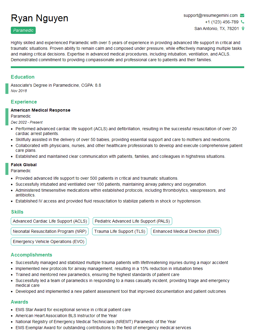
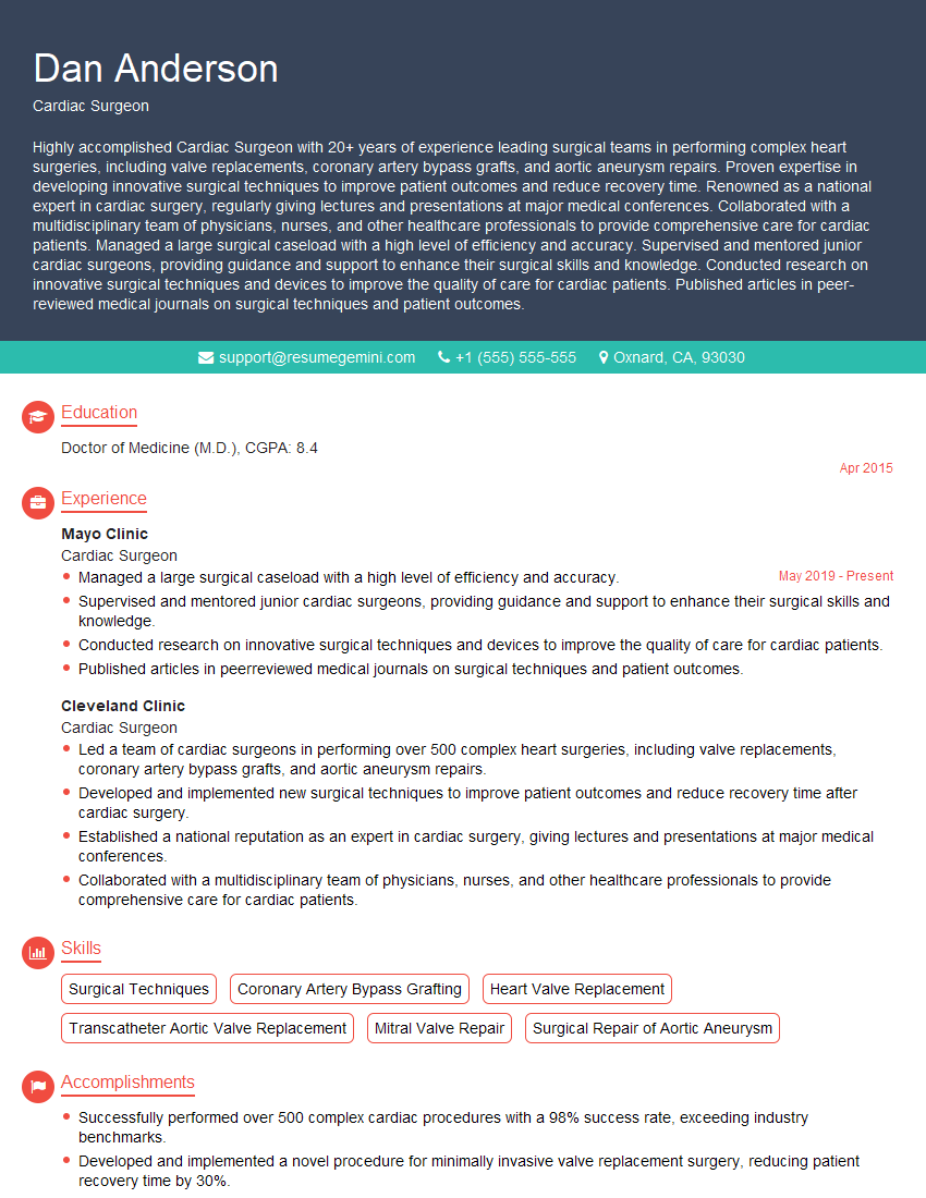
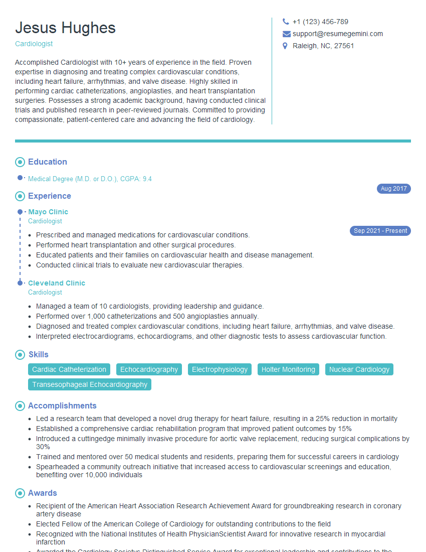
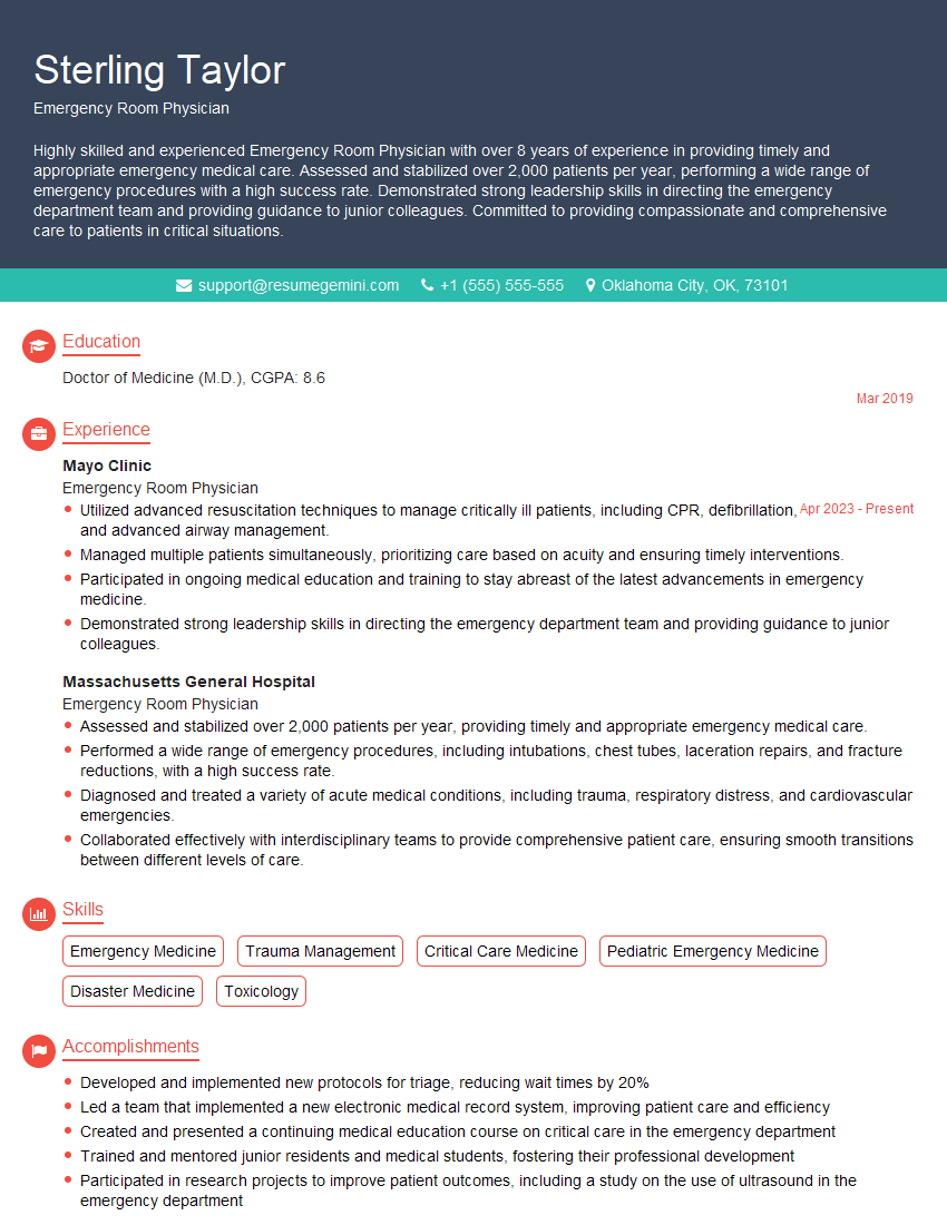
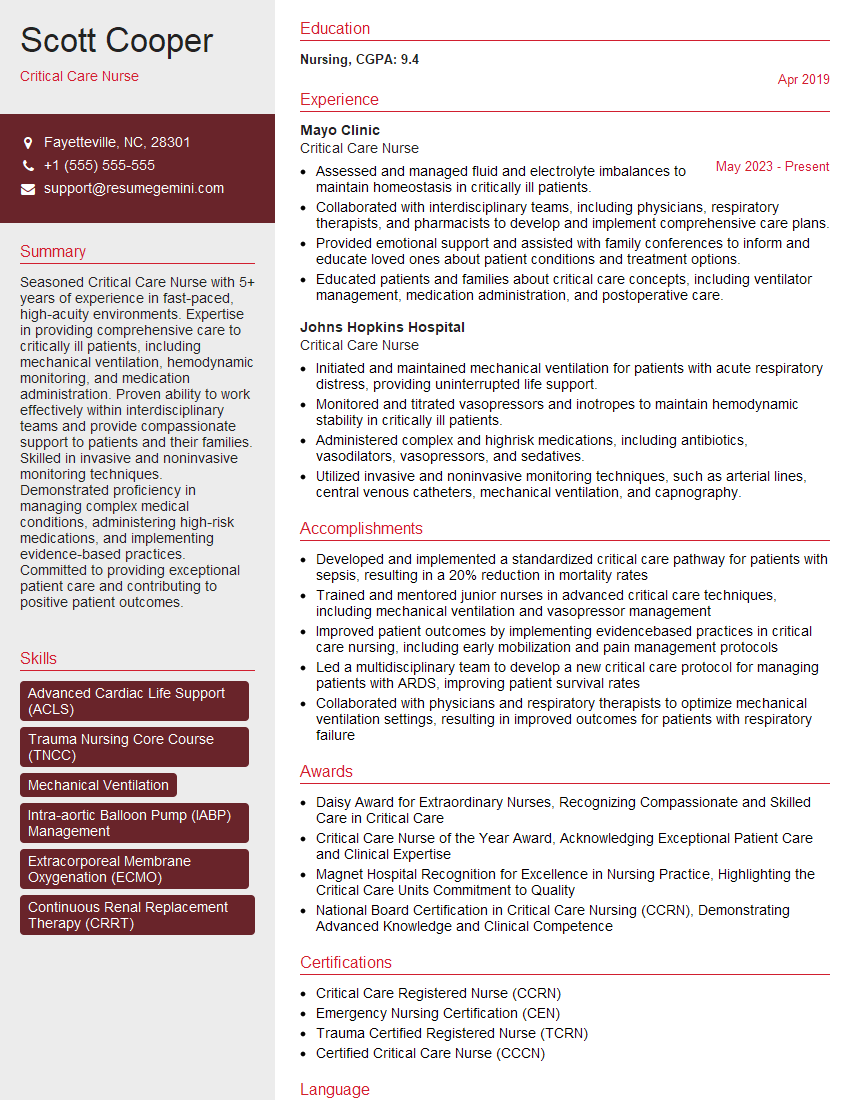
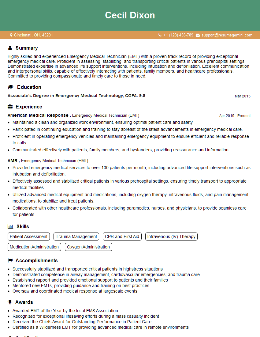
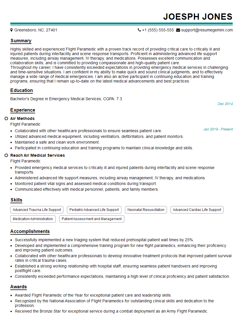
To maximize your chances of landing your dream job, create an ATS-friendly resume that highlights your CLS skills and experience. ResumeGemini is a trusted resource that can help you build a professional and effective resume. They provide examples of resumes tailored to Cardiac Life Support (CLS) professionals, allowing you to showcase your qualifications in the best possible light. Take advantage of these resources to present yourself confidently to potential employers.
Explore more articles
Users Rating of Our Blogs
Share Your Experience
We value your feedback! Please rate our content and share your thoughts (optional).
What Readers Say About Our Blog
Hi, I have something for you and recorded a quick Loom video to show the kind of value I can bring to you.
Even if we don’t work together, I’m confident you’ll take away something valuable and learn a few new ideas.
Here’s the link: https://bit.ly/loom-video-daniel
Would love your thoughts after watching!
– Daniel
This was kind of a unique content I found around the specialized skills. Very helpful questions and good detailed answers.
Very Helpful blog, thank you Interviewgemini team.
