Are you ready to stand out in your next interview? Understanding and preparing for Charcot Foot Management interview questions is a game-changer. In this blog, we’ve compiled key questions and expert advice to help you showcase your skills with confidence and precision. Let’s get started on your journey to acing the interview.
Questions Asked in Charcot Foot Management Interview
Q 1. Describe the pathophysiology of Charcot neuroarthropathy.
Charcot neuroarthropathy, often called Charcot foot, is a debilitating condition characterized by bone destruction and joint deformity in the foot and ankle. Its pathophysiology stems from a disruption of the normal proprioceptive feedback loop due to peripheral neuropathy, most commonly associated with diabetes. This neuropathy impairs the ability to sense pain, temperature, and position in the foot.
Without the protective sensation of pain, repeated micro-trauma goes unnoticed leading to repetitive stress injuries. This triggers an inflammatory response, activating osteoclasts (bone-resorbing cells) which break down bone tissue faster than osteoblasts (bone-forming cells) can replace it. This imbalance results in the characteristic bone destruction and joint instability seen in Charcot foot. The process involves stages of inflammation, fragmentation, and then consolidation, leading to significant deformity and potential for ulceration and infection.
Imagine it like this: Normally, your foot acts as a sophisticated shock absorber. But with neuropathy, you’re essentially walking around on a very fragile structure without realizing it, leading to damage. The body tries to ‘repair’ the damage, but does so in a disorganized and destructive manner.
Q 2. What are the common risk factors for developing Charcot foot?
Several factors significantly increase the risk of developing Charcot foot. The most prominent risk factor is peripheral neuropathy, frequently a complication of long-standing diabetes. This is because the lack of sensation is critical in the pathophysiological mechanism. Other risk factors include:
- Diabetes Mellitus: The most common cause.
- Alcoholism: Can lead to nutritional deficiencies and peripheral neuropathy.
- Syphilis: Historically a significant cause, less prevalent now.
- Spinal Cord Injury: Can cause sensory loss in the lower extremities.
- Rheumatoid Arthritis: Though less frequently associated.
- Other Neurological Disorders: Conditions causing peripheral nerve damage.
- Obesity: Increases stress on the joints.
- Poorly Controlled Blood Sugar (HbA1c): High blood glucose levels exacerbate nerve damage.
It’s important to note that having one or more of these risk factors doesn’t guarantee the development of Charcot foot, but it substantially increases the likelihood.
Q 3. Explain the different stages of Charcot foot.
Charcot foot progression is typically described in stages, although the exact staging systems vary. A commonly used simplified model divides it into three phases:
- Acute Stage (Stage 1): Characterized by significant inflammation, swelling, redness, warmth, and increased local temperature. This phase is often very painful despite the underlying neuropathy, and there’s marked bone destruction evident on imaging.
- Subacute Stage (Stage 2): The inflammation subsides. Bone fragmentation continues, but at a slower pace. The clinical presentation might be less dramatic, with less swelling and erythema. However, significant deformity is becoming more apparent.
- Chronic Stage (Stage 3): The inflammatory process resolves entirely. Bone healing occurs, but often results in a deformed and unstable foot. The risk of ulceration and infection is significantly elevated in this stage.
Understanding these stages helps guide treatment strategies. Early intervention during the acute phase is crucial to minimize long-term deformity.
Q 4. How do you clinically diagnose Charcot foot?
Diagnosing Charcot foot relies on a combination of clinical examination and imaging. Clinically, physicians look for the classic clinical triad of edema, erythema (redness), and increased skin temperature, in addition to the presence of peripheral neuropathy and the characteristic deformity. Pain, while expected, might not always be reported due to neuropathy. Physical examination assesses range of motion, stability, and presence of any ulcers or infections. Other clinical signs include altered gait and loss of ankle reflexes.
However, it’s crucial to remember that these clinical features can be subtle or absent, particularly in individuals with limited sensory perception due to advanced neuropathy. Therefore, imaging is essential for confirming the diagnosis.
Q 5. What imaging modalities are used to diagnose Charcot foot, and what are their limitations?
Imaging plays a crucial role in diagnosing and monitoring Charcot foot. The most commonly used modality is X-ray, which demonstrates the characteristic bone changes. Early changes might show soft tissue swelling, while later stages show bone fragmentation, resorption, and the eventual deformity. However, X-rays have limitations; they might miss early bone changes, and don’t show the soft tissue components as well as other modalities.
MRI (Magnetic Resonance Imaging) offers better visualization of soft tissue abnormalities and early bone changes that might be missed on X-ray. It can show bone marrow edema and subtle fractures. However, it is relatively expensive and not always readily available.
Bone scan, using technetium-99m, shows areas of increased metabolic activity and is helpful in identifying early bone changes, although it may not always distinguish Charcot changes from other inflammatory conditions.
Each modality has its strengths and weaknesses, and the choice depends on clinical context, availability, and the urgency of the situation. Frequently, a combination of imaging techniques is used to obtain a comprehensive picture.
Q 6. What is the role of total contact casting in Charcot foot management?
Total contact casting (TCC) is a cornerstone of non-surgical Charcot foot management. Its primary purpose is to offload pressure from the affected foot and ankle, minimizing further bone damage and promoting healing. TCC provides uniform support across the entire plantar surface of the foot, distributing weight evenly and preventing stress concentration on any particular area. This is achieved by using a custom-molded cast that fills all the spaces and contours of the foot.
The immobilization provided by TCC also helps to reduce inflammation and promote bone consolidation. It needs to be meticulously applied and regularly monitored for proper fit, to ensure optimal offloading. Improperly applied casts can lead to increased pressure, skin breakdown, and ulceration.
Think of TCC as a protective boot for the damaged foot, giving it the rest it needs to heal. It’s a crucial tool in preventing further damage and promoting healing in the acute phase. Typically, TCC is employed for several weeks to months, depending on the clinical response.
Q 7. Describe the indications and contraindications for surgical intervention in Charcot foot.
Surgical intervention in Charcot foot is usually reserved for cases that fail to respond adequately to conservative management, such as TCC, or for situations where significant deformity poses a risk of ulceration, infection, or functional impairment. The main indications for surgery include:
- Persistent deformity resistant to non-surgical treatment: When conservative measures fail to stabilize the foot and ankle.
- Recurrent ulceration or infection: Surgery might be needed to correct deformity and remove necrotic tissue.
- Severe instability leading to significant gait disturbances: Surgical stabilization can improve ambulation and reduce the risk of falls.
- Prevention of further deformity: For example, arthrodesis (fusion) of joints to stabilize a collapsing structure.
Contraindications for surgery include:
- Active infection: Surgery should be deferred until the infection is adequately controlled.
- Poorly controlled diabetes: Patients with poor glycemic control have a higher risk of complications.
- Severe peripheral vascular disease: Inadequate blood supply increases the risk of wound healing problems.
- Patient’s overall health status: Significant comorbidities may make surgery high-risk.
Surgical options range from simple procedures like debridement to complex reconstructions involving arthrodesis or even amputation. The choice of surgical procedure is individualized based on the patient’s specific situation and the extent of the deformity.
Q 8. What are the different surgical techniques used to treat Charcot foot?
Surgical intervention in Charcot foot is usually reserved for cases where conservative management has failed to control the deformity or when significant infection is present. The specific technique depends on the severity and location of the deformity.
- Arthrodesis: This involves fusing one or more joints in the foot to stabilize the bone and correct deformity. For example, a triple arthrodesis fuses the subtalar, talonavicular, and calcaneocuboid joints. This is a common procedure for severe midfoot collapse.
- Osteotomy: This procedure involves cutting and reshaping bones to correct deformities and improve alignment. It might be used to address a specific bone malalignment contributing to the Charcot foot deformity.
- Excisional Arthroplasty: In cases of severe joint destruction, the damaged joint surfaces might be removed, allowing for some degree of mobility, although stability may be compromised. This is less common than arthrodesis.
- Debridement: This surgical procedure focuses on removing infected tissue to control infection, often used in conjunction with other surgical techniques in cases of Charcot with osteomyelitis.
The choice of surgical technique is highly individualized and requires careful consideration of the patient’s overall health, the extent of the deformity, and the presence of infection. Pre-operative planning often involves advanced imaging techniques like CT scans and MRI to accurately assess the bone structure and soft tissue involvement.
Q 9. Discuss the role of offloading in Charcot foot management.
Offloading is absolutely crucial in Charcot foot management. It refers to the reduction of pressure on the affected foot to prevent further bone destruction and promote healing. Imagine a fractured bone – you wouldn’t put weight on it, and the same principle applies here. The intense inflammation characteristic of Charcot foot causes bone resorption and deformity. By relieving pressure, we allow the bones to rest and heal. Early and aggressive offloading is key to minimizing deformity and preventing complications. Without effective offloading, the cycle of inflammation and bone destruction continues, leading to significant functional impairment.
Think of offloading as giving the foot a ‘break’ from the constant stress of weight-bearing. This is achieved by avoiding weight-bearing altogether or by significantly reducing the amount of weight applied to the affected area. Failure to adequately offload is a major reason for poor outcomes in Charcot foot.
Q 10. What types of offloading devices are available?
Various offloading devices cater to different needs and severity levels. The choice depends on patient factors, such as mobility and tolerance, alongside the severity of the Charcot foot.
- Total Contact Casts (TCCs): These are commonly used in the acute phase and offer a high degree of offloading. They fully immobilize the foot and distribute pressure evenly across the sole.
- Custom-molded shoes/orthoses: These are individually crafted to accommodate the specific foot shape and deformity. They provide customized pressure relief and support.
- Removable Cast Walkers (RCWs): These combine the benefits of a cast with increased mobility. The patient can remove the device for hygiene and wound care.
- Knee-high walkers/crutches: Non-weight-bearing options offering complete pressure relief.
- Specialised footwear: Examples include rocker-bottom shoes which adjust the angle of weight bearing and reduce pressure points, and extra-depth shoes to accommodate swelling.
The transition between these devices often occurs gradually as the condition improves. It’s a collaborative process with regular assessment and adjustment of the offloading strategy.
Q 11. How do you manage infection in a Charcot foot?
Infection is a significant complication in Charcot foot, often caused by breaks in the skin that are easily overlooked. This can rapidly escalate to osteomyelitis (bone infection). Management involves a multi-faceted approach:
- Antibiotics: Appropriate intravenous antibiotics are crucial, guided by culture and sensitivity testing to ensure effective coverage against the infecting organism.
- Surgical Debridement: This may be necessary to remove infected bone and soft tissues. This is often performed repeatedly until all infected tissue is removed.
- Wound Care: Meticulous wound care, potentially including negative pressure wound therapy (NPWT) or hyperbaric oxygen therapy (HBO), to promote healing and prevent further infection.
- Continued Offloading: Essential to promote healing and prevent further trauma to the area.
It is crucial to monitor patients closely for signs of infection, such as increased pain, swelling, redness, warmth, and purulent drainage. Early diagnosis and aggressive intervention are key to preventing serious complications and limb loss. We often need to collaborate closely with infectious disease specialists and wound care specialists.
Q 12. What are the common complications of Charcot foot?
Charcot foot can lead to a number of serious complications, significantly impacting quality of life. Some common complications include:
- Infection (Osteomyelitis): As discussed earlier, infection is a major concern, potentially leading to sepsis and limb amputation.
- Deformity and Instability: The progressive bone destruction leads to foot deformities, such as rocker-bottom foot or claw toes, resulting in pain, gait disturbances, and difficulty with mobility.
- Non-union Fractures: Delayed healing or non-union of fractures is more common in patients with Charcot foot due to the ongoing inflammatory process.
- Ulcers: Pressure points created by deformity may lead to recurrent skin breakdown and ulcers, increasing the risk of infection.
- Amputation: In severe cases, despite optimal management, amputation might be necessary to prevent life-threatening complications.
- Pain: Chronic neuropathic pain is a common symptom, even after the acute phase has subsided.
Preventing these complications necessitates early diagnosis, aggressive offloading, and meticulous management of infection.
Q 13. How do you prevent Charcot foot in patients with diabetes?
Preventing Charcot foot in diabetic patients is paramount, focusing on minimizing risk factors.
- Strict Glycemic Control: Maintaining optimal blood glucose levels through diet, exercise, and medication is crucial in reducing the risk of neuropathy and Charcot foot.
- Regular Foot Exams: Routine foot examinations by a healthcare professional to detect early signs of neuropathy, deformity, or infection.
- Proper Footwear: Wearing properly fitted shoes and avoiding barefoot walking is vital to prevent foot injuries and pressure points. Custom orthotics can offer additional support and cushioning.
- Neuropathy Management: Treating existing neuropathy with medication or other interventions is important to reduce the risk of Charcot arthropathy.
- Smoking Cessation: Smoking significantly impairs wound healing and increases the risk of infection, therefore it’s essential to encourage patients to quit.
- Patient Education: Educating patients about foot care, including proper hygiene, moisturization, and regular inspections, is critical.
A proactive approach to risk factor management is vital in preventing this devastating complication of diabetes.
Q 14. Explain the importance of patient education in Charcot foot management.
Patient education is the cornerstone of successful Charcot foot management. Patients need to understand the seriousness of the condition, the importance of compliance with treatment, and how to recognize and manage potential complications.
We need to empower patients to actively participate in their care. This includes explaining the rationale behind each treatment modality (e.g., offloading, medication, surgery), demonstrating proper foot care techniques, and providing clear instructions on recognizing warning signs of infection. Regular follow-up appointments and open communication are essential to ensure adherence to the treatment plan and promptly address any issues that arise. We often use visual aids and printed materials to reinforce key messages and improve patient understanding and engagement in their long-term care.
Effective patient education can significantly improve outcomes, reduce complications, and enhance quality of life for individuals affected by Charcot foot. A patient who understands their condition and actively participates in its management has a far better chance of a positive outcome.
Q 15. Describe the role of multidisciplinary team approach in Charcot foot management.
Managing Charcot foot, a debilitating complication of diabetes, requires a truly collaborative effort. A multidisciplinary team approach is not just beneficial—it’s essential for optimal patient outcomes. Think of it like a finely tuned orchestra, where each instrument (healthcare professional) plays a crucial role in creating harmonious care.
Diabetologist/Endocrinologist: Manages the underlying diabetes, crucial to slowing disease progression.
Orthopedic Surgeon/Podiatrist: Provides surgical intervention if needed (e.g., arthrodesis, osteotomy) and manages bone deformities.
Infectious Disease Specialist: Addresses infections, a common and serious complication in Charcot foot.
Wound Care Specialist: Manages ulcers and promotes healing, often employing advanced wound care techniques.
Physical Therapist: Develops and implements a customized rehabilitation plan, focusing on weight-bearing, range of motion, and gait training.
Certified Diabetes Educator: Educates the patient about diabetes management and self-care, emphasizing prevention.
Regular team meetings ensure coordinated care, minimizing conflicting advice and maximizing the effectiveness of the treatment plan. For example, the physical therapist might communicate directly with the surgeon about a patient’s progress to adjust the treatment approach accordingly. This collaborative model significantly improves patient compliance and promotes better long-term results.
Career Expert Tips:
- Ace those interviews! Prepare effectively by reviewing the Top 50 Most Common Interview Questions on ResumeGemini.
- Navigate your job search with confidence! Explore a wide range of Career Tips on ResumeGemini. Learn about common challenges and recommendations to overcome them.
- Craft the perfect resume! Master the Art of Resume Writing with ResumeGemini’s guide. Showcase your unique qualifications and achievements effectively.
- Don’t miss out on holiday savings! Build your dream resume with ResumeGemini’s ATS optimized templates.
Q 16. What are the long-term goals of Charcot foot management?
The long-term goals of Charcot foot management are multifaceted, aiming to improve both the patient’s quality of life and prevent further complications. We strive for:
Pain Control: Minimizing pain and discomfort to allow for normal function and mobility.
Prevention of Ulceration and Infection: Protecting the foot from further injury and preventing devastating infections, which can lead to amputation.
Wound Healing: Promoting the healing of any existing wounds through advanced wound care and proper offloading.
Preservation of Limb: The ultimate goal is to save the limb and prevent amputation whenever possible.
Improved Mobility and Function: Restoring the patient’s ability to walk and perform daily activities without significant limitations.
Improved Quality of Life: Enhancing the patient’s overall well-being and independence.
Achieving these goals often requires a long-term commitment, with regular follow-up appointments and ongoing management of the underlying diabetes.
Q 17. How do you assess the effectiveness of treatment for Charcot foot?
Assessing the effectiveness of Charcot foot treatment is a complex process that involves several key indicators.
Clinical Examination: Regular assessment of pain levels, wound size and healing, presence of infection, and any deformity progression.
Imaging Studies: X-rays and other imaging techniques (e.g., MRI) are used to monitor bone healing, assess the extent of deformity, and detect any new fractures or infections.
Biomechanical Assessment: This evaluates how the patient’s foot and ankle function during weight-bearing activities and helps to guide the choice of orthotics and other interventions.
Functional Outcomes: Measuring the patient’s ability to walk, stand, and perform daily activities provides a valuable assessment of their functional status.
Patient-Reported Outcomes: Questionnaires and surveys assess the patient’s pain levels, quality of life, and satisfaction with treatment.
For example, a reduction in wound size, improved range of motion, and decreased pain scores suggest effective treatment. Conversely, worsening deformity, recurrent infection, or continued pain indicate a need for adjustments to the treatment plan.
Q 18. Describe the use of biomechanical assessment in Charcot foot management.
Biomechanical assessment plays a vital role in Charcot foot management. It helps us understand how the foot and ankle move and bear weight, which is crucial for preventing further damage and promoting healing. Think of it like a car’s alignment—if the wheels aren’t aligned properly, the car won’t drive smoothly, and parts may wear out prematurely. Similarly, if the foot’s biomechanics are faulty, it can lead to further damage in Charcot foot.
This assessment involves:
Gait Analysis: Observing how the patient walks to identify abnormalities in their gait pattern.
Pressure Measurement: Using pressure sensors to determine how weight is distributed across the foot. This helps identify areas of high pressure that could lead to ulceration.
Range of Motion Assessment: Measuring the flexibility of the foot and ankle joints.
Static Postural Analysis: Analyzing the patient’s foot and ankle alignment while standing.
The information gathered is used to design custom orthotics or other supportive devices to correct biomechanical abnormalities, offload pressure points, and improve gait. For instance, a patient with excessive midfoot plantar pressure might benefit from a custom-molded orthotic with a rocker-bottom sole to redistribute weight.
Q 19. What are the criteria for successful Charcot foot management?
Success in Charcot foot management is defined by a combination of factors, focusing on both the short-term and long-term outcomes. There’s no single metric, but rather a constellation of indicators:
Absence of Active Inflammation: The inflammatory process, a hallmark of Charcot arthropathy, must be resolved.
Healed Wounds: Any ulcers or open wounds should be completely healed.
Absence of Infection: No evidence of infection (bacterial, fungal, or otherwise).
Stable Deformity: Minimal or no progression of the bone deformities, often achieved through appropriate offloading and, in some cases, surgical intervention.
Improved Function: The patient should experience significant improvement in their ability to walk, stand, and perform daily activities.
Pain Control: Reduction in pain to a manageable level.
Prevention of Recurrence: Strategies are implemented to minimize the risk of future Charcot episodes.
Ultimately, successful management means that the patient can maintain an acceptable quality of life with minimal pain and risk of limb loss, and they have the knowledge and resources to manage their condition proactively.
Q 20. Discuss the role of advanced wound care techniques in Charcot foot management.
Advanced wound care techniques are integral to Charcot foot management, given the high risk of ulceration and infection. These techniques go beyond basic wound dressings and aim to create an optimal healing environment. Think of it like providing fertile ground for a plant to thrive, rather than just planting it and hoping for the best.
These techniques include:
Debridement: Removal of necrotic (dead) tissue to allow for healthy granulation tissue formation.
Negative Pressure Wound Therapy (NPWT): Applying controlled negative pressure to the wound to promote healing and remove excess fluid and debris.
Growth Factor Therapy: Using growth factors to stimulate cell regeneration and accelerate healing.
Bioengineered Skin Substitutes: Applying artificial skin to cover wounds and promote healing.
Hyperbaric Oxygen Therapy: Increasing oxygen levels in the tissues to improve healing and fight infection.
The specific techniques used depend on the wound’s characteristics and severity. For instance, a small, superficial wound might only require a simple dressing, while a deep, infected ulcer might require a combination of debridement, NPWT, and antibiotics. Close monitoring is crucial to assess the effectiveness of the chosen treatment and make necessary adjustments.
Q 21. How do you differentiate Charcot foot from other foot conditions?
Differentiating Charcot foot from other foot conditions requires a careful clinical evaluation, incorporating patient history, physical examination, and imaging studies. The key lies in recognizing the characteristic features of Charcot neuroarthropathy.
Here’s how we distinguish it:
History of Diabetes: Charcot foot is strongly associated with diabetes, especially poorly controlled diabetes. Conditions like rheumatoid arthritis or osteoarthritis can also cause foot deformities, but lack the diabetic connection.
Insensitivity to Pain: Diabetic neuropathy often leads to reduced sensation in the foot, allowing for unnoticed injuries and progressive joint destruction that is not seen in other foot conditions which are often accompanied by pain.
Warmth, Erythema, and Edema: The early stages of Charcot foot often present with warmth, redness, and swelling in the foot. This inflammatory response is less pronounced in other conditions.
Radiographic Findings: X-rays play a crucial role. Charcot foot shows characteristic changes, including fragmentation of bone, joint destruction, and bone resorption. Other conditions like stress fractures might show some bone changes, but lack the diffuse and severe destruction seen in Charcot arthropathy.
Clinical Presentation: While other conditions such as cellulitis can appear similar, the gradual onset and progressive deformity differentiate Charcot arthropathy. Cellulitis, for instance, presents as an acute inflammatory process.
It’s important to emphasize that a thorough examination is necessary. Sometimes, further investigations may be needed, such as MRI or bone scans, to establish a definitive diagnosis and exclude other conditions.
Q 22. What are the early warning signs of Charcot foot?
Early detection of Charcot foot is crucial for effective management. The initial signs can be subtle and often mimic other foot problems, making early diagnosis challenging. Patients may experience a warm, swollen, red, and possibly numb foot. They might also report increased foot temperature, redness, and swelling, often localized to a specific area. Importantly, there may be a lack of pain, which is often alarmingly unusual with an injury of this magnitude and severity. This painless nature is one of the most distinctive early warning signs. Pain reduction is due to neuropathy and the damage being done to the nerves in the foot. Think of it like a broken bone, but without the usual agonizing pain. This lack of pain can lead to delayed diagnosis, allowing the condition to progress rapidly.
Other early signs include changes in skin color or temperature; skin changes are typically localized to one area of the foot and can be seen as reddish, bluish, or discolored skin compared to the other areas of the foot. The foot may also feel remarkably loose, less stable, and more pliable than usual. Any unexplained swelling or deformity of the foot should also raise suspicion. These early symptoms frequently occur in patients with poorly controlled diabetes.
Q 23. What is the role of medication in Charcot foot management?
Medication plays a vital, albeit often supportive, role in Charcot foot management. While there’s no single medication that cures Charcot foot, several medications can help manage associated conditions and reduce complications. The primary goals of medication are to manage pain, improve blood glucose control (crucial for diabetic patients), and address potential infections. Analgesics, such as NSAIDs, may be used to control pain, but their usage should be approached cautiously given the potential for gastrointestinal side effects.
In diabetic patients, optimizing blood glucose levels using insulin or oral hypoglycemic agents is essential. Good glucose control can significantly reduce the risk of future Charcot episodes and promote healing. Antibiotics are critical if an infection develops, which is a frequent complication of Charcot arthropathy. Finally, medications to manage underlying conditions like hypertension or dyslipidemia are also important, as these may have an impact on overall vascular health and wound healing.
Q 24. How do you monitor for complications during and after treatment of Charcot foot?
Monitoring for complications during and after Charcot foot treatment is vital to prevent permanent disability. Regular clinical assessments are critical to catch problems early. This includes repeated physical examinations focusing on the affected foot, noting any changes in swelling, temperature, redness, skin integrity, and the presence of any signs of infection (pus, increased pain, fever). We utilize advanced imaging techniques such as X-rays and MRI scans to monitor the progression of the bone changes and assess the effectiveness of treatment. These images allow us to observe the extent of the joint destruction and any development of pressure sores.
Regular blood tests can help monitor for signs of infection, evaluate overall health markers, and ensure blood glucose levels are within the therapeutic range for diabetic patients. Neurological examinations help to track the progress of any existing neuropathy. Throughout the healing process, which can take many months, ongoing monitoring is crucial to identify potential issues like non-union fractures or infections. A prompt and appropriate intervention is necessary to prevent further complications.
Q 25. Discuss the importance of maintaining adequate blood glucose control in preventing Charcot foot.
Maintaining adequate blood glucose control is paramount in preventing Charcot foot, particularly in individuals with diabetes. Charcot neuroarthropathy results from a combination of neuropathy (nerve damage) and neuroischemia (reduced blood flow), both significantly influenced by hyperglycemia. High blood sugar levels damage nerves and blood vessels in the feet, leading to the diminished sensation and compromised blood supply that characterises Charcot foot. This makes the feet vulnerable to injury without the individual realizing the extent of the damage.
Tight blood glucose control, therefore, helps to protect nerves and blood vessels, thus lowering the risk of Charcot foot. Patients must achieve and maintain optimal HbA1c levels. This often involves a comprehensive management plan involving diet, exercise, and appropriate medication. Education and support for patients to manage their diabetes effectively are essential in reducing the incidence of this debilitating complication.
Q 26. Explain your experience with specific cases of Charcot foot and the challenges you faced.
I’ve encountered many challenging cases of Charcot foot. One patient presented with a severely deformed foot and an extensive ulcer, already showing signs of infection. Getting him to follow through with the prescribed offloading strategies was exceptionally difficult due to the intense pain initially, and managing his expectation of functional recovery took time and patience. Another case highlighted the difficulty in distinguishing Charcot foot from cellulitis. The patient initially presented with swelling, redness, and inflammation, which could have been either condition. We had to use advanced imaging and close clinical monitoring to make the correct diagnosis to start appropriate treatment. In both cases, prompt collaboration with the multidisciplinary team was essential for optimal outcomes. These experiences have refined my approach to diagnosis, treatment, and patient education.
Q 27. What are your preferred methods for communicating with patients and their families regarding Charcot foot?
Effective communication is the cornerstone of successful Charcot foot management. I prioritize clear, empathetic communication with patients and their families, tailoring my approach to their individual needs and understanding. I use plain language, avoiding medical jargon, and ensure they understand their diagnosis, treatment plan, and potential outcomes. I encourage questions, address concerns openly, and offer repeated explanations as needed. I utilize visual aids like diagrams and photographs to illustrate the condition and its progression. For patients with impaired cognitive function, I involve caregivers actively in the communication process.
Furthermore, I involve patients in the decision-making process, emphasizing shared-decision making. This empowers them, encouraging adherence to the treatment plan. Regular follow-up appointments, both in person and via phone calls, ensure we monitor progress and address any emerging concerns proactively. Finally, I provide written materials summarizing key information, including contact details for additional support and resources.
Q 28. Describe your experience working in a multidisciplinary team setting for Charcot foot management.
Multidisciplinary team work is crucial in managing Charcot foot. Our team typically includes endocrinologists, podiatrists, orthotists, physical therapists, infectious disease specialists, and vascular surgeons. Each member contributes unique expertise to provide holistic patient care. The endocrinologist manages diabetes control, the podiatrist assesses foot health and wound care, the orthotists create custom footwear and braces for offloading, and physical therapists design rehabilitation programs to improve mobility. The infectious disease specialist manages any infections, and the vascular surgeon intervenes if circulatory issues arise. Regular team meetings, case discussions, and shared patient records are essential for effective coordination and optimal outcomes.
This collaborative approach ensures comprehensive management tailored to the individual patient’s needs, addressing not only the immediate problem of the Charcot foot but also the underlying medical conditions. The multidisciplinary approach makes it possible to identify and address potential complications rapidly, enhancing treatment outcomes and minimizing the risk of long-term disability.
Key Topics to Learn for Charcot Foot Management Interview
- Pathophysiology of Charcot Foot: Understanding the underlying mechanisms of bone destruction, joint instability, and ulceration is crucial. This includes the role of neuropathy and vascular compromise.
- Clinical Presentation and Diagnosis: Learn to differentiate Charcot foot from other foot pathologies. Master the skills of physical examination, imaging interpretation (X-ray, MRI, etc.), and laboratory test analysis relevant to Charcot foot.
- Non-surgical Management: Be prepared to discuss various offloading strategies (e.g., total contact casts, custom-molded footwear), wound care techniques, and the role of pharmacological interventions (e.g., pain management).
- Surgical Management: Familiarize yourself with surgical options, such as arthrodesis, osteotomy, and amputation. Understand indications, contraindications, and potential complications for each procedure.
- Patient Education and Rehabilitation: Discuss the importance of patient education in preventing recurrence and promoting long-term foot health. Outline key components of a comprehensive rehabilitation program.
- Complications and their Management: Be ready to discuss common complications like infection, ulceration, and limb loss, including preventative measures and treatment strategies.
- Long-term Monitoring and Follow-up: Understand the importance of regular follow-up appointments for patients with Charcot foot to monitor disease progression and intervene promptly.
- Case Studies and Problem Solving: Practice analyzing hypothetical cases involving different presentations, complications, and treatment options for Charcot Foot.
Next Steps
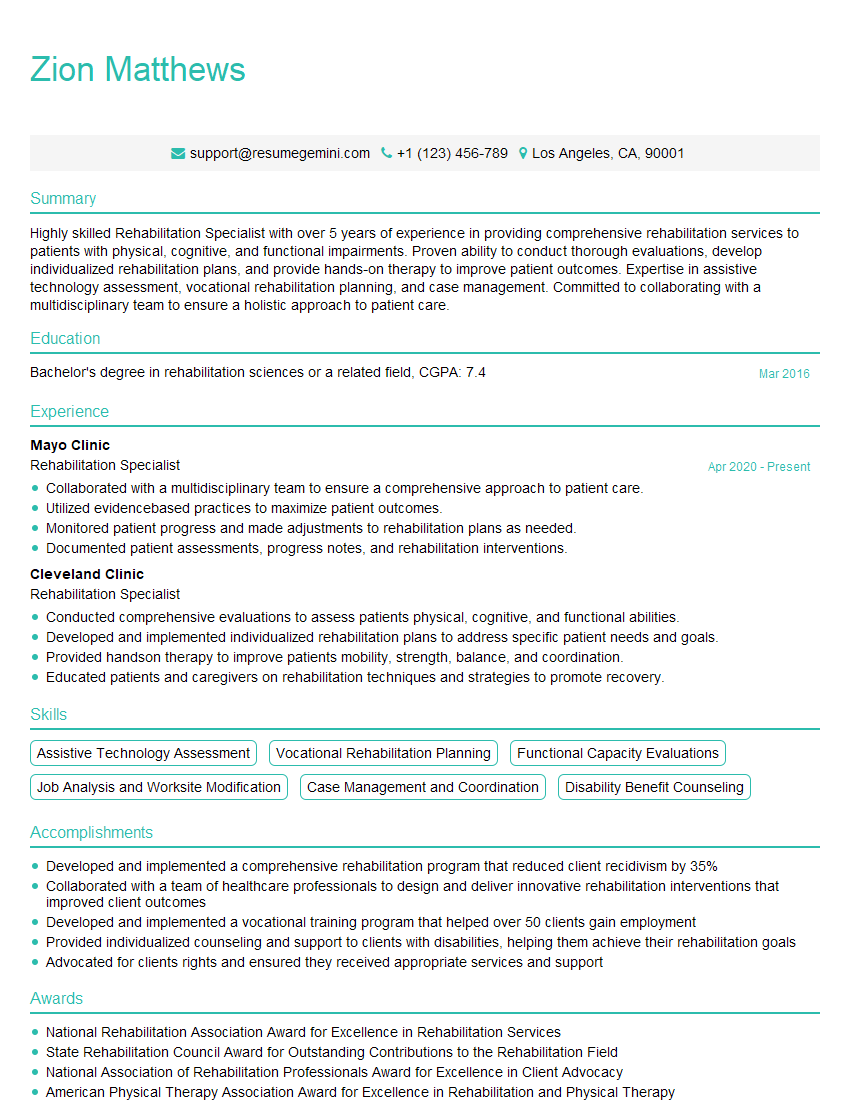
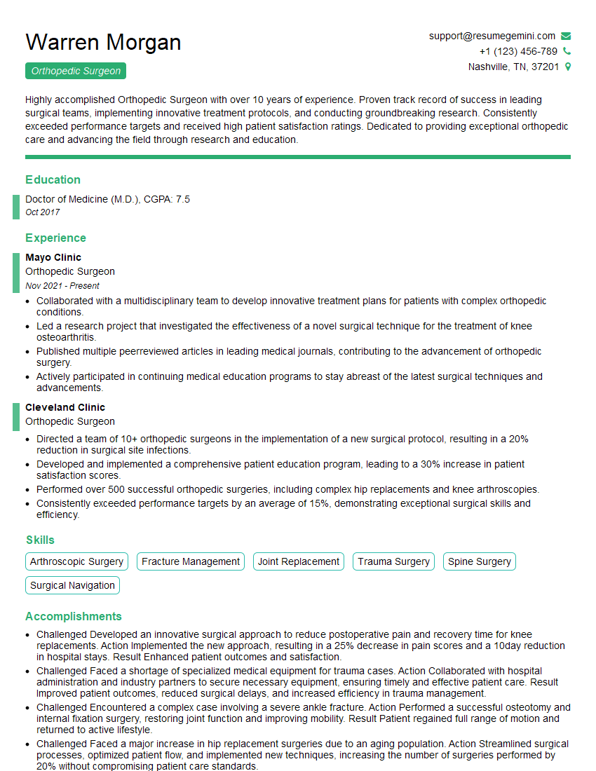
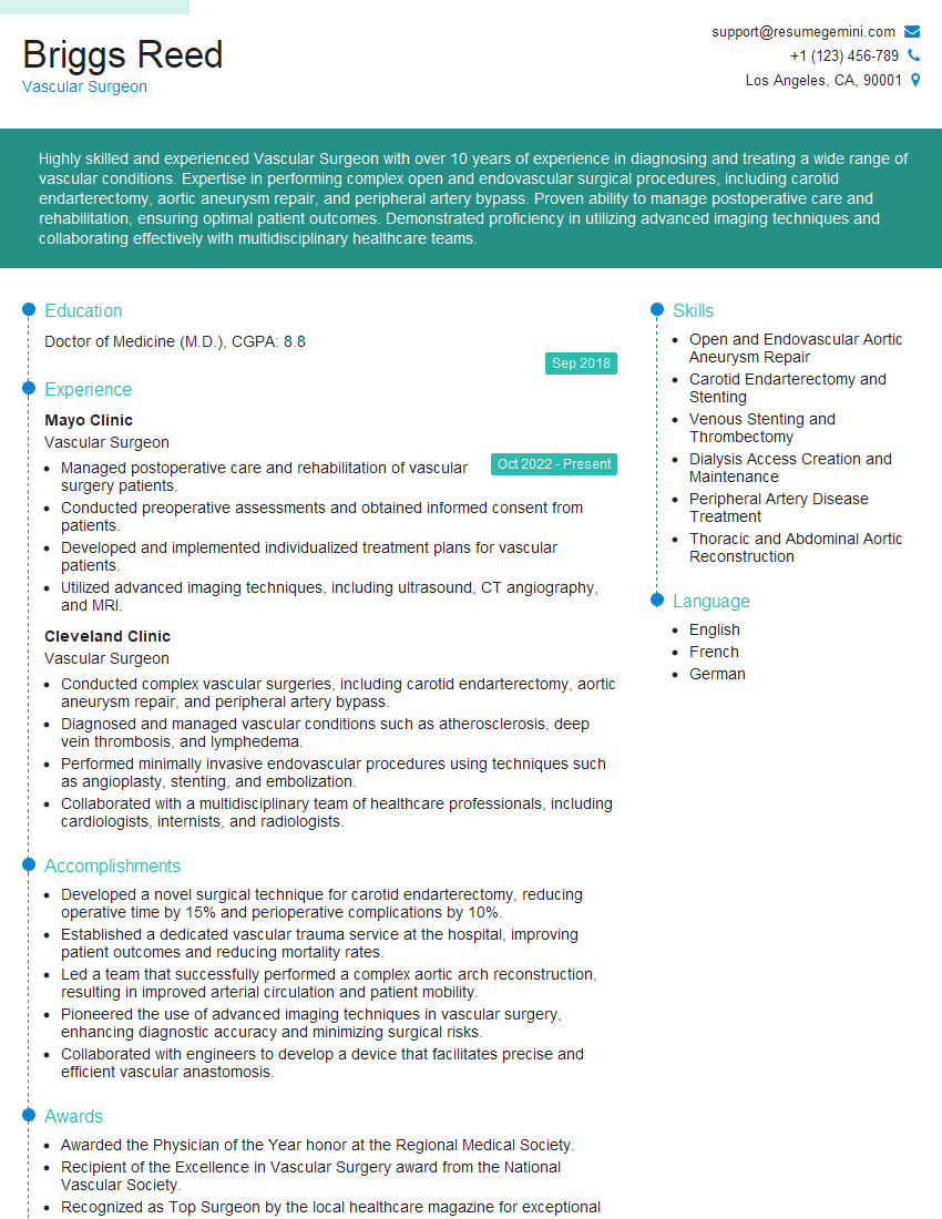
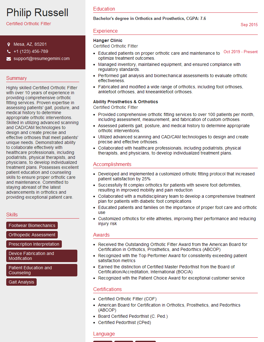
Mastering Charcot Foot Management significantly enhances your value as a healthcare professional, opening doors to specialized roles and advanced career opportunities. To maximize your job prospects, it’s vital to present your skills effectively. Creating an ATS-friendly resume is essential for getting your application noticed. ResumeGemini is a trusted resource to help you build a professional and impactful resume that highlights your expertise in Charcot Foot Management. Examples of resumes tailored to this specialization are available to guide you. Invest in crafting a compelling resume – it’s your first impression with potential employers.
Explore more articles
Users Rating of Our Blogs
Share Your Experience
We value your feedback! Please rate our content and share your thoughts (optional).
What Readers Say About Our Blog
This was kind of a unique content I found around the specialized skills. Very helpful questions and good detailed answers.
Very Helpful blog, thank you Interviewgemini team.
