Every successful interview starts with knowing what to expect. In this blog, we’ll take you through the top Cystoscopy interview questions, breaking them down with expert tips to help you deliver impactful answers. Step into your next interview fully prepared and ready to succeed.
Questions Asked in Cystoscopy Interview
Q 1. Describe the steps involved in a standard cystoscopy procedure.
A standard cystoscopy involves several key steps, all aimed at visualizing the urethra and bladder. Think of it like a detailed internal inspection. First, the patient is positioned comfortably, usually lying on their back with their legs elevated in stirrups. Next, a topical anesthetic gel is applied to the urethral opening to numb the area and minimize discomfort. Then, the cystoscope, a thin, flexible tube with a camera and light, is gently inserted into the urethra. The scope is advanced carefully into the bladder, allowing the physician to view the bladder lining, looking for abnormalities like tumors, stones, or inflammation. The bladder can be filled with sterile fluid (distension) to provide a better view. Images are captured, and biopsies may be taken if necessary. Finally, the cystoscope is carefully removed, and the patient is monitored for any adverse effects.
- Preparation: Patient positioning, topical anesthesia.
- Insertion: Gentle insertion of the cystoscope into the urethra and bladder.
- Visualization: Examination of the bladder lining for any abnormalities.
- Biopsy (if needed): Collection of tissue samples for further analysis.
- Removal: Careful removal of the cystoscope.
- Post-procedure monitoring: Observing the patient for any complications.
Q 2. What are the different types of cystoscope used and when would you choose one over another?
Cystoscopes come in different varieties depending on the procedure’s needs. We mainly categorize them as rigid or flexible. Rigid cystoscopes, thicker and less maneuverable, offer a superior optical quality for clearer images, ideal for procedures like stone removal or precise tissue sampling. Imagine it as a powerful magnifying glass for a specific location. Flexible cystoscopes, thinner and more bendable, are excellent for navigating complex anatomy, making them perfect for less invasive examinations and those requiring exploration of the ureters. It’s like having a flexible camera probe that can explore the entire system. The choice depends entirely on the clinical scenario. A rigid scope would be preferred for a straightforward stone removal, while a flexible scope might be chosen if a detailed exploration of the upper urinary tract is necessary.
Q 3. Explain the process of inserting and maneuvering a cystoscope.
Inserting and maneuvering a cystoscope requires a delicate touch and precision. The process begins with lubrication of the urethral meatus with a sterile lubricant. The scope is then gently inserted, often with the patient instructed to bear down as if they are urinating, which helps relax the urethral sphincter. Slow, steady advancement is crucial. Resistance indicates the need for readjustment, possibly a slightly different angle or a pause to allow for better patient relaxation. Once in the bladder, the scope can be manipulated to view different areas, using its directional capabilities and the fluid distention to explore the bladder wall. Remember, patient comfort is paramount; the process needs to be explained step-by-step and any discomfort must be addressed immediately.
Q 4. How do you ensure patient comfort and safety during a cystoscopy?
Patient comfort and safety are always the top priority. This starts with pre-procedure education, where we explain the procedure, expectations, and potential discomfort in detail. Topical anesthetic gels effectively reduce pain, while intravenous sedation can provide relaxation and minimize discomfort. Continuously monitoring vital signs throughout the procedure helps detect any adverse effects promptly. Open communication is also key, encouraging the patient to communicate any pain or discomfort. Post-procedure care involves managing any potential complications like bleeding or infection. For example, for a particularly anxious patient, pre-procedure anxiolytic medication might be considered.
Q 5. What are the potential complications associated with cystoscopy?
While cystoscopy is generally safe, potential complications exist. These include bleeding, infection (urinary tract infection), perforation of the bladder or urethra (though rare), and pain. Rarely, complications like fluid overload (from distension fluid) can occur. The risk factors for complications include pre-existing medical conditions, history of pelvic inflammatory disease, and the use of anticoagulants. Proper technique and careful monitoring are essential to minimize these risks.
Q 6. How do you manage a complication such as bleeding during cystoscopy?
Managing bleeding during cystoscopy depends on its severity. Minor bleeding usually stops spontaneously after the procedure. However, if significant bleeding occurs, several management strategies can be employed. These include direct pressure using a sponge or applying a hemostatic agent to the bleeding site during the procedure itself. In case of persistent bleeding, further investigation and potentially interventions like cauterization or surgical repair might be necessary. Monitoring of hemoglobin levels and blood pressure post-procedure is essential to assess and address any ongoing blood loss. For example, we might consider keeping the patient for observation and administering IV fluids.
Q 7. What are the pre-operative and post-operative instructions for a patient undergoing cystoscopy?
Pre-operative instructions typically involve advising the patient to fast for a certain period before the procedure, to avoid potential complications during sedation. They should also inform us of any medications they are currently taking, especially anticoagulants, as these may need to be adjusted before cystoscopy. Post-operative instructions include drinking plenty of fluids to flush out the urinary tract, monitoring for signs of infection (fever, pain, burning), and following any specific medication instructions. It’s also crucial to inform the patient about expected symptoms, such as mild discomfort or burning during urination, and when to seek immediate medical attention for more severe complications.
Q 8. Describe your experience with different types of irrigation solutions used in cystoscopy.
Choosing the right irrigation solution during cystoscopy is crucial for maintaining clear visualization and patient safety. The most commonly used is sterile saline, its isotonic nature minimizing the risk of fluid overload or electrolyte imbalances. However, its low viscosity can sometimes lead to reduced visibility during procedures with significant bleeding. In such cases, we might use glycine, a hypotonic solution that helps to wash away blood and debris, improving visualization. Glycine is preferred due to its relative safety if some absorption occurs, compared to other solutions like sorbitol which have higher risks of hemolysis. The choice depends on the procedure’s complexity and anticipated bleeding. For example, a simple cystoscopy for hematuria investigation will likely use saline, while a more complex transurethral resection of a bladder tumor might necessitate glycine.
Less frequently, we might use water, but only with extreme caution and in short bursts, as the absorption of water can lead to significant fluid overload and electrolyte disturbances, specifically hyponatremia. The use of any other solutions, or solutions with preservatives or additives is generally avoided as there’s a risk of toxicity or allergic reactions.
Q 9. How do you identify and handle abnormal findings during a cystoscopy?
Identifying abnormal findings during cystoscopy relies on a systematic approach and a keen eye for detail. We start by systematically inspecting the entire bladder wall, paying close attention to the mucosa’s color, texture, and vascularity. Normal bladder mucosa is smooth, pink, and glistening. Deviations from this, such as redness (inflammation), pallor (ischemia), irregularities (tumors), or bleeding, are key indicators of abnormality. We also observe the ureteral orifices for signs of obstruction or leakage. The presence of stones, foreign bodies, or masses is also carefully noted. For example, a suspicious lesion – a bulge or ulceration – would require careful observation noting its location, size, and appearance.
Handling abnormal findings involves documenting their precise location, size, and characteristics, ideally with photographic documentation. This is essential for comparison with future cystoscopies. We then carefully assess the findings in clinical context and consider further steps. This might include taking biopsies, performing additional investigations such as urine cytology or further imaging (CT scan, MRI). A suspicious lesion would necessitate referral to urology for further management and possible surgical intervention.
Q 10. Explain the process of obtaining biopsies during a cystoscopy.
Obtaining biopsies during cystoscopy allows for histological examination of suspicious lesions, vital for diagnosing bladder cancer or other conditions. The process begins with identifying the target area to be biopsied. A cold cup biopsy forceps is introduced through the cystoscope, and the tissue sample is carefully grasped and excised by opening and closing the jaws of the forceps. It’s important to take multiple biopsies to ensure adequate sampling representing the entire lesion. The tissue sample is then placed in a formalin-filled container for processing and pathological analysis. The procedure can cause mild bleeding, typically controlled by continuous irrigation and sometimes the use of electrocautery for hemostasis.
Proper technique is vital. It’s important to take biopsies from the most suspicious areas. The depth of the biopsy will vary depending on the target but should be performed carefully to avoid perforation and reduce discomfort. The procedure is usually well tolerated by patients and is generally performed without local anesthesia unless the patient experiences discomfort.
Q 11. How do you manage a patient with a known allergy during cystoscopy?
Managing a patient with a known allergy during cystoscopy requires meticulous pre-procedural planning to prevent any adverse reactions. A thorough allergy history is crucial, especially regarding latex, iodine-based contrast agents (if used), and various medications (anesthetics, antibiotics). If a patient has a latex allergy, we utilize latex-free equipment throughout the procedure. If there is an allergy to iodine contrast agents, alternatives may need to be explored if imaging of the urinary tract is necessary. Any medication allergies should be addressed in advance by either avoiding the medication or having alternative management strategies readily available. In some instances, pre-medication with antihistamines might be considered to minimize the risk of a reaction. It’s crucial to have a plan and have emergency medications and resuscitation equipment readily available in case of an acute reaction. The patient’s allergy should be clearly documented in the patient’s record, and the treatment team should be well-informed.
Q 12. What are the key differences between flexible and rigid cystoscopy?
Flexible and rigid cystoscopy differ significantly in their design, capabilities, and applications. Rigid cystoscopes offer a wider field of view and are better suited for procedures that require precise manipulation, such as transurethral resection of bladder tumors (TURBT). They are usually larger in diameter than flexible scopes, which implies that it requires a larger urethra and that there is more patient discomfort during the procedure. They typically require general or spinal anesthesia.
Flexible cystoscopes are smaller, more maneuverable, and are often preferred for diagnostic purposes and less invasive procedures. Their smaller size improves patient comfort, and they allow for examination of the urethra, bladder and even the ureters, depending on the length of the scope. Flexible cystoscopes can be used with topical anesthesia, making them suitable for outpatient settings. The choice between the two depends entirely on the clinical indication. A rigid scope is needed for surgical procedures, while a flexible scope is adequate for diagnostic cystoscopy.
Q 13. How do you interpret cystoscopic images?
Interpreting cystoscopic images requires a thorough understanding of normal bladder anatomy and pathology. We systematically examine the bladder wall, noting the color, texture, and vascularity of the mucosa. Any deviation from the normal appearance, such as lesions, inflammation, stones, or foreign bodies, needs to be carefully documented. The location, size, shape, and characteristics of any abnormalities are carefully assessed. We look for patterns of inflammation, or the presence of lesions that might indicate malignancy, such as papillary lesions, or flat, ulcerative lesions. The images are evaluated in conjunction with the patient’s history and other diagnostic results.
For example, the presence of a mass with irregular borders and telangiectasia might be highly suggestive of bladder cancer, necessitating further investigation. The process is also influenced by experience and knowledge of bladder pathologies. We often need to compare the images obtained from different perspectives to be sure we are looking at the totality of the lesion.
Q 14. What is your experience with cystoscopy in different patient populations (e.g., pediatric, geriatric)?
My experience with cystoscopy spans various patient populations, each requiring a tailored approach. In pediatric patients, smaller instruments and a gentle technique are employed to minimize trauma and anxiety. The procedure may require general anesthesia or sedation, given the limitations in patient cooperation. Geriatric patients might present with comorbidities requiring careful assessment and management before and during the procedure. The procedure may need to be adjusted according to their physiological status, including their tolerance to anesthesia. We also need to consider their physical limitations in terms of mobility and general health. Careful attention to fluid management is vital in both pediatric and geriatric populations to prevent fluid overload or dehydration. For instance, a pediatric patient would be given less fluid, whereas a geriatric patient might have a higher risk of complications if too much fluid is administered. The overall approach adapts to the specific needs and vulnerabilities of each age group, emphasizing patient safety and comfort.
Q 15. Describe your understanding of sterilization and disinfection protocols for cystoscopes.
Sterilization and disinfection of cystoscopes are crucial to prevent infections. The process involves a multi-step approach prioritizing both high-level disinfection and, ideally, sterilization. High-level disinfection uses chemicals to kill most microorganisms, while sterilization eliminates all forms of microbial life.
High-Level Disinfection: Typically involves immersion in a liquid chemical sterilant, such as glutaraldehyde or ortho-phthalaldehyde (OPA), for a specified duration according to the manufacturer’s instructions. This is often followed by thorough rinsing with sterile water.
Sterilization: Steam sterilization (autoclaving) is the gold standard for cystoscopes if the instrument is compatible. This involves exposing the scope to high-pressure saturated steam at a specific temperature for a designated time. Other methods such as ethylene oxide sterilization might be used for scopes that cannot withstand autoclaving, but these methods require extensive aeration to remove residual chemicals.
Critical Steps: Proper cleaning before sterilization is vital. This involves meticulous removal of all organic debris and blood using enzymatic cleaners and brushes designed for the instrument’s delicate components. Any damage to the scope, such as cracks or leaks, necessitates immediate replacement to maintain sterility and patient safety.
Monitoring and Documentation: Sterilization processes require rigorous monitoring using biological indicators (spores) and chemical indicators (strips that change color indicating exposure to sterilization parameters) to confirm the effectiveness of each cycle. Meticulous documentation of these processes is crucial for quality control and infection prevention.
Career Expert Tips:
- Ace those interviews! Prepare effectively by reviewing the Top 50 Most Common Interview Questions on ResumeGemini.
- Navigate your job search with confidence! Explore a wide range of Career Tips on ResumeGemini. Learn about common challenges and recommendations to overcome them.
- Craft the perfect resume! Master the Art of Resume Writing with ResumeGemini’s guide. Showcase your unique qualifications and achievements effectively.
- Don’t miss out on holiday savings! Build your dream resume with ResumeGemini’s ATS optimized templates.
Q 16. How do you maintain the equipment used in cystoscopy?
Maintaining cystoscopy equipment goes beyond sterilization. It encompasses a holistic approach to ensure longevity, optimal functionality, and patient safety.
- Regular Cleaning and Inspection: After each use, the scope undergoes thorough cleaning as described above, paying close attention to the lens system and the working channels. Regular visual inspection checks for any damage or wear and tear.
- Calibration and Testing: Regular calibration of any associated equipment, such as the light source and camera system, ensures optimal image quality. Regular testing of the irrigation and fluid management systems is also essential.
- Preventative Maintenance: Manufacturers usually provide recommended maintenance schedules. These might include periodic servicing by qualified biomedical technicians to lubricate moving parts, check seals, and replace worn components.
- Storage: Proper storage prevents damage and contamination. Cystoscopes should be stored in a clean, dry, and dust-free environment.
- Staff Training: Thorough training of all personnel involved in handling and maintaining the equipment is crucial. This includes proper cleaning, disinfection, sterilization techniques, and troubleshooting common issues.
Regular maintenance ensures the equipment operates efficiently, provides high-quality images, and minimizes the risk of complications during procedures. A malfunctioning scope could lead to procedure delays, incomplete visualization, or even patient injury. Regular maintenance minimizes this risk significantly.
Q 17. What are the limitations of cystoscopy?
While cystoscopy is a valuable diagnostic and therapeutic tool, it has limitations:
- Limited Visualization: It might not visualize all areas of the urinary tract, especially areas with significant inflammation, obstruction, or strictures. Small lesions or very early changes might be missed.
- Patient Discomfort: The procedure can be uncomfortable, and some patients may experience pain, burning, or bleeding. Appropriate sedation or analgesia might be required.
- Complications: Although rare, complications such as bladder perforation, infection, bleeding, and ureteral trauma can occur.
- Inability to Biopsy Large Masses: While small biopsies can be taken, cystoscopy is not ideal for obtaining large tissue samples for histological examination. Other methods may be required.
- False Negatives: Early cancers or small lesions can be missed, especially in the presence of inflammatory conditions. This is why combining cystoscopy with other imaging modalities and tests is common.
Understanding these limitations is essential for proper patient selection, managing expectations, and choosing the most appropriate diagnostic and therapeutic approach. For example, if we suspect a large bladder tumor, we’d likely integrate cystoscopy with CT urography or MRI for a more complete assessment.
Q 18. How do you document the findings of a cystoscopy?
Documentation of cystoscopy findings is essential for continuity of care and legal protection. It should be thorough, accurate, and easily understandable.
Essential Elements:
- Patient Demographics: Name, date of birth, medical record number.
- Date and Time of Procedure:
- Indication for Cystoscopy: Reason for performing the procedure (e.g., hematuria, dysuria, suspected bladder cancer).
- Technique Used: Rigid or flexible cystoscopy.
- Findings: Detailed description of the bladder mucosa, including color, texture, presence of lesions, masses, or calculi. Specific locations and measurements should be noted. For lesions, a thorough description of its morphology, size, and location is crucial.
- Biopsy Information: If biopsies were taken, record the number of biopsies, location, and any special handling instructions.
- Treatment Performed: If any procedures, such as resection of a bladder tumor or removal of foreign bodies were performed during the cystoscopy, details should be included.
- Complications: Any complications encountered during the procedure should be recorded in detail.
- Impression/Diagnosis: Summary of the findings and the likely diagnosis based on the findings.
- Physician Signature and Credentials:
High-quality images (cystograms or videos) supplement the written report, providing visual confirmation of the findings.
Q 19. What is your experience with electronic health record (EHR) systems related to cystoscopy?
My experience with EHR systems in the context of cystoscopy is extensive. We utilize the EHR for scheduling, pre-operative instructions, documentation of the procedure, and subsequent follow-up.
Benefits: EHR systems streamline the entire process. Automated reminders improve efficiency. The digital integration of images and reports enhances communication among healthcare professionals and reduces the risks associated with lost or misplaced paperwork. Templates for procedure notes reduce variability and ensure thorough documentation. EHRs also facilitate the tracking of patients for follow-up, ensuring timely management of any identified issues. Data analytics capabilities within the EHR provide valuable insights into procedure frequency, complications, and outcomes, enabling improvements in patient care and the overall efficiency of our practice.
Challenges: While EHRs offer many benefits, challenges can include navigating complex software systems, maintaining data security and privacy, and ensuring that the electronic documentation is legible and sufficiently detailed. Ensuring proper training of staff to effectively utilize the EHR remains a continuous process.
Q 20. How do you communicate the findings of a cystoscopy to the patient and the physician?
Communicating cystoscopy findings requires clear and empathetic communication, tailored to the patient’s level of understanding and emotional state. For the patient, I explain the procedure in simple terms, emphasizing that I carefully examined their bladder. I provide a summary of my findings in language they can understand, avoiding medical jargon. I answer their questions patiently and address their concerns, allowing for time to process the information.
Positive Findings: If the cystoscopy reveals no abnormalities, I reassure the patient about their health.
Concerning Findings: If there are concerning findings, such as a suspicious lesion or mass, I deliver this news with sensitivity and empathy. I explain the next steps, which might include further investigations or treatment. I offer support and answer questions, connecting them with appropriate resources for emotional support and follow-up care.
Physician Communication: For the referring physician, I use a formal report incorporating the information outlined in the previous question. Clarity, conciseness, and accuracy are paramount. I highlight any significant findings, recommendations for further management, and potential differential diagnoses. If necessary, I follow up with a verbal discussion to answer any questions and address any concerns the physician might have.
Q 21. Describe a challenging cystoscopy case you encountered and how you handled it.
One challenging case involved a patient with a history of recurrent urinary tract infections and a suspected stricture. Initial cystoscopy attempts with a standard flexible scope were unsuccessful due to severe anatomical distortion from the inflammation. The scope couldn’t navigate the narrowed areas of the urethra and bladder neck. This presented a significant challenge in visualizing the bladder mucosa properly.
My approach involved:
- Switching to a smaller-diameter, more flexible ureteroscope: This allowed me to navigate the stricture more easily.
- Utilizing hydro-distention: This technique involved gently inflating the bladder with sterile fluid to help dilate the narrowed passage.
- Careful manipulation and patience: I proceeded slowly and cautiously to avoid causing further trauma to the already inflamed tissues. This required more time than a routine cystoscopy.
- Close collaboration with the urology team: We discussed the findings and alternative approaches, including the possibility of intervention such as urethral dilation or surgical correction.
Through this careful and methodical approach, we successfully visualized the bladder and ruled out any other significant pathology beyond the stricture. While the case was more challenging than a routine cystoscopy, the outcome was successful in providing the necessary diagnosis and enabling further management.
Q 22. What are your knowledge of different types of urinary catheters used in conjunction with cystoscopy?
Several types of urinary catheters are used in conjunction with cystoscopy, primarily for purposes of bladder drainage and visualization. The choice depends on the specific clinical situation and the anticipated duration of the procedure.
- Foley Catheter: This is the most common type. A balloon at the tip allows it to remain securely in place for urine drainage, even after cystoscopy. The balloon is inflated with sterile water post-insertion. Its use before and after cystoscopy helps maintain a clear view during the procedure and allows for post-procedural monitoring.
- Coude Catheter: This catheter has a curved tip, making it useful for navigating difficult-to-catheterize urethras, particularly in patients with prostatic enlargement or urethral strictures. This is less frequently used with cystoscopy itself, but might be employed prior if access is difficult.
- Three-way Catheter: This catheter has three lumens – one for drainage, one for inflation, and one for irrigation during cystoscopy. This enables continuous irrigation of the bladder during the procedure, which helps maintain a clear field of view and removes debris.
- Suprapubic Catheter: In cases where urethral catheterization is impossible or contraindicated (e.g., urethral trauma), a suprapubic catheter is inserted directly into the bladder through a small incision in the lower abdomen. This would not typically be used in conjunction *during* a cystoscopy, but it might be required post-op.
Proper catheter selection and insertion are crucial to prevent complications like infection and trauma.
Q 23. How do you ensure proper fluid balance during a lengthy cystoscopy procedure?
Maintaining proper fluid balance during a lengthy cystoscopy is vital to prevent complications like hyponatremia (low sodium) or fluid overload. This is particularly important with prolonged procedures involving bladder irrigation.
We monitor fluid intake and output meticulously. The irrigation fluid used (typically sterile saline) is measured precisely. We track the amount infused and the amount drained. The patient’s blood pressure, heart rate, and electrolyte levels are monitored closely throughout the procedure. Any discrepancies between infused and drained fluid are carefully accounted for. For instance, if there is significant fluid retention (e.g., due to bleeding), the surgeon must be alerted to make appropriate adjustments. Post-procedure, we assess for any fluid imbalances and take corrective measures if necessary. In some cases, particularly if significant irrigation was used, electrolyte levels are checked several hours later as well.
Think of it like managing a delicate balance – too much fluid is just as problematic as too little.
Q 24. What are the ethical considerations related to cystoscopy?
Ethical considerations in cystoscopy are centered around informed consent, patient autonomy, and maintaining patient dignity and privacy.
- Informed Consent: The patient must be fully informed about the procedure, including its purpose, risks, benefits, and alternatives. This includes potential complications like infection, bleeding, or perforation. They must understand and voluntarily agree to the procedure.
- Patient Autonomy: The patient has the right to refuse the procedure at any time, even after initial consent. Their wishes must be respected.
- Privacy and Confidentiality: The patient’s medical information must be kept strictly confidential. Strict adherence to HIPAA regulations (or the equivalent in other countries) is crucial. All images and records are treated with the utmost care.
- Competent Personnel: The procedure must only be performed by trained and qualified healthcare professionals.
Maintaining a respectful and empathetic approach throughout the process is essential, ensuring the patient feels safe and comfortable.
Q 25. How do you handle a situation where the cystoscope becomes obstructed during the procedure?
Obstruction of the cystoscope during a procedure is a serious complication. The first step involves assessing the situation calmly. Gentle, careful manipulation may dislodge the obstruction if it is due to debris or tissue. Irrigating the cystoscope with sterile saline can sometimes help clear the blockage.
If simple maneuvers fail, imaging such as fluoroscopy may be necessary to identify the nature and location of the obstruction. In severe cases, it might be necessary to remove the cystoscope to avoid causing damage to the urethra or bladder. In some cases, the blockage may require surgical intervention. Documentation of the event and subsequent steps is crucial. Post-procedure analysis will clarify whether changes to technique are needed in future cases.
It is a critical situation; quick thinking and a systematic approach are paramount. This situation highlights the importance of thorough preparation and a well-equipped procedure room.
Q 26. What is your knowledge of different types of bladder stones and their management?
Bladder stones vary in composition, size, and number. Their management depends on these factors and the patient’s overall health.
- Composition: Stones can be composed of calcium oxalate, uric acid, struvite, or cystine, each requiring slightly different management strategies.
- Size and Number: Small stones may pass spontaneously, while larger stones require intervention. Multiple stones might necessitate different approaches than a single large stone.
- Management: Cystoscopy itself can be used to remove smaller stones. Larger stones may be fragmented using laser lithotripsy (described in the next question), and then removed using a cystoscope or basket. In some cases, extracorporeal shock wave lithotripsy (ESWL) may be a preferred alternative. If the stone is particularly large or complex, open surgery might be necessary.
Pre-operative imaging, such as an abdominal X-ray or CT scan, is vital in assessing stone size, number, and composition to determine the best treatment strategy. Each case requires a personalized approach. This is not just ‘stone removal,’ but meticulous assessment and tailored plans.
Q 27. Describe your experience with cystoscopic laser surgery
I have extensive experience with cystoscopic laser surgery. This minimally invasive technique uses lasers to precisely target and ablate tissue within the bladder. Different laser types, such as holmium:YAG or potassium-titanyl-phosphate (KTP), are used depending on the specific application.
We use this technology frequently for treating bladder tumors, removing bladder stones (as mentioned previously), and managing benign prostatic hyperplasia (BPH) via laser resection or vaporization. The advantages of laser surgery include precise tissue ablation, reduced bleeding, and a shorter recovery time compared to open surgery. The precision is key; it avoids excessive tissue damage while effectively treating the target.
Before a laser procedure, we carefully assess the patient’s condition and perform a thorough cystoscopic evaluation to precisely plan the surgical approach. Following the procedure, strict monitoring and post-operative care are vital to ensure proper healing and to manage any potential complications.
Q 28. What is your understanding of the role of imaging (ultrasound, CT) in relation to cystoscopy?
Imaging plays a crucial role before, during, and sometimes after cystoscopy. It provides valuable information to guide the procedure and assess its effectiveness.
- Pre-operative Imaging: Ultrasound or CT scans of the abdomen and pelvis help assess bladder size, identify stones or masses, and evaluate the presence of any abnormalities that might impact the procedure. It helps tailor the procedure to the individual case.
- Intra-operative Imaging (Fluoroscopy): Fluoroscopy can be used during the procedure, particularly if there are issues with navigating the cystoscope or handling complications as discussed earlier.
- Post-operative Imaging: Imaging can be used after cystoscopy to evaluate the results of the procedure, such as confirming the removal of stones or assessing the extent of tumor resection. It ensures we’ve achieved our objectives.
Imaging techniques provide a non-invasive way to gain important visual data which greatly enhances the safety and effectiveness of cystoscopy procedures. It’s an essential part of a comprehensive approach.
Key Topics to Learn for Cystoscopy Interview
- Instrumentation and Setup: Understanding the cystoscopy equipment, its components, and the proper setup procedure for a safe and effective examination.
- Patient Preparation and Positioning: Mastering the techniques for preparing patients (both physically and psychologically) and positioning them for optimal visualization during the procedure.
- Cystoscopic Techniques: Developing proficiency in inserting and maneuvering the cystoscope, navigating the urethra and bladder, and obtaining clear images.
- Identifying Normal Anatomy: Accurately recognizing the normal appearance of the urethra, bladder, and surrounding structures.
- Recognizing and Interpreting Pathologies: Developing the ability to identify common abnormalities such as stones, tumors, inflammation, and strictures through cystoscopic examination.
- Biopsy and Treatment Techniques: Understanding the procedures for obtaining biopsies and performing minor therapeutic interventions during cystoscopy (e.g., removing stones, treating bleeding).
- Post-Procedure Care and Patient Management: Knowing the essential post-procedure instructions and addressing potential complications.
- Sterilization and Infection Control: Understanding and adhering to strict protocols for sterilization and infection control to maintain a sterile environment and prevent infections.
- Documentation and Reporting: Accurately documenting the procedure, findings, and any interventions performed in the patient’s medical record.
- Complications and their Management: Being prepared to discuss potential complications of cystoscopy and the appropriate management strategies.
Next Steps
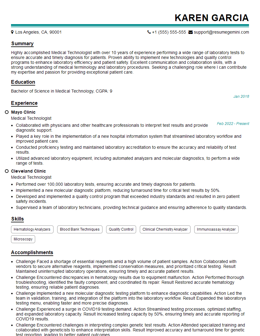
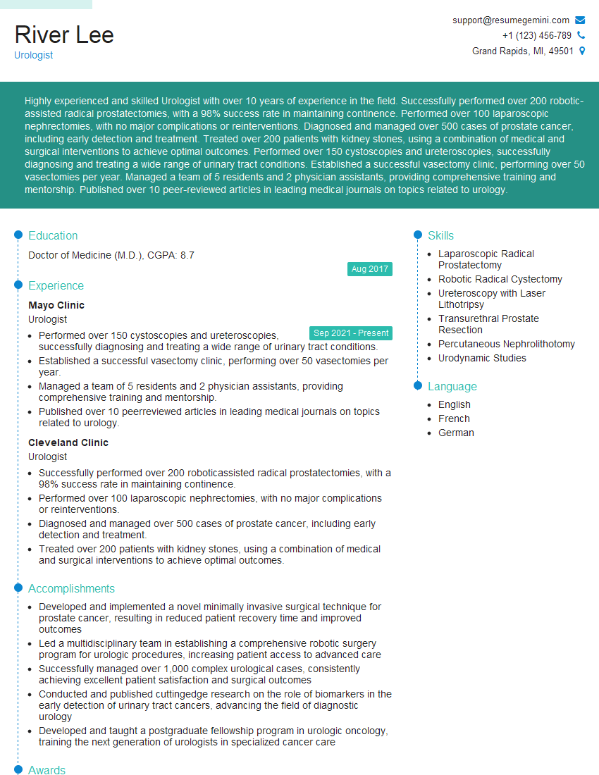
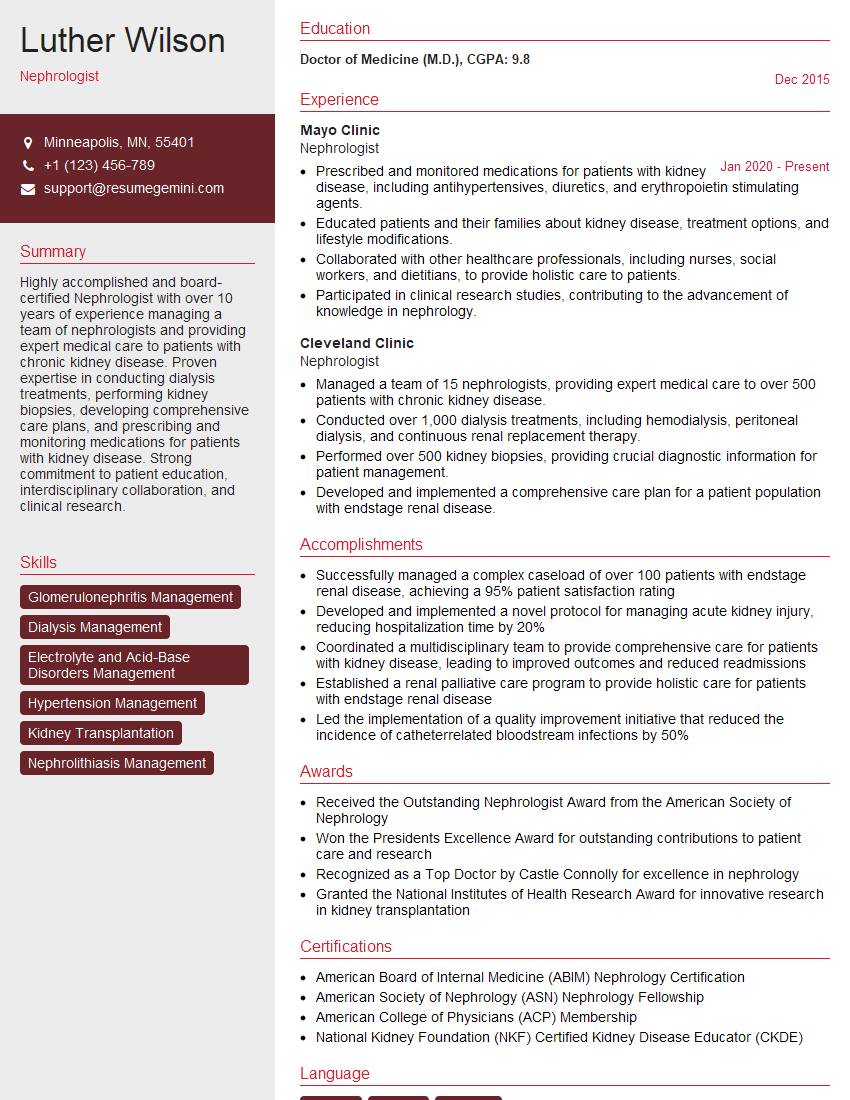
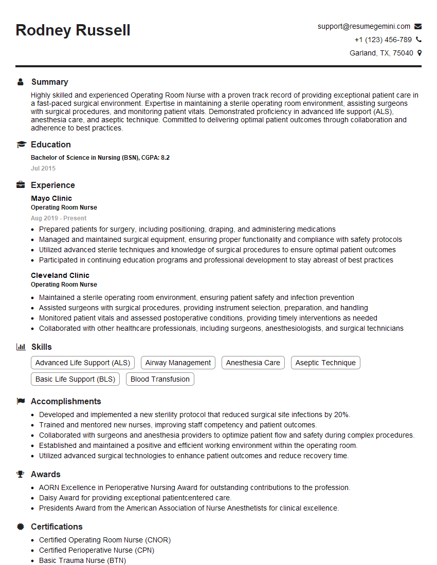
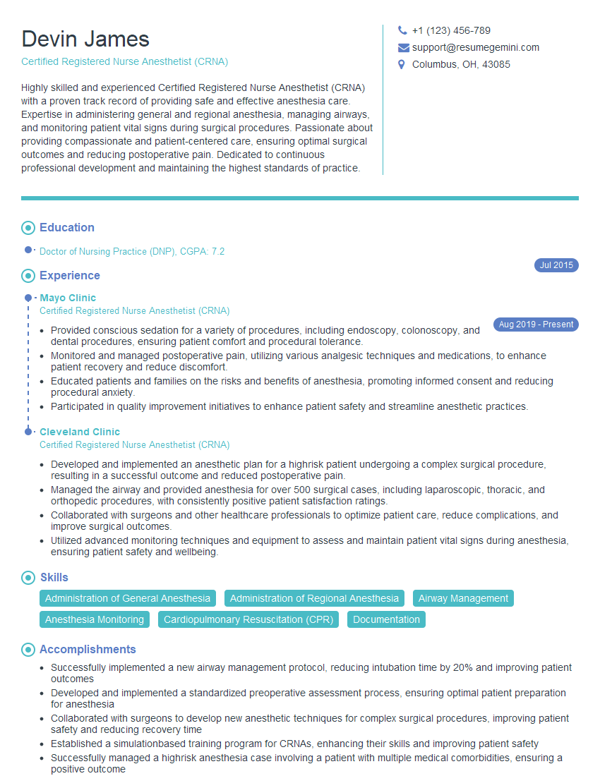
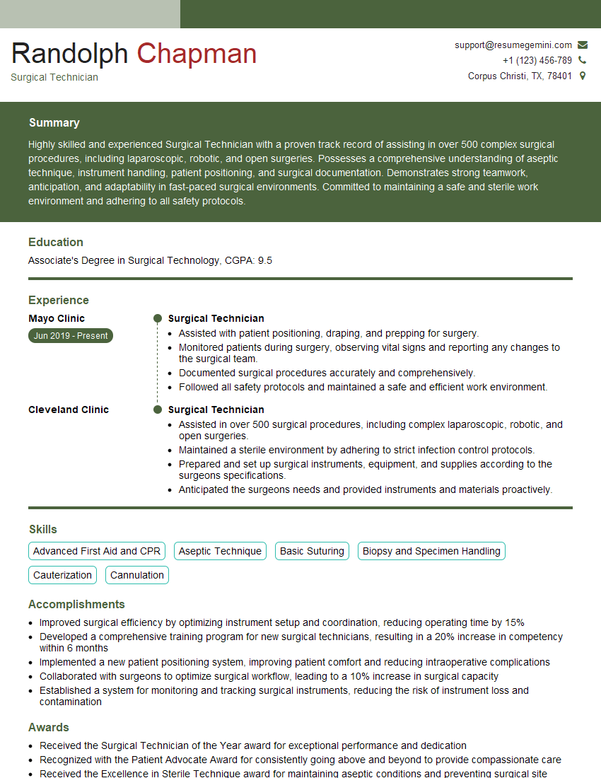
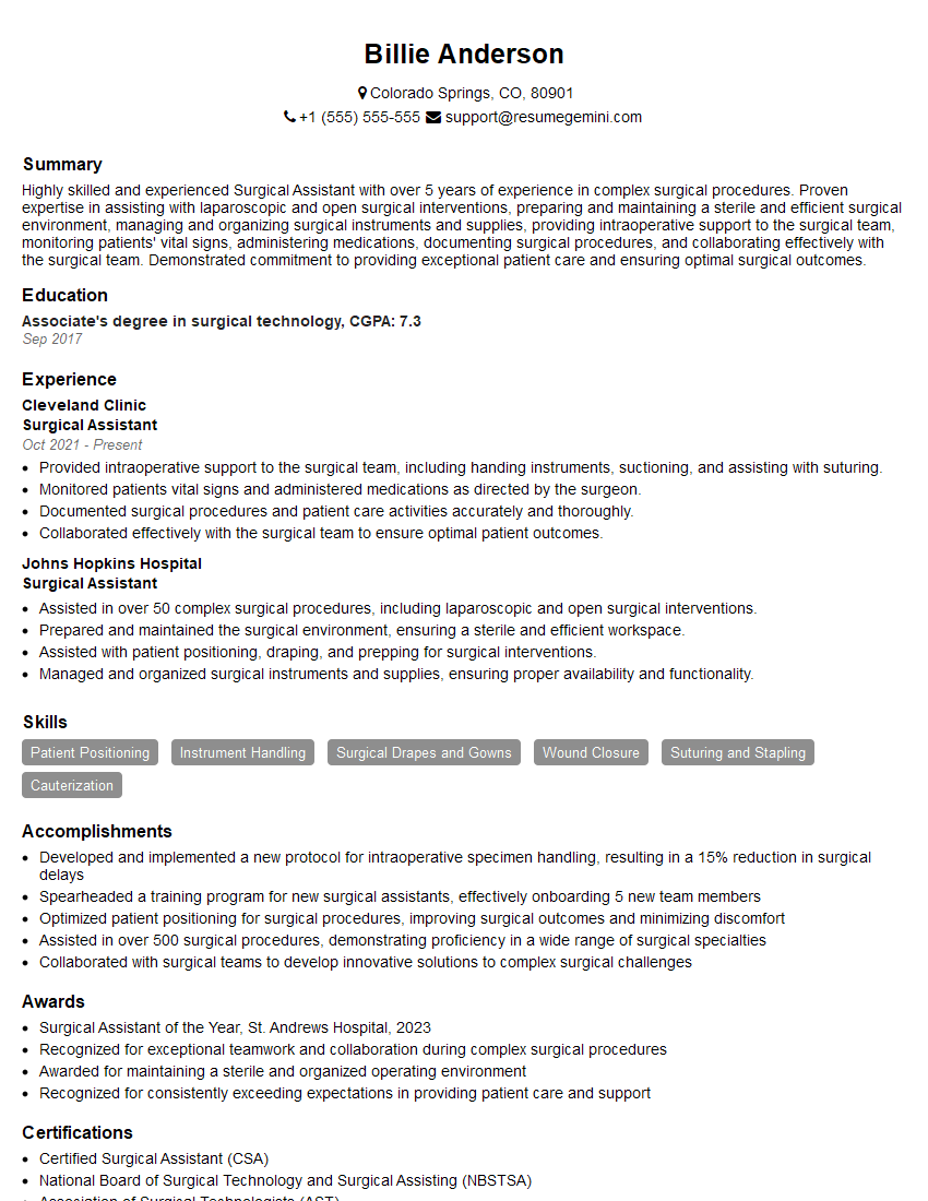
Mastering cystoscopy is crucial for career advancement in urology and related fields. A strong understanding of the procedure and its applications will significantly enhance your professional profile and open doors to exciting opportunities. To maximize your job prospects, it’s essential to create an ATS-friendly resume that effectively showcases your skills and experience. We highly recommend using ResumeGemini to build a professional and impactful resume. ResumeGemini provides you with the tools and resources to create a compelling document, and examples of resumes tailored to Cystoscopy are available to guide you. Take the next step in your career journey today!
Explore more articles
Users Rating of Our Blogs
Share Your Experience
We value your feedback! Please rate our content and share your thoughts (optional).
What Readers Say About Our Blog
To the interviewgemini.com Webmaster.
Very helpful and content specific questions to help prepare me for my interview!
Thank you
To the interviewgemini.com Webmaster.
This was kind of a unique content I found around the specialized skills. Very helpful questions and good detailed answers.
Very Helpful blog, thank you Interviewgemini team.
