The thought of an interview can be nerve-wracking, but the right preparation can make all the difference. Explore this comprehensive guide to Endoscopic Cancer Screening and Surveillance interview questions and gain the confidence you need to showcase your abilities and secure the role.
Questions Asked in Endoscopic Cancer Screening and Surveillance Interview
Q 1. Describe the different types of endoscopic procedures used in cancer screening.
Endoscopic procedures are minimally invasive techniques used to visualize the gastrointestinal tract and other internal organs. In cancer screening, the most common procedures are colonoscopy and esophagogastroduodenoscopy (EGD).
- Colonoscopy: A long, flexible tube with a camera is inserted into the rectum to examine the entire colon and rectum. This allows for visualization of polyps and lesions which can be biopsied or removed.
- Esophagogastroduodenoscopy (EGD): Similar to colonoscopy, but the endoscope is passed through the mouth to examine the esophagus, stomach, and duodenum. This is crucial for detecting cancers of these regions.
- Capsule endoscopy: A small, disposable camera is swallowed, allowing for visualization of the small bowel, which is difficult to access with traditional endoscopy. This is used less frequently in screening, more for diagnosis of obscure gastrointestinal bleeding.
- Endoscopic ultrasound (EUS): Combines endoscopy with ultrasound to provide detailed images of the layers of the gastrointestinal wall, enabling better assessment of tumor depth and spread. This is primarily used for staging, not initial screening.
The choice of procedure depends on the suspected location and type of cancer.
Q 2. Explain the indications for colonoscopy, endoscopy, and other endoscopic procedures.
Indications for these procedures vary, but generally involve:
- Colonoscopy: Screening for colorectal cancer in individuals at average risk (starting at age 45, or earlier with family history or other risk factors), investigation of gastrointestinal symptoms such as bleeding, changes in bowel habits, abdominal pain, surveillance after polypectomy or colorectal cancer resection, and evaluation of inflammatory bowel disease.
- Endoscopy (EGD): Screening for esophageal and gastric cancers in high-risk individuals (e.g., those with Barrett’s esophagus), investigation of dysphagia (difficulty swallowing), heartburn, abdominal pain, upper gastrointestinal bleeding, and follow-up after treatment for gastric cancer.
- Other endoscopic procedures: Capsule endoscopy is indicated for the investigation of obscure gastrointestinal bleeding when other methods have failed. EUS is used to stage gastrointestinal cancers and assess the extent of tumor involvement.
Each procedure has specific indications based on patient history, symptoms, and risk factors. A thorough discussion with a gastroenterologist is essential to determine the appropriate endoscopic approach.
Q 3. What are the common complications associated with endoscopic procedures?
While generally safe, endoscopic procedures carry potential complications, including:
- Bleeding: Minor bleeding is common, but significant bleeding requiring transfusion is rare. This is more likely after polypectomy.
- Perforation: A hole in the gastrointestinal tract, a serious complication requiring immediate surgical intervention. Risk is higher with certain procedures, like colonoscopy in patients with diverticulitis or previous abdominal surgery.
- Infection: Infection at the puncture site or more serious sepsis is possible, especially in immunocompromised patients.
- Pancreatitis: A rare but serious complication associated with ERCP (endoscopic retrograde cholangiopancreatography), a procedure to examine the bile and pancreatic ducts.
- Adverse reactions to sedation: Sedation is commonly used during these procedures, and complications such as respiratory depression or hypotension can occur.
The risk of complications varies depending on the procedure, the patient’s health status, and the skill of the endoscopist. These risks are generally low and often outweighed by the benefits of early cancer detection.
Q 4. How do you manage a perforation during a colonoscopy?
Management of a colonoscopy perforation is a surgical emergency. Immediate actions include:
- Stabilize the patient: This involves monitoring vital signs, fluid resuscitation, and supportive care.
- Abdominal imaging: CT scan to confirm the location and extent of the perforation.
- Surgical consultation: Immediate surgical consultation is crucial to determine the best course of action, which may involve primary repair of the perforation, resection of the affected bowel segment, or creation of a colostomy.
- Broad-spectrum antibiotics: To prevent peritonitis (infection of the abdominal cavity).
- Intensive care monitoring: Close monitoring in an intensive care unit (ICU) is necessary due to the risk of sepsis and other complications.
The outcome depends on several factors, including the size and location of the perforation, the patient’s overall health, and the promptness of intervention. Early recognition and rapid management are crucial to improve patient survival.
Q 5. What are the current guidelines for colorectal cancer screening?
Current guidelines for colorectal cancer screening vary slightly depending on the organization (e.g., American Cancer Society, US Preventive Services Task Force), but generally recommend:
- Average-risk individuals: Screening should begin at age 45 with colonoscopy every 10 years, or other options such as flexible sigmoidoscopy every 5 years, or stool-based tests (FIT, FOBT) annually or every 1-2 years.
- Individuals with family history or other risk factors: Screening should begin earlier and more frequently, often with colonoscopy.
- Specific risk factors include: Family history of colorectal cancer or adenomatous polyposis, inflammatory bowel disease (ulcerative colitis, Crohn’s disease), personal history of colorectal cancer or adenomas.
The choice of screening method is individualized based on factors such as patient preferences, risk factors, and access to healthcare. It’s important to consult with a healthcare provider to determine the best screening approach.
Q 6. Discuss the role of polypectomy in colorectal cancer prevention.
Polypectomy, the surgical removal of polyps during colonoscopy, plays a pivotal role in colorectal cancer prevention. Most colorectal cancers arise from adenomatous polyps (adenomas). Removing these polyps prevents them from progressing to cancer.
The procedure is relatively simple and safe, typically performed during a routine colonoscopy. Polypectomy reduces the risk of developing colorectal cancer significantly, especially for larger and advanced adenomas.
Regular surveillance colonoscopies after polypectomy are crucial to detect any remaining or new polyps, ensuring early detection and removal.
Q 7. Explain the significance of polyp size and histology in determining the risk of malignancy.
Both polyp size and histology (tissue type) are crucial in determining the risk of malignancy.
- Size: Larger polyps have a higher risk of containing cancer or having cancerous potential. Polyps smaller than 1 cm have a low risk, while those larger than 1 cm have an increasingly higher risk.
- Histology: The microscopic appearance of the polyp determines its type. Adenomas are precancerous polyps, with different subtypes (tubular, villous, tubulovillous) carrying varying risks. Villous adenomas have a significantly higher risk of malignancy compared to tubular adenomas. The presence of high-grade dysplasia (abnormal cell growth) is a strong indicator of a high risk of cancer.
For example, a large (e.g., >2 cm) villous adenoma with high-grade dysplasia carries a substantially higher risk of malignancy than a small (<1 cm) tubular adenoma with low-grade dysplasia. These factors influence the recommendations for surveillance after polypectomy and the need for additional procedures.
Q 8. Describe the different types of polyps found during colonoscopy.
Colonoscopy allows visualization of the colon’s lining, often revealing polyps. These growths vary significantly in size, shape, and risk of cancer. They’re broadly classified based on their microscopic appearance (histology) and macroscopic features.
- Hyperplastic Polyps: These are the most common type, typically small and benign (non-cancerous). They’re usually found in the right colon and have a characteristic serrated appearance. Think of them as tiny, harmless bumps.
- Adenomatous Polyps (Adenomas): These are precancerous polyps, meaning they have the potential to develop into colorectal cancer if left untreated. They’re categorized by their architecture:
- Tubular adenomas: These are the most common type of adenoma, resembling a small tube.
- Villous adenomas: These have a finger-like, frond-like projection. They tend to be larger and carry a higher risk of cancer than tubular adenomas.
- Tubulovillous adenomas: These polyps exhibit features of both tubular and villous adenomas.
- Sessile serrated adenomas (SSA): These polyps have a flat, sessile (broad-based) growth pattern and a serrated (saw-tooth) surface. They are increasingly recognized as important precursors to colorectal cancer, particularly in the right colon.
- Inflammatory polyps: These are associated with chronic inflammatory bowel disease like ulcerative colitis or Crohn’s disease.
The size and histological features of a polyp dictate management. Small hyperplastic polyps usually don’t require removal, while adenomas, especially larger ones, necessitate removal to prevent cancer development.
Q 9. How do you interpret endoscopic findings and correlate them with pathology reports?
Interpreting endoscopic findings involves a meticulous comparison between what’s visualized during the procedure and the subsequent pathology report from the laboratory. This process is crucial for accurate diagnosis and appropriate management of colonic lesions.
For example, during colonoscopy, a polyp might appear as a 1cm sessile lesion with a slightly irregular surface. The endoscopist would note its size, location, and appearance. The polyp is then removed and sent for histopathological analysis. The pathology report will then specify the type of polyp (e.g., tubular adenoma, hyperplastic polyp), its size, the presence of dysplasia (precancerous changes), and depth of invasion (if cancerous).
Discrepancies can occur. For instance, a polyp might appear benign endoscopically but show low-grade dysplasia on pathology, requiring closer surveillance or further intervention. Conversely, a polyp might look concerning endoscopically but turn out to be benign on histology. This highlights the importance of a detailed and accurate description of endoscopic findings to aid in pathological interpretation.
Q 10. What are the indications for endoscopic mucosal resection (EMR)?
Endoscopic mucosal resection (EMR) is a technique used to remove larger, flat lesions or polyps that are difficult to completely remove with standard polypectomy techniques (snare resection). The indications for EMR include:
- Large sessile polyps: Polyps that are flat and broad-based, often exceeding 2 cm in size.
- Villous adenomas: Due to their high risk of malignancy.
- Adenomas with high-grade dysplasia or early cancer: EMR provides complete removal of the lesion with minimal risk.
- Early colorectal cancer (T1 lesions): EMR offers a curative option for selected early-stage cancers, avoiding the need for surgery.
- Other lesions: EMR can also be used for various other lesions such as inflammatory lesions and certain types of lymphoma.
EMR provides a minimally invasive alternative to surgical resection for these lesions, reducing the need for extensive surgery.
Q 11. Explain the procedure for endoscopic submucosal dissection (ESD).
Endoscopic submucosal dissection (ESD) is an advanced endoscopic technique used to resect larger, laterally spreading lesions, such as early colorectal cancer or large adenomas. It’s more complex than EMR. The procedure involves:
- Injection of submucosal saline: This elevates the lesion, creating a space between the lesion and the muscularis propria (the underlying muscle layer).
- Precise incision: The endoscopist makes an incision around the lesion using a specialized knife, creating a circumferential cut.
- Dissection: The endoscopist uses a specialized dissecting knife (such as a hook knife or a flex knife) to carefully dissect the lesion from the underlying submucosa.
- En bloc resection: The lesion is removed en bloc (in one piece), aiming for complete resection with clear margins.
- Hemostasis: Bleeding is controlled using various techniques like clipping, coagulation, or injection of hemostatic agents.
ESD is technically demanding and requires specialized training. It’s preferred when EMR is not suitable because of the size or nature of the lesion.
Q 12. Describe the techniques for advanced endoscopic procedures such as argon plasma coagulation (APC).
Argon plasma coagulation (APC) is a non-contact energy source used in advanced endoscopy. It uses argon gas to deliver radiofrequency energy to the tissue surface. This leads to localized coagulation (heat-induced tissue destruction) and hemostasis (stopping bleeding).
Technique: Argon gas is channeled through a small probe, and radiofrequency energy is applied, creating a plasma arc that contacts the tissue surface. The depth of coagulation can be controlled by altering the gas flow and power settings. APC is useful for:
- Hemostasis: Controlling bleeding from small vessels or friable (easily bleeding) tissue.
- Ablation of lesions: Removing small polyps or superficial lesions.
- Treating angiodysplasia: Cauterizing abnormal blood vessels responsible for bleeding.
APC offers a precise and minimally invasive approach to these tasks.
Q 13. What are the different methods for hemostasis during endoscopic procedures?
Hemostasis (stopping bleeding) is crucial during endoscopic procedures. Several methods are available, chosen based on the bleeding source and severity:
- Mechanical methods: These include clipping (using hemostatic clips to occlude bleeding vessels) or applying pressure with a sponge.
- Thermal methods: These involve using heat to coagulate (seal) blood vessels. Examples include monopolar or bipolar electrocoagulation, and APC, discussed previously.
- Injection methods: Hemostatic agents such as epinephrine or thrombin can be injected into the bleeding site to promote coagulation.
- Combination methods: Often, a combination of these methods is used for optimal hemostasis.
The choice of technique depends on the specific circumstances. For example, a small bleeding vessel might be managed with simple coagulation, while a larger bleeding source might require a combination of clipping and injection.
Q 14. How do you handle difficult intubations during endoscopy?
Difficult intubations during endoscopy can be challenging and require experience and a systematic approach. Factors contributing to difficulty include anatomical variations, bowel pathology, and patient factors.
Strategies to manage difficult intubations:
- Careful pre-procedural assessment: A thorough review of the patient’s history, including previous endoscopic procedures and any known anatomical abnormalities, helps anticipate potential difficulties.
- Appropriate bowel preparation: Thorough bowel preparation helps minimize fecal residue and improve visualization.
- Use of advanced techniques: Experienced endoscopists can use advanced techniques such as using a pediatric endoscope, utilizing different insertion angles, or employing guidewires to navigate difficult areas.
- Collaboration and consultation: If unsuccessful, consulting with a more experienced endoscopist or a gastroenterologist specialized in difficult intubations is essential. In some cases, alternative imaging modalities such as CT or MRI might be considered.
- Abandonment if necessary: If the procedure cannot be safely performed, it’s crucial to abandon it to avoid causing harm to the patient.
Patient safety is paramount. If repeated attempts to intubate are unsuccessful, the procedure should be stopped to avoid complications such as perforation.
Q 15. What are the safety precautions associated with sedation during endoscopy?
Sedation during endoscopy, while enhancing patient comfort and tolerance, necessitates stringent safety precautions. The primary goal is to minimize risks while ensuring adequate procedural sedation. This involves careful patient selection, meticulous monitoring, and having a skilled team ready to manage complications.
- Patient Selection: Patients with significant respiratory or cardiac disease, or those with a history of difficult airway management, may require more careful consideration or alternative approaches. A thorough review of medical history and medication use is essential.
- Monitoring: Continuous monitoring of vital signs (heart rate, blood pressure, oxygen saturation, respiratory rate) is crucial. Pulse oximetry is standard, and capnography is frequently used to assess ventilation. A dedicated nurse or anesthesia professional is often part of the team to constantly observe the patient’s response to sedation.
- Sedation Techniques: A variety of sedatives, often in combination (e.g., propofol, fentanyl, midazolam), are used, carefully titrated to the individual patient’s response and the procedural needs. The goal is to achieve moderate sedation—where the patient is comfortable and relaxed but can still respond to verbal stimuli. Deep sedation requires additional expertise and often involves anesthesiologists.
- Reversal Agents: Having readily available reversal agents like flumazenil (for benzodiazepines) and naloxone (for opioids) is essential for managing any adverse effects or oversedation. Knowing when and how to administer these agents is critical.
- Post-procedure Care: Close observation is needed post-procedure to ensure the patient’s safe recovery from sedation, including monitoring for respiratory depression, hypotension, and nausea/vomiting. Patients are typically discharged only when fully alert and stable.
For instance, a patient with a history of sleep apnea might require a modified sedation approach, potentially using lower doses or avoiding certain medications. Careful monitoring in such cases is paramount to prevent respiratory compromise.
Career Expert Tips:
- Ace those interviews! Prepare effectively by reviewing the Top 50 Most Common Interview Questions on ResumeGemini.
- Navigate your job search with confidence! Explore a wide range of Career Tips on ResumeGemini. Learn about common challenges and recommendations to overcome them.
- Craft the perfect resume! Master the Art of Resume Writing with ResumeGemini’s guide. Showcase your unique qualifications and achievements effectively.
- Don’t miss out on holiday savings! Build your dream resume with ResumeGemini’s ATS optimized templates.
Q 16. Discuss the use of chromoendoscopy in detecting early cancers.
Chromoendoscopy enhances the visualization of subtle mucosal changes, increasing the detection rate of early cancers. It involves applying dyes (like indigo carmine, methylene blue, or crystal violet) to the mucosal surface, which highlights abnormal areas based on their different absorption or retention of the dye.
For example, cancerous or precancerous lesions might appear darker or lighter than surrounding normal tissue after dye application. This allows for the easier identification of lesions that might otherwise be missed during standard white light endoscopy. The improved visualization enables more accurate targeting of biopsies, leading to early diagnosis and treatment.
The choice of dye depends on the specific application and target lesion. Indigo carmine is frequently used for colorectal lesions, while methylene blue is sometimes employed in upper gastrointestinal endoscopy. The technique can be used with both colonoscopes and endoscopes used to examine the upper GI tract.
Q 17. Explain the role of narrow band imaging (NBI) in endoscopic examinations.
Narrow band imaging (NBI) is an advanced endoscopic technique that uses specific wavelengths of light to enhance the visualization of mucosal microvasculature and surface patterns. This allows for better differentiation between normal and abnormal tissue, particularly in detecting early neoplastic lesions.
Unlike chromoendoscopy which relies on dye application, NBI uses specialized filters to highlight the surface structures. This gives endoscopists improved resolution of the mucosal capillaries and surface architecture. Abnormal tissue often exhibits altered microvascular patterns, such as irregular or dilated capillaries, which are readily apparent with NBI.
For instance, in a colonoscopy, NBI can help identify subtle adenomas (precancerous polyps) or early colorectal cancers that might appear normal under standard white light. It also aids in the differentiation between benign and malignant lesions, improving the accuracy of biopsy targeting and reducing the number of unnecessary biopsies. The increased sensitivity can improve early cancer detection rates significantly.
Q 18. Describe the importance of bowel preparation for colonoscopy.
Adequate bowel preparation is crucial for a successful colonoscopy. This involves cleaning the colon to remove fecal matter and gas, ensuring clear visualization of the entire colonic mucosa. Without proper bowel preparation, the view is obscured, potentially leading to missed polyps or lesions, and resulting in an incomplete examination.
The preparation usually involves a low-residue diet for several days before the procedure, followed by a bowel cleansing regimen. This typically includes oral laxatives (such as polyethylene glycol solutions) that induce bowel movements. The goal is to achieve clear, watery stool, indicating a clean colon. The quality of bowel preparation is graded by the endoscopist, and inadequate preparation can necessitate rescheduling the procedure.
For example, a poorly prepared colon could result in missing a small but significant adenoma that could later develop into colorectal cancer. Thorough bowel preparation is critical for the accuracy and efficacy of the procedure. Careful adherence to the instructions for bowel preparation as given by the physician is essential for optimal results.
Q 19. What are the contraindications for colonoscopy?
Several contraindications exist for colonoscopy. These are situations where the procedure might be too risky or impractical, requiring alternative approaches.
- Severe active inflammatory bowel disease: The procedure may exacerbate inflammation and cause complications.
- Toxic megacolon: A severely dilated colon is at risk of perforation.
- Recent bowel perforation or surgery: Increased risk of complications related to perforation or injury.
- Uncontrolled bleeding disorders: Increased risk of bleeding during the procedure or biopsy.
- Severe cardiovascular instability: Risks associated with sedation and positioning during the procedure are amplified.
- Inability to tolerate bowel preparation: Poor bowel preparation will compromise the diagnostic value of the examination.
For example, a patient with a recent history of ischemic colitis (inflammation of the colon) would likely have colonoscopy postponed to allow the inflammation to resolve to minimize risks.
Q 20. What are the limitations of endoscopic techniques?
While endoscopic techniques are highly valuable, they have certain limitations.
- Accessibility: Some areas of the GI tract might be difficult to access due to anatomical variations or strictures (narrowing of the gut).
- Sampling limitations: Biopsies are limited to superficial tissue; deeper invasion by cancer might be underestimated.
- Observer variability: The interpretation of findings can vary between endoscopists, especially regarding subtle lesions.
- False negative results: Small or flat lesions can be missed, especially in cases of poor bowel preparation.
- Procedural risks: Although rare, complications like perforation, bleeding, and infection can occur.
For example, a small, flat lesion might appear unremarkable during endoscopy, yet histology reveals cancer. Regular surveillance, even with negative initial findings, is often necessary.
Q 21. Discuss the role of endoscopic ultrasound (EUS) in cancer staging.
Endoscopic ultrasound (EUS) plays a crucial role in cancer staging, providing detailed information about the depth of tumor invasion and the presence of regional lymph node involvement. This is critical for treatment planning and prognosis.
EUS combines endoscopy with ultrasound technology. A specialized endoscope with an ultrasound probe at its tip is introduced into the GI tract. The ultrasound waves generate images of the layers of the GI wall and surrounding structures, enabling the precise assessment of tumor depth (T stage) and lymph node status (N stage). This is essential in the TNM staging system (Tumor, Node, Metastasis) used to classify cancers.
For example, in a patient with a suspected pancreatic cancer, EUS can determine whether the tumor is confined to the pancreas or has invaded adjacent structures. It can also detect the presence of regional lymph node metastases, which significantly impacts treatment choices and overall prognosis. EUS-guided fine-needle aspiration (FNA) can further allow for cytological confirmation of malignancy and potentially molecular analysis.
Q 22. Describe the role of endoscopic retrograde cholangiopancreatography (ERCP).
ERCP, or endoscopic retrograde cholangiopancreatography, is a sophisticated procedure combining endoscopy and fluoroscopy to diagnose and treat conditions affecting the biliary and pancreatic ducts. Think of it as a detective’s tool, allowing us to visually inspect and even intervene in these often-hidden areas.
During an ERCP, a thin, flexible endoscope is passed through the mouth and into the duodenum (the first part of the small intestine). Once there, a contrast dye is injected, and X-ray fluoroscopy is used to visualize the bile ducts and pancreatic duct. This allows us to identify blockages caused by gallstones, tumors, or strictures (narrowing). Crucially, the procedure isn’t just diagnostic; it’s also therapeutic. We can use specialized instruments passed through the endoscope to remove gallstones (using a basket or balloon), place stents to relieve blockages, or take biopsies for further analysis. For instance, a patient experiencing persistent jaundice and elevated liver enzymes might undergo ERCP to determine the cause and receive appropriate treatment like stone removal or stent placement.
Q 23. How do you manage a patient with a bleeding polyp during colonoscopy?
Managing a bleeding polyp during colonoscopy requires a calm, systematic approach prioritizing patient safety. The initial step is careful assessment. We need to determine the location, size, and type of polyp, as well as the severity of the bleeding. Simple, non-bleeding polyps can often be safely removed with a snare or forceps, and the bleeding site cauterized with heat or clips if needed. However, larger or actively bleeding polyps present greater challenges. For instance, if a large polyp is aggressively bleeding, epinephrine injection might be necessary to help constrict blood vessels and reduce bleeding. More extensive bleeding might require specialized techniques like argon plasma coagulation or even surgical intervention if the endoscopy doesn’t control the bleeding completely.
Post-procedure, close monitoring of the patient’s vital signs and stool output is vital to detect any further bleeding. If needed, blood transfusions or other supportive care might be required. It’s always a team effort involving nurses, surgical colleagues, and radiologists to ensure optimal patient management. Each case is unique and the approach needs to be tailored according to the specific situation.
Q 24. Explain the process of obtaining tissue biopsies during endoscopy.
Obtaining tissue biopsies during endoscopy is crucial for accurate diagnosis. It involves using specialized forceps or needles to collect small tissue samples from suspicious areas, like polyps or ulcers. The process is typically straightforward, yet requires precision and a good understanding of anatomy. The endoscopist navigates the endoscope to the target area, often using magnified views and chromoendoscopy (dye enhancement for improved visualization) to precisely target the abnormality. Once the target is identified, we insert the biopsy forceps, carefully grasp a small piece of tissue, and gently withdraw it. Multiple biopsies might be taken from different locations to ensure a representative sample.
Each biopsy is labeled carefully and handled meticulously to avoid any contamination or degradation, which is paramount for accurate pathology results. These biopsies are then sent to the pathology lab for microscopic examination, which assists in confirming the diagnosis, staging the cancer, and guiding treatment planning.
Q 25. What are your strategies for communicating findings to patients and their families?
Communicating findings to patients and their families is a crucial aspect of my work, demanding sensitivity and clarity. I always prefer a face-to-face discussion in a private setting, ensuring ample time is available for questions and emotional support. The language I use is adapted to the individual’s comprehension level, avoiding medical jargon where possible. If there’s a diagnosis of cancer, I explain it clearly, emphasizing the type, stage, and treatment options available. I make sure they understand the next steps, potential side effects of treatments, and prognosis.
I often encourage patients to bring a trusted family member or friend for emotional support during the discussion. Follow-up appointments and referrals to specialists, support groups, or genetic counselors are also arranged as needed, providing comprehensive care that goes beyond the initial consultation.
Q 26. How do you maintain proper infection control during endoscopic procedures?
Maintaining proper infection control during endoscopic procedures is paramount. It’s a high-priority area involving strict adherence to established guidelines. Our facility employs stringent protocols, encompassing meticulous cleaning and high-level disinfection of endoscopes before and after each procedure. We utilize automated endoscope reprocessors (AERs) that ensure effective cleaning and sterilization according to manufacturer guidelines. The use of appropriate personal protective equipment (PPE), including gloves, gowns, masks, and eye protection, is mandatory for all personnel involved.
Hand hygiene is rigorously practiced before and after each patient interaction. Environmental disinfection of the procedure room is also performed regularly, focusing on high-touch surfaces. We also adhere to guidelines for proper waste disposal and handling of potentially infectious materials. Consistent monitoring and regular staff training are essential to maintaining these infection control protocols effectively. Ignoring this would place patients at significant risk for infections such as Clostridium difficile, and various viral infections.
Q 27. Discuss your experience with electronic health records (EHR) in the context of endoscopy.
Electronic health records (EHRs) have fundamentally transformed endoscopy. They have enhanced efficiency and improved patient care in many ways. For instance, EHRs allow for seamless documentation of the procedure, including images, reports, and biopsy results. This digital record is readily accessible to healthcare providers involved in the patient’s care, leading to better communication and coordination among specialists. EHRs have also automated aspects of scheduling, appointment reminders, and patient communication.
Moreover, EHRs offer valuable tools for quality assurance and data analysis. We can track procedure volume, complications, and outcomes, aiding in benchmarking and identifying areas for improvement. However, the efficient use of EHRs also requires careful attention to ensure data security and patient privacy, issues that our institution takes extremely seriously.
Q 28. How do you stay updated on the latest advancements in endoscopic cancer screening?
Staying abreast of advancements in endoscopic cancer screening requires a multi-pronged approach. I actively participate in continuing medical education (CME) activities, attending conferences and workshops, both in-person and virtual, which offer updates on the latest techniques, technologies, and guidelines. I regularly review peer-reviewed journals and medical literature, focusing on high-impact publications to keep my knowledge current.
Participation in professional organizations such as the American Society for Gastrointestinal Endoscopy (ASGE) provides access to their guidelines and resources, which provide summaries of new evidence and recommendations for optimal practice. Staying connected with colleagues through professional networks and collaboration allows for information sharing and discussion of novel approaches. Ultimately, ongoing learning is crucial to ensure that I deliver the highest quality of care, informed by the most up-to-date evidence and best practice guidelines.
Key Topics to Learn for Endoscopic Cancer Screening and Surveillance Interview
- Endoscopic Techniques: Mastering the principles and practical application of colonoscopy, endoscopy, and other relevant procedures. This includes understanding indications, contraindications, and potential complications.
- Polyp Detection and Classification: Develop expertise in identifying and classifying different types of polyps (e.g., adenomas, hyperplastic polyps) based on their morphology and histology. Understand the implications of each type for cancer risk.
- Biopsy Techniques and Interpretation: Gain a strong understanding of appropriate biopsy techniques and the interpretation of biopsy results, including the recognition of dysplasia and carcinoma.
- Image Interpretation and Reporting: Practice interpreting endoscopic images and writing clear, concise, and accurate reports that effectively communicate findings to referring physicians.
- Surveillance Protocols: Familiarize yourself with established guidelines and protocols for endoscopic surveillance of patients at high risk for colorectal cancer and other cancers detected through endoscopy.
- Patient Management and Communication: Develop your skills in patient communication, including pre-procedure counseling, post-procedure instructions, and managing patient expectations and anxieties.
- Advanced Endoscopic Techniques: Explore advanced procedures such as endoscopic mucosal resection (EMR) and endoscopic submucosal dissection (ESD), including their indications, contraindications, and procedural steps.
- Quality Assurance and Improvement: Understand the importance of quality assurance measures in endoscopic procedures and how to contribute to continuous improvement in patient care.
- Risk Stratification and Prevention: Learn how to effectively stratify patients based on their risk factors for colorectal cancer and other cancers, and how to implement preventive strategies.
- Ethical and Legal Considerations: Understand the ethical and legal responsibilities related to endoscopic cancer screening and surveillance, including informed consent and patient confidentiality.
Next Steps
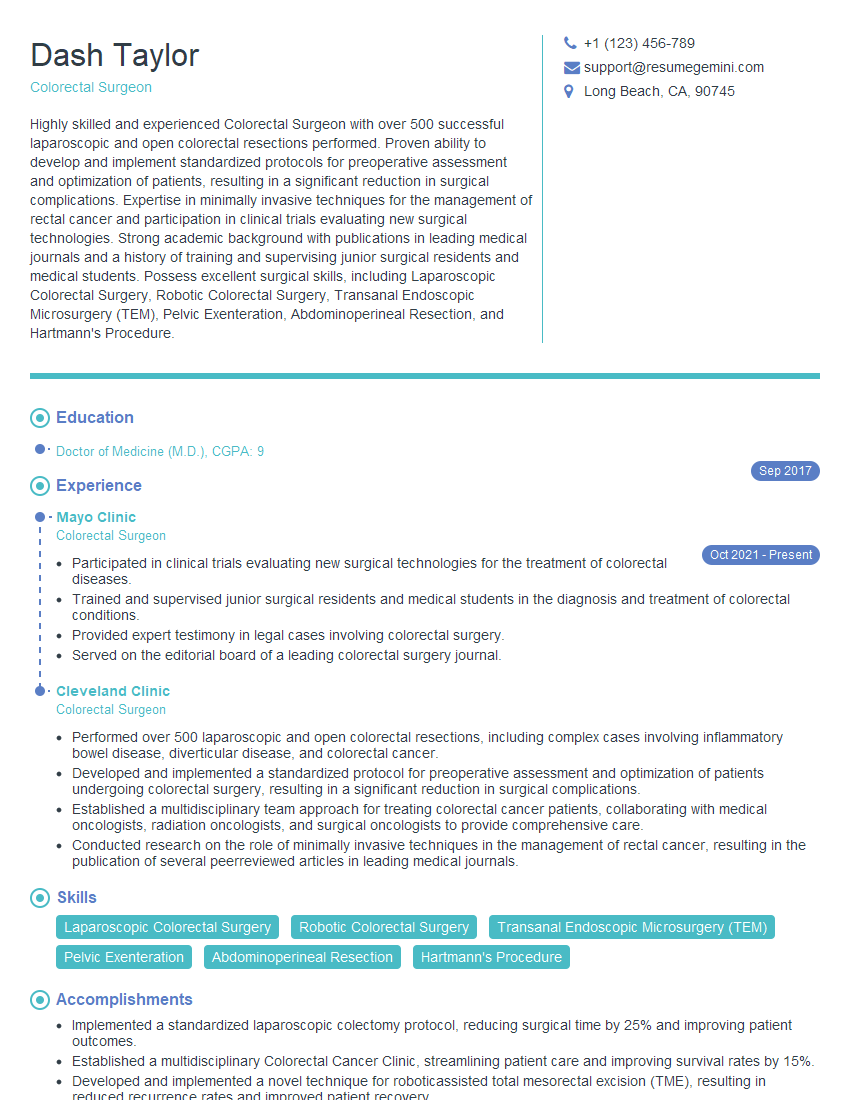
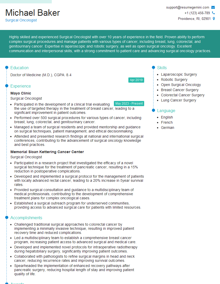
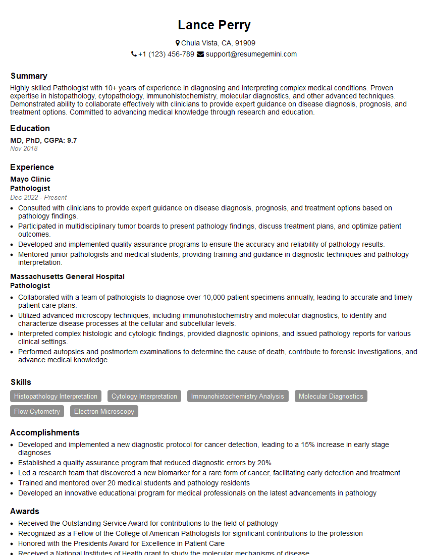
Mastering Endoscopic Cancer Screening and Surveillance opens doors to exciting career opportunities with significant impact on patient lives. Demonstrating this expertise effectively is crucial for career advancement. A well-crafted, ATS-friendly resume is your first step to securing interviews. ResumeGemini can significantly enhance your resume-building experience by providing the tools and resources you need to showcase your skills and experience effectively. ResumeGemini offers examples of resumes tailored to Endoscopic Cancer Screening and Surveillance, helping you create a compelling document that highlights your unique qualifications. Take the next step towards your dream career today!
Explore more articles
Users Rating of Our Blogs
Share Your Experience
We value your feedback! Please rate our content and share your thoughts (optional).
What Readers Say About Our Blog
Hi, I have something for you and recorded a quick Loom video to show the kind of value I can bring to you.
Even if we don’t work together, I’m confident you’ll take away something valuable and learn a few new ideas.
Here’s the link: https://bit.ly/loom-video-daniel
Would love your thoughts after watching!
– Daniel
This was kind of a unique content I found around the specialized skills. Very helpful questions and good detailed answers.
Very Helpful blog, thank you Interviewgemini team.


