Every successful interview starts with knowing what to expect. In this blog, we’ll take you through the top Fracture Repair interview questions, breaking them down with expert tips to help you deliver impactful answers. Step into your next interview fully prepared and ready to succeed.
Questions Asked in Fracture Repair Interview
Q 1. Describe the different types of bone fractures.
Bone fractures are classified in various ways, considering the type of break, the bone’s integrity, and the surrounding soft tissues. A common classification system considers the fracture’s appearance on an X-ray.
- Complete vs. Incomplete: A complete fracture breaks the bone entirely, while an incomplete fracture (like a greenstick fracture common in children) only partially breaks the bone.
- Closed vs. Open (Compound): A closed fracture doesn’t break the skin, whereas an open fracture does, increasing the risk of infection.
- Comminuted: The bone is broken into more than two fragments.
- Transverse: The fracture line runs across the bone at a right angle.
- Oblique: The fracture line runs diagonally across the bone.
- Spiral: The fracture line spirals around the bone, often indicating a twisting injury.
- Segmental: The bone is broken in two places, resulting in a floating segment.
- Avulsion: A fragment of bone is pulled away from the main bone by a tendon or ligament.
- Impacted: One end of the broken bone is driven into the other.
- Stress Fracture: A tiny crack in the bone caused by overuse or repetitive stress.
Understanding these fracture types is crucial for choosing the appropriate treatment strategy. For example, a simple transverse fracture might heal with casting, while a complex comminuted fracture may require surgery.
Q 2. Explain the process of bone healing.
Bone healing is a remarkable process involving several stages. It’s like a carefully orchestrated construction project, where the body rebuilds the fractured bone.
- Hematoma Formation: Immediately after the fracture, bleeding occurs, forming a blood clot (hematoma) at the fracture site. This is the foundation for repair.
- Inflammation and Cellular Proliferation: Inflammation is crucial, bringing immune cells and growth factors that initiate the healing cascade. Cells responsible for bone formation (osteoblasts) and bone resorption (osteoclasts) are activated.
- Callus Formation: A soft callus, composed of cartilage and fibrous tissue, forms, bridging the fracture gap. This callus provides stability while the bone repairs.
- Ossification: The soft callus gradually hardens (ossifies) into a bony callus, becoming progressively stronger.
- Remodeling: The bony callus is remodeled over time, shaping the bone back to its original form and strength. Excess bone is resorbed, and new bone is laid down.
The entire process can take weeks or months, depending on factors like the type of fracture, the patient’s age and overall health, and the presence of complications.
Q 3. What are the different methods of fracture fixation?
Fracture fixation aims to stabilize the broken bone, promoting optimal healing. Several methods exist, each with its own advantages and disadvantages.
- Casting/Splinting: Non-surgical method providing immobilization for simple fractures. Often used for stable fractures in appropriate anatomical locations.
- External Fixation: Metal pins or screws are inserted through the skin and attached to an external frame. Used for complex fractures, especially those with significant soft tissue damage.
- Internal Fixation: Surgical procedure involving plates, screws, rods, or intramedullary nails placed directly onto the bone. This offers better stability than external fixation but requires surgery.
The choice of fixation method depends greatly on the fracture type, the patient’s overall health, and the surgeon’s expertise.
Q 4. What are the advantages and disadvantages of each fracture fixation method?
Each fracture fixation method has its strengths and weaknesses:
- Casting/Splinting:
- Advantages: Non-invasive, relatively inexpensive, simple to apply.
- Disadvantages: Limited stability, potential for malunion (incorrect healing), risk of skin breakdown, requires prolonged immobilization.
- External Fixation:
- Advantages: Allows for early mobilization, good for complex fractures and soft tissue injuries, allows for wound inspection.
- Disadvantages: Pins can cause infection, more noticeable than internal fixation, requires regular adjustments.
- Internal Fixation:
- Advantages: Excellent stability, allows for early mobilization, less visible than external fixation.
- Disadvantages: Invasive procedure, risk of infection, implant failure is possible, requires surgery.
The best approach always depends on the specific clinical scenario and patient-related factors.
Q 5. How do you select the appropriate implant for a given fracture?
Selecting the appropriate implant is a critical decision, based on several factors:
- Fracture Type and Location: A simple fracture in a stable area might only need screws, while a complex, unstable fracture could need a plate and screws or an intramedullary nail.
- Bone Quality: Osteoporotic bones require implants designed for weaker bone stock.
- Patient Factors: Age, overall health, and activity level influence the implant selection. A younger, active patient may benefit from a more robust implant.
- Surgical Approach: The surgical technique influences which type of implant is most feasible and effective.
- Implant Biocompatibility: The implant should be compatible with the patient’s body, minimizing the risk of adverse reactions.
Often, the decision involves a thorough preoperative assessment, including imaging studies and a careful consideration of all these factors by an experienced orthopedic surgeon.
Q 6. Describe the factors influencing fracture healing.
Many factors influence fracture healing, some promoting healing and others hindering it.
- Patient Factors: Age (children heal faster), overall health (diabetes or smoking can impede healing), nutrition (sufficient calcium and vitamin D are crucial).
- Fracture Characteristics: Type of fracture, degree of displacement, comminution, and the presence of infection all impact healing time and outcome.
- Implant Factors: Proper implant placement and stability are critical for successful healing. Implant failure can impede healing.
- Soft Tissue Condition: Adequate blood supply to the fracture site is essential. Soft tissue damage can negatively affect healing.
- Infection: Infection is a major complication that can significantly delay or prevent healing.
Optimizing these factors—through proper surgical technique, postoperative care, and addressing underlying medical conditions—improves the chances of successful fracture healing.
Q 7. What are the complications associated with fracture repair?
Complications following fracture repair are not uncommon and can range from minor to severe.
- Infection: Particularly significant in open fractures and those requiring surgery. Can lead to implant failure, nonunion, and even life-threatening sepsis.
- Nonunion: Failure of the fracture to heal completely. Requires further intervention, such as bone grafting or revision surgery.
- Malunion: Healing of the fracture in an incorrect position, leading to deformity and functional impairment.
- Delayed Union: Slower than expected healing, often requiring more time for complete consolidation.
- Implant Failure: Loosening, breakage, or other issues related to the implant. Might necessitate implant removal or revision surgery.
- Nerve or Vessel Injury: Can occur during the fracture or the surgical procedure, leading to neurological deficits or circulatory problems.
- Compartment Syndrome: A serious condition in which swelling within a confined muscle compartment compromises blood supply. Requires immediate surgical decompression.
Careful surgical technique, appropriate post-operative care, and diligent monitoring of the patient can minimize the risk of these complications.
Q 8. How do you manage complications associated with fracture repair?
Managing complications in fracture repair requires a proactive and multi-faceted approach. Early identification is key. Common complications include infection, malunion (incorrect healing), nonunion (failure to heal), delayed union (slow healing), and hardware failure.
Infection: This is a serious complication. Treatment involves aggressive debridement (surgical removal of infected tissue), intravenous antibiotics, and potentially implant removal. Prevention is crucial, employing strict sterile techniques during surgery and diligent post-operative wound care.
Malunion/Nonunion: These often necessitate revision surgery, which might involve bone grafting, internal fixation adjustments, or external fixation to correct alignment and promote healing. Regular clinical and radiological follow-up is vital for early detection.
Delayed Union: This can sometimes be managed conservatively with improved immobilization, electrical stimulation, or medications that promote bone growth. Surgical intervention may be necessary if conservative measures fail.
Hardware Failure: This can involve breakage, loosening, or migration of implants. Surgical removal or revision surgery might be necessary. Careful implant selection and surgical technique are crucial for prevention.
A patient’s overall health, age, and the nature of the fracture also influence complication management. For example, smokers are at higher risk of complications due to impaired healing.
Q 9. Explain the principles of biomechanics as they relate to fracture repair.
Biomechanics plays a crucial role in fracture repair, focusing on the forces acting on the bone and how they affect healing. Successful repair depends on achieving and maintaining stable fracture reduction (proper alignment of the broken bone fragments). This requires understanding:
Load Sharing: The fixation method (plates, screws, external fixator) must distribute forces across the fracture site, preventing excessive stress on the healing bone. Think of it like distributing weight evenly across a bridge to prevent collapse.
Fracture Stability: The fixation device must provide sufficient stability to prevent movement at the fracture site, allowing for optimal healing. Insufficient stability can lead to delayed or nonunion.
Bone Mechanics: Understanding bone density, geometry, and material properties is vital to predict how the bone will respond to stress during the healing process. Factors like age and bone quality affect healing time and the risk of complications.
Stress Shielding: This occurs when the implant bears almost all the load, preventing the bone from participating in the healing process. It’s like carrying a heavy load in a backpack – you’re not strengthening your arm muscles. Modern implants are designed to minimize this effect.
Biomechanical principles guide implant design, surgical planning, and post-operative management, ensuring optimal conditions for fracture healing.
Q 10. Describe the role of imaging in fracture repair.
Imaging is indispensable in fracture repair, providing crucial information at every stage. It allows for accurate diagnosis, treatment planning, assessment of reduction, and monitoring of healing.
Initial Assessment: X-rays are the primary imaging modality, showing the fracture location, type, and displacement. CT scans can provide more detailed 3D images, especially for complex fractures.
Fracture Reduction Assessment: Post-operative X-rays verify the accuracy of the reduction and the positioning of implants. Any malalignment can be detected and addressed.
Monitoring Healing: Serial X-rays are used to monitor callus formation (new bone growth) over time. This helps assess healing progress and identify complications like delayed or nonunion.
Advanced Imaging: MRI and bone scans may be used in specific situations, such as assessing soft tissue injuries or evaluating bone viability.
Imaging is crucial not only for initial management but also for long-term follow-up, ensuring successful fracture repair and detecting potential problems early.
Q 11. What are the different types of bone grafts?
Bone grafts are used to supplement bone healing, providing structural support and cells that aid in the regeneration process. They can be categorized into several types:
Autografts: Bone taken from the patient’s own body (e.g., iliac crest). These are considered the ‘gold standard’ due to their excellent osteoconductive and osteoinductive properties (ability to promote bone growth). However, they have limitations such as donor site morbidity.
Allografts: Bone from a deceased donor. These are processed to minimize the risk of disease transmission. Readily available but have lower osteoinductive potential compared to autografts.
Xenografts: Bone from another species (e.g., bovine). These are processed and used for their osteoconductive properties, offering an alternative when autografts or allografts are not feasible.
Synthetic Bone Grafts: Materials such as calcium phosphate ceramics mimic the composition of bone and provide a scaffold for bone regeneration. These are readily available and easy to use.
The choice of graft depends on the specific clinical scenario, considering factors like bone defect size, patient’s overall health, and cost-effectiveness.
Q 12. What are the indications and contraindications for bone grafting?
The decision to use bone grafting depends on various factors.
Indications: Bone grafting is indicated when there is a significant bone defect preventing fracture union (nonunion), large bone loss due to trauma or infection, or when there is insufficient bone stock for internal fixation.
Contraindications: Contraindications include active infection at the fracture site or the graft donor site, poor patient health (e.g., uncontrolled diabetes, immunosuppression), inadequate blood supply to the fracture site, and uncontrolled bleeding disorders.
A careful assessment of the patient’s overall condition and the specific characteristics of the fracture are essential to determine the suitability of bone grafting.
Q 13. How do you assess fracture reduction?
Assessing fracture reduction involves determining how well the broken bone fragments are aligned and stabilized. It’s crucial for ensuring proper healing. This assessment utilizes several methods:
Clinical Examination: Assessing the limb’s length, alignment, and rotation. This provides a preliminary assessment but relies on palpation and visual inspection, which are not always precise.
Imaging: X-rays are the cornerstone. They provide objective evidence of alignment and the relationship between the bone fragments and the internal fixation devices (if used). Specific measurements are taken to quantify reduction.
Fluoroscopy (Real-time X-ray): During surgery, fluoroscopy allows for real-time visualization of bone fragments, assisting in achieving and maintaining accurate reduction.
Quantitative Measurements: Software analysis of radiographic images provides precise measurements of angular displacement, shortening, and rotational malalignment.
Accurate reduction is paramount for preventing complications and achieving optimal healing. Poor reduction can lead to malunion, nonunion, or functional impairment.
Q 14. Describe the use of external fixation devices.
External fixation devices are used to stabilize fractures, particularly complex ones, and in situations where internal fixation is not suitable. They consist of pins or screws inserted into the bone, which are connected to an external frame. This frame provides stability by transferring forces from the limb to the frame, bypassing the fracture site.
Advantages: External fixation offers several advantages, including minimal invasiveness (smaller incisions), excellent fracture stability, ability to manage soft tissue injuries, and the ease of performing wound care.
Disadvantages: Disadvantages include the potential for pin-site infection, increased risk of stiffness, and the device’s bulkiness, which can be cumbersome for patients.
Applications: External fixation is often used in polytrauma patients, open fractures with significant soft tissue damage, or fractures that are difficult to stabilize with internal fixation. It can also be used temporarily to stabilize a fracture before definitive surgery.
Types: Various types of external fixators exist, including unilateral, bilateral, and hybrid frames, each designed to address different fracture patterns and patient needs.
The decision to use external fixation involves carefully weighing the benefits against potential risks and considering the patient’s specific circumstances.
Q 15. Describe the use of internal fixation devices.
Internal fixation devices are surgically implanted instruments used to stabilize fractured bones, promoting healing and restoring function. Think of them as internal scaffolding for the bone. They bypass the need for external casts or splints, allowing for earlier mobilization and potentially faster recovery. These devices come in various forms, each designed for specific fracture types and locations.
- Plates and Screws: These are commonly used for fractures of long bones like the femur or tibia. Plates provide structural support along the bone, while screws secure the fractured fragments together.
- Intramedullary Rods: These are long rods inserted into the hollow medullary cavity of long bones, providing axial stability. They’re often used for femur fractures.
- Pins and Wires: Smaller devices used for stabilizing smaller bones or fragments, often in conjunction with other fixation methods.
For example, a severely comminuted (shattered) tibia fracture might require a plate and multiple screws to hold the bone fragments in alignment. Conversely, a simple, stable fracture of the femur might be adequately managed with an intramedullary rod.
Career Expert Tips:
- Ace those interviews! Prepare effectively by reviewing the Top 50 Most Common Interview Questions on ResumeGemini.
- Navigate your job search with confidence! Explore a wide range of Career Tips on ResumeGemini. Learn about common challenges and recommendations to overcome them.
- Craft the perfect resume! Master the Art of Resume Writing with ResumeGemini’s guide. Showcase your unique qualifications and achievements effectively.
- Don’t miss out on holiday savings! Build your dream resume with ResumeGemini’s ATS optimized templates.
Q 16. What are the indications for surgical versus non-surgical management of fractures?
The decision between surgical and non-surgical fracture management depends on several factors, including the fracture type, location, severity, patient’s overall health, and associated injuries. Non-surgical management, typically involving casting or splinting, is preferred for stable fractures with minimal displacement.
- Indications for Non-surgical Management: Simple, minimally displaced fractures; fractures in elderly patients with significant comorbidities where the risks of surgery outweigh the benefits; certain fractures in specific locations (e.g., some hand fractures).
- Indications for Surgical Management: Open fractures (where the bone protrudes through the skin); severely displaced fractures requiring anatomical reduction; unstable fractures at risk for non-union; polytrauma cases with multiple fractures; fractures involving joints; fractures in young, active patients requiring rapid return to function.
Consider a patient with a simple, minimally displaced wrist fracture. A cast might be sufficient. However, a patient with a severely comminuted femur fracture requiring anatomical alignment for optimal healing would almost certainly require surgical intervention (ORIF – Open Reduction and Internal Fixation).
Q 17. How do you manage a patient with a complex fracture?
Managing a complex fracture requires a multidisciplinary approach. ‘Complex’ can refer to severe comminution, significant displacement, associated soft tissue injuries, or involvement of vital structures. The initial management focuses on stabilization and resuscitation, addressing life-threatening issues first.
- Imaging and Assessment: Detailed imaging (X-rays, CT scans) is crucial for accurate assessment of the fracture pattern, degree of displacement, and involvement of adjacent structures.
- Wound Management: For open fractures, meticulous wound debridement and prophylactic antibiotics are vital to prevent infection.
- Fracture Reduction: This involves restoring the bone fragments to their anatomical position, which may be achieved through closed manipulation (without surgery) or open reduction (surgical intervention).
- Fixation: Appropriate internal fixation devices (plates, screws, rods, etc.) are selected based on the fracture pattern and patient-specific factors. External fixation may also be used in certain cases.
- Post-operative Care: This involves pain management, early mobilization (as tolerated), physical therapy, and regular follow-up to monitor healing progress. Infection control is paramount.
For example, a patient with a severely comminuted tibia fracture with significant soft tissue damage might require a staged approach, with initial external fixation to stabilize the fracture, followed by definitive internal fixation once the soft tissues have healed sufficiently. Throughout, close monitoring for infection and complications is vital.
Q 18. What are the different types of bone plates and screws?
Bone plates and screws vary in design, material, and size to accommodate diverse fracture patterns and anatomical locations. The choice depends on the specific fracture characteristics and surgeon preference.
- Types of Plates: Dynamic Compression Plates (DCPs), Locking Compression Plates (LCPs), Reconstruction Plates, Buttress Plates.
- Screw Types: Cortical screws (for dense cortical bone), Cancellous screws (for less dense cancellous bone), Self-tapping screws, Locking screws.
LCPs are increasingly popular due to their ability to provide stable fixation even with imperfect bone contact, making them suitable for osteoporotic bone or comminuted fractures. DCPs rely on bone compression for stability.
Q 19. Describe the surgical techniques for plating and screwing.
Plating and screwing techniques involve a precise surgical approach guided by meticulous planning based on pre-operative imaging. The procedure generally follows these steps:
- Surgical Exposure: An incision is made to expose the fracture site.
- Reduction: The bone fragments are manipulated into their anatomical position, often aided by specialized instruments.
- Plate Placement: The plate is secured to the bone using screws, ensuring proper alignment and stability.
- Screw Insertion: Screws are strategically placed to provide optimal fixation, avoiding vital structures.
- Wound Closure: The incision is closed in layers, with meticulous hemostasis (control of bleeding).
The specific surgical technique varies depending on the fracture type, location, and the chosen implants. Advanced imaging guidance techniques such as intraoperative fluoroscopy are frequently employed to ensure accurate placement of implants.
Q 20. How do you manage infection in fracture repair?
Infection in fracture repair is a serious complication that can lead to non-union, delayed union, implant failure, and even life-threatening sepsis. Prevention is key.
- Prophylactic Antibiotics: Administered before and during surgery for open fractures and high-risk cases.
- Meticulous Surgical Technique: Minimizing contamination during surgery is crucial.
- Wound Care: Proper wound management and regular assessment for signs of infection.
- Early Detection and Treatment: Any signs of infection (fever, pain, swelling, drainage) require prompt investigation and treatment with appropriate antibiotics and potentially surgical debridement (removal of infected tissue).
If infection develops, aggressive treatment is necessary, which might include surgical removal of infected bone and/or implant material, prolonged antibiotic therapy, and possibly bone grafting.
Q 21. How do you manage non-union of fractures?
Non-union refers to the failure of a fracture to heal within a reasonable timeframe. Management depends on several factors, including the cause (e.g., infection, poor blood supply, interposition of soft tissue), fracture location, and patient factors.
- Conservative Treatment: May involve bone stimulation techniques (e.g., ultrasound, electromagnetic fields), medication to enhance bone healing, and prolonged immobilization.
- Surgical Treatment: This involves addressing any underlying cause (e.g., infection removal, soft tissue removal), bone grafting (to provide additional bone for healing), and/or the use of internal fixation devices to provide stability.
For example, a non-union of the tibia caused by infection might require surgical debridement of infected bone and tissue, followed by bone grafting and internal fixation to promote healing. The choice of treatment strategy is highly individualized and determined by the specific case characteristics.
Q 22. How do you assess fracture union?
Assessing fracture union involves a multi-faceted approach, combining clinical examination with imaging techniques. We look for evidence of healing at the fracture site, which progresses through several stages. Initially, we might see a hematoma formation followed by callus formation – a visible bridge of new bone tissue. Later, the callus matures and remodels, gradually becoming indistinguishable from the original bone.
Clinically, we assess pain levels, range of motion, and the presence of any instability at the fracture site. Imaging plays a crucial role. X-rays are the mainstay, showing the alignment of fracture fragments and the presence of callus. More advanced techniques like CT scans provide detailed three-dimensional views, especially helpful in complex fractures. We also look for signs of complications, like infection or delayed union.
For instance, in a tibia fracture, we would expect to see evidence of bridging callus on X-ray within 6-8 weeks. A lack of callus or significant malalignment could indicate a need for intervention. Ultimately, assessing fracture union requires a blend of clinical judgment and careful image interpretation.
Q 23. What are the different types of bone cements?
Bone cements are biocompatible materials used in orthopedic surgery to fill bone defects, augment fixation, or deliver drugs. They are primarily composed of a polymer powder and a liquid monomer that, when mixed, undergo a polymerization reaction to form a hard, solid mass. Different types of bone cements exist, each with its own properties and applications:
- Polymethylmethacrylate (PMMA): This is the most commonly used bone cement. It’s known for its rapid setting time, high compressive strength, and ease of use. However, it lacks osteoconductivity (the ability to promote bone growth) and can generate heat during polymerization, potentially damaging surrounding tissues.
- Calcium Phosphate Cements: These cements are bioactive, meaning they interact with the body’s natural bone formation processes. They’re osteoconductive and resorbable, gradually being replaced by new bone tissue. However, they generally have lower compressive strength and longer setting times compared to PMMA.
- Hybrid Cements: Combining features of PMMA and calcium phosphate cements, these materials aim to leverage the strengths of both types. They often offer improved biocompatibility and osteoconductivity compared to PMMA while maintaining reasonably good mechanical properties.
The choice of bone cement depends on the specific clinical situation. For instance, PMMA might be preferred for its strength in a load-bearing fracture, while a calcium phosphate cement could be better suited for a situation requiring enhanced bone regeneration.
Q 24. What is the role of rehabilitation in fracture repair?
Rehabilitation plays a crucial role in fracture repair, going beyond simply allowing the bone to heal. It aims to restore function, minimize complications, and improve the patient’s quality of life. A well-designed rehabilitation program begins early, even before the bone is fully healed. It typically involves:
- Pain Management: Addressing pain is paramount to allow early mobilization and participation in therapy.
- Range of Motion Exercises: These exercises help prevent stiffness and contractures in the affected joint.
- Strengthening Exercises: Once allowed, gradually increasing the strength of the muscles surrounding the fracture is key to regaining function.
- Functional Training: This focuses on activities of daily living, enabling patients to perform tasks independently.
Think of rehabilitation as a bridge connecting the healing bone to restored function. Without proper rehabilitation, even a perfectly healed fracture might result in limited mobility and persistent pain. For example, after a hip fracture, rehabilitation focuses on regaining gait, balance, and preventing falls, crucial for independent living.
Q 25. Describe your experience with different fracture fixation techniques.
My experience encompasses a wide range of fracture fixation techniques, from simple to complex procedures. I’m proficient in:
- Closed Reduction and Casting: This technique, suitable for stable fractures, involves manually realigning the bones and immobilizing them with a cast or splint. I’ve successfully treated numerous distal radius fractures using this method.
- Open Reduction and Internal Fixation (ORIF): This involves surgically exposing the fracture, realigning the fragments, and using implants like plates, screws, or intramedullary nails to provide stable fixation. I’ve performed numerous ORIF procedures on complex femur and tibia fractures.
- External Fixation: This technique uses pins inserted through the skin and bone, connected to an external frame. It’s useful for severely comminuted fractures or those with significant soft tissue damage. I’ve employed external fixation in cases of open tibial fractures.
The choice of technique depends on various factors including fracture type, location, patient’s overall health, and the presence of complications. The goal is always to achieve stable fracture fixation and facilitate optimal healing while minimizing risk and discomfort.
Q 26. How do you stay updated on the latest advancements in fracture repair?
Staying updated in the dynamic field of fracture repair requires a multi-pronged approach. I actively participate in:
- Professional Societies and Conferences: Attending meetings like those of the American Academy of Orthopaedic Surgeons (AAOS) exposes me to cutting-edge research and new treatment modalities.
- Peer-Reviewed Journals and Publications: Regularly reviewing journals such as the Journal of Bone and Joint Surgery and Clinical Orthopaedics and Related Research helps me stay abreast of advancements in techniques and technologies.
- Continuing Medical Education (CME): I participate in CME courses to maintain my knowledge and skills. This ensures I remain updated with best practices and the latest guidelines.
- Collaborative Networks: Engaging in discussions and collaborations with other orthopedic surgeons and specialists broadens my perspectives and knowledge base.
Continuous learning is critical in this field to provide my patients with the best possible care.
Q 27. What are your strengths and weaknesses in fracture repair?
My strengths lie in my meticulous surgical technique, my ability to effectively communicate with patients, and my thorough approach to pre-operative planning. I strive for the optimal balance between minimally invasive techniques and achieving durable fracture fixation. I’m also adept at managing complex cases and complications.
One area I am constantly working to improve is time management, especially in the operating room. The complexity of some procedures can lead to extended surgical times, and I am focused on streamlining my workflow to optimize efficiency without compromising the quality of care.
Q 28. Describe a challenging case you encountered in fracture repair and how you managed it.
One particularly challenging case involved a young athlete who sustained a high-energy, comminuted fracture of the tibia and fibula in a motorcycle accident. The fracture was open, with significant soft tissue damage and considerable bone loss. Initial management focused on stabilizing the patient, controlling bleeding, and preventing infection. We performed extensive debridement to remove contaminated tissue and then utilized a combination of external fixation and bone grafting. The bone graft was crucial to fill the significant bone defect and encourage healing. The patient required multiple surgeries and a long period of rehabilitation, but ultimately achieved a good functional outcome, returning to most of his pre-injury activities. This case reinforced the importance of a multidisciplinary approach, involving surgeons, nurses, physical therapists, and infectious disease specialists to ensure optimal patient care.
Key Topics to Learn for Fracture Repair Interview
- Fracture Classification: Understand different classification systems (e.g., AO/OTA, Gustilo-Anderson) and their implications for treatment planning.
- Fracture Healing: Master the biological processes involved in bone healing, including inflammation, callus formation, and remodeling. Consider the impact of various factors on healing time.
- Imaging Techniques: Demonstrate proficiency in interpreting radiographs, CT scans, and other imaging modalities used to assess fractures.
- Treatment Modalities: Be prepared to discuss various treatment options, including conservative management (casting, splinting), surgical techniques (open reduction and internal fixation, external fixation), and post-operative care.
- Biomechanics of Fracture Fixation: Explain the principles behind different fixation methods and their biomechanical properties. Understand the concepts of stability and load-sharing.
- Complications: Discuss potential complications associated with fractures and their management, such as nonunion, malunion, infection, and nerve injury.
- Implant Selection: Understand the factors influencing the selection of appropriate implants for different fracture types and patient characteristics.
- Patient Assessment and Management: Be ready to discuss the importance of a comprehensive patient assessment, including medical history, physical examination, and functional evaluation.
- Current Research and Advancements: Stay updated on the latest advancements in fracture repair techniques and technologies. Demonstrate awareness of ongoing research in the field.
Next Steps
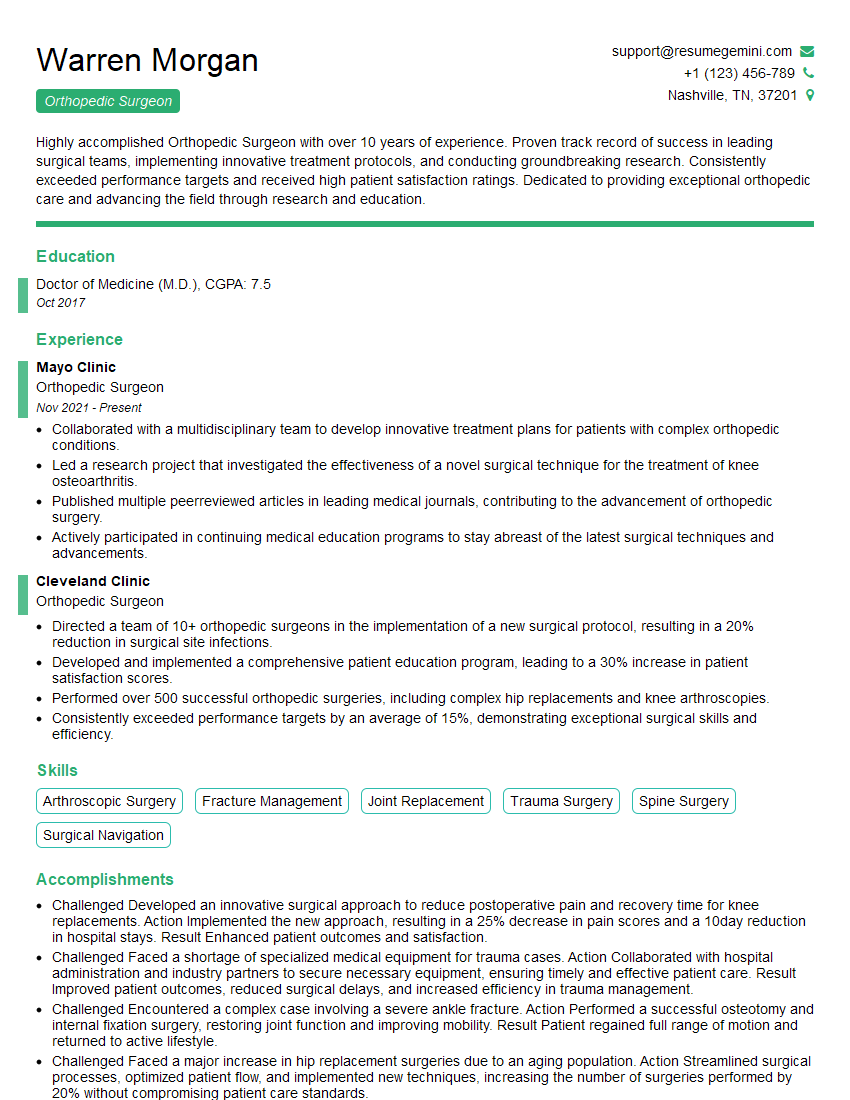
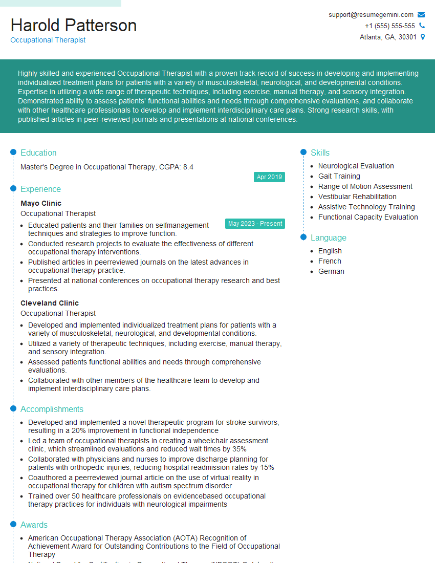
Mastering Fracture Repair is crucial for career advancement in orthopedics and related fields. A strong understanding of these concepts will significantly enhance your interview performance and open doors to exciting opportunities. To maximize your chances of success, invest time in creating a professional and ATS-friendly resume that highlights your skills and experience. ResumeGemini is a trusted resource that can help you build a compelling resume, ensuring your qualifications stand out to potential employers. Examples of resumes tailored to Fracture Repair are available to guide you through the process.
Explore more articles
Users Rating of Our Blogs
Share Your Experience
We value your feedback! Please rate our content and share your thoughts (optional).
What Readers Say About Our Blog
To the interviewgemini.com Webmaster.
Very helpful and content specific questions to help prepare me for my interview!
Thank you
To the interviewgemini.com Webmaster.
This was kind of a unique content I found around the specialized skills. Very helpful questions and good detailed answers.
Very Helpful blog, thank you Interviewgemini team.
