Preparation is the key to success in any interview. In this post, we’ll explore crucial Hallux Valgus Repair interview questions and equip you with strategies to craft impactful answers. Whether you’re a beginner or a pro, these tips will elevate your preparation.
Questions Asked in Hallux Valgus Repair Interview
Q 1. Describe the different surgical techniques used in Hallux Valgus repair.
Hallux valgus repair, or bunion surgery, encompasses a variety of surgical techniques aimed at correcting the deformity of the big toe joint. The choice of technique depends on several factors, including the severity of the deformity, the patient’s age, activity level, and overall health. Common procedures include osteotomies (bone cuts), soft tissue procedures, and arthrodesis (joint fusion).
- Osteotomies: These involve reshaping the bones of the big toe to realign the joint. Different types include Chevron, Scarf, Akin, and base wedge osteotomies.
- Soft Tissue Procedures: These address soft tissue imbalances contributing to the bunion. Examples include capsular releases and tendon transfers.
- Arthrodesis: This involves fusing the bones of the big toe joint to eliminate movement. It’s typically reserved for severe arthritis or failed prior surgeries.
Each technique has its own nuances and indications, which we’ll explore further.
Q 2. What are the indications and contraindications for each technique?
The choice of surgical technique is highly individualized.
- Osteotomies (Chevron, Scarf, Akin): Indicated for moderate deformities with relatively good joint cartilage. Contraindicated in severe arthritis, significant bone loss, or inadequate bone quality.
- Soft Tissue Procedures: Often used in conjunction with osteotomies, especially in cases with significant soft tissue contractures or to address associated conditions like hallux limitus (limited big toe movement). Contraindicated when the underlying bony deformity is severe.
- Arthrodesis: Indicated for severe arthritis, failed prior surgeries, and cases where joint preservation is not possible. Contraindicated in young, active patients who require significant range of motion in the big toe.
For example, a young, active patient with a moderate bunion and good joint cartilage would likely be a candidate for a Chevron or Akin osteotomy. An older patient with severe arthritis might be better suited for arthrodesis.
Q 3. Explain the principles of Akin osteotomy.
The Akin osteotomy involves a distal (farther from the body) osteotomy of the proximal phalanx (the bone of the big toe closest to the foot). A small wedge of bone is removed, and the bone is then rotated to correct the angle of the big toe.
The principle lies in correcting the angulation of the proximal phalanx to realign it with the metatarsal bone (long bone of the foot). This realignment reduces the prominence of the bunion and improves the overall alignment of the toe. A small screw or pin is commonly used to hold the bone fragments in the correct position while it heals. This technique is often preferred for patients with a relatively flexible deformity and well-preserved cartilage.
Q 4. What are the advantages and disadvantages of a Chevron osteotomy?
The Chevron osteotomy is a more proximal (closer to the body) osteotomy of the first metatarsal. A V-shaped cut is made, and the bone fragments are realigned and fixed.
- Advantages: Relatively simple technique, less bone resection needed compared to some other osteotomies, suitable for moderate deformities.
- Disadvantages: Potential for malunion (improper bone healing), increased risk of shortening the metatarsal, less correction of the intermetatarsal angle (the angle between the big and second toe bones) compared to other techniques. It might not be ideal for very severe deformities or those with significant joint arthritis.
For instance, a Chevron osteotomy may be a good option for a patient with a moderate bunion and good bone quality, but it might not be suitable for a patient with significant arthritis or a very severe deformity.
Q 5. How do you manage post-operative pain and swelling?
Post-operative pain and swelling are common after Hallux Valgus repair. Management strategies are crucial for patient comfort and successful recovery.
- Pain Management: This usually involves a combination of oral analgesics (pain relievers) such as NSAIDs (nonsteroidal anti-inflammatory drugs) and, in some cases, opioids for more severe pain. Regular ice packs to the affected area can also help reduce inflammation and pain.
- Swelling Management: Elevation of the foot above the heart helps reduce swelling. Compression bandages and intermittent pneumatic compression devices can further assist in managing swelling. Regular physical therapy is also implemented to improve range of motion and reduce swelling.
It’s important to carefully monitor pain levels and swelling, adjusting medication and other interventions as needed. Open communication with the patient is crucial throughout the healing process.
Q 6. What are the common complications associated with Hallux Valgus repair?
While Hallux Valgus repair is generally successful, several complications can arise.
- Infection: A serious complication requiring prompt treatment with antibiotics and potentially surgical debridement (removal of infected tissue).
- Nonunion/Malunion: Failure of the bone fragments to heal properly or heal in an incorrect position, requiring revision surgery.
- Stiffness: Limited range of motion in the big toe joint, often addressed with physical therapy.
- Recurrence: Return of the bunion deformity, potentially requiring further surgery.
- Nerve Damage: Numbness or tingling in the toes, which may resolve spontaneously or require further intervention.
- Hardware Problems: Problems with screws or pins used for fixation, such as loosening or breakage.
Careful surgical technique, meticulous post-operative care, and early recognition of potential problems are crucial to minimizing these risks.
Q 7. How do you address infection following Hallux Valgus surgery?
Infection after Hallux Valgus surgery is a serious concern that requires immediate attention. Management involves a multi-pronged approach.
- Wound Assessment: Thorough examination of the surgical site to assess the extent of the infection.
- Antibiotic Therapy: Intravenous antibiotics are usually administered to effectively target the infection. The specific antibiotics used will depend on the type of bacteria identified via culture.
- Surgical Debridement: In more severe cases, surgical debridement may be necessary to remove infected tissue and allow for adequate drainage. This may involve reopening the incision and thoroughly cleaning the wound.
- Wound Care: Regular wound dressing changes are essential to prevent further infection and promote healing.
Prompt diagnosis and aggressive treatment are crucial to prevent the spread of infection and potential life-threatening complications.
Q 8. Describe your approach to pre-operative patient education.
Pre-operative patient education for Hallux Valgus repair is crucial for setting realistic expectations and ensuring a smooth recovery. My approach involves a multi-faceted strategy focusing on clear communication and individualized attention.
First, I thoroughly explain the condition, demonstrating its impact on the foot’s biomechanics using models and imaging. This helps patients understand why they’re experiencing pain and limitations. I then discuss the surgical options available, detailing the pros and cons of each procedure – including minimally invasive options like Chevron osteotomy or Akin osteotomy versus more extensive procedures like a Lapidus bunionectomy. I make sure to highlight the potential risks and complications associated with any surgery, emphasizing the importance of realistic expectations regarding pain management, recovery time, and potential limitations.
A critical aspect is empowering the patient to actively participate in their recovery. We discuss post-operative care in detail, including pain management strategies, physical therapy expectations, and the need for appropriate footwear. I provide detailed written instructions and visual aids to ensure clear understanding. Finally, I encourage questions and provide ample time for discussion to address any concerns the patient may have.
For example, I often use visual aids like anatomical models to show the deformity and explain how the surgery will correct it. This enhances understanding and reduces anxiety. This personalized approach ensures the patient feels informed and confident throughout the process, leading to better compliance and a smoother recovery.
Q 9. How do you assess the patient’s functional outcome post-surgery?
Assessing functional outcome post-Hallux Valgus surgery involves a comprehensive evaluation that goes beyond just pain reduction. We utilize a multi-modal approach to gauge success.
- Pain Scales: Visual Analog Scales (VAS) or Numerical Rating Scales (NRS) are used to quantify pain levels at rest and during activity.
- Range of Motion: We measure the range of motion in the metatarsophalangeal (MTP) joint of the great toe to assess joint flexibility.
- Gait Analysis:Observing the patient’s gait provides insights into their ability to walk comfortably and without limping. We assess stride length, cadence, and any deviations in the foot’s mechanics.
- Functional Outcome Scores: Standardized questionnaires like the American Orthopaedic Foot and Ankle Society (AOFAS) Hallux Metatarsophalangeal-Interphalangeal (MTP-IP) score are used to objectively measure the patient’s functional status and overall satisfaction. This score assesses pain, function, and quality of life.
- Imaging: Post-operative X-rays are analyzed to evaluate the bony alignment and assess the success of the osteotomy or other surgical correction.
By combining these methods, we get a complete picture of the patient’s functional recovery and ensure their surgery was successful in restoring their mobility and quality of life.
Q 10. What are the different types of implants used in Hallux Valgus surgery?
The choice of implant in Hallux Valgus surgery depends on the specific surgical technique and the surgeon’s preference. However, several common implants are used:
- Screws: Bioabsorbable screws are often used to fix the osteotomy in place. These screws gradually dissolve over time, eliminating the need for a second surgery for removal. Titanium screws, which are non-absorbable, are also commonly used. Their advantage is increased strength.
- Plates: While less common for simpler osteotomies, plates can provide additional stability, especially in complex cases or in patients with osteoporotic bone. They are typically made of titanium.
- K-wires (Kirschner wires): These are thin, smooth wires temporarily used for fixation, often removed after a few weeks.
- Pins: Similar to K-wires, pins provide temporary stabilization. The difference is mainly in diameter.
- Interlocking Nails: Used in more complex cases, these offer superior stability and strength.
The selection of an implant is a crucial aspect of the surgical planning. The chosen implant must match the patient’s overall health condition, the type of correction performed, and the surgeon’s experience and surgical preferences.
Q 11. Explain the use of screws vs. plates in Hallux Valgus repair.
The decision to use screws versus plates in Hallux Valgus repair is primarily determined by the type of osteotomy performed and the surgeon’s preference. Simpler osteotomies, such as a Chevron or Akin osteotomy, usually require only screws for fixation. Screws provide adequate stability for these procedures because the bone fragments are relatively small and the forces acting upon them are relatively low.
Plates, on the other hand, are generally reserved for more complex procedures or those with increased risk of non-union (failure of the bone fragments to heal), such as a Scarf or Lapidus bunionectomy. Plates provide additional strength and rigidity, holding the bone fragments firmly together. They are particularly useful in situations where there’s a large gap between the bone fragments or when the bone quality is compromised.
The choice is ultimately a clinical judgment made on a case-by-case basis, balancing the risks and benefits of each approach. In some cases, a combination of screws and plates might be used for optimum stability.
Q 12. How do you manage non-union after Hallux Valgus surgery?
Non-union after Hallux Valgus surgery, while uncommon with proper surgical technique, can be a challenging complication. Management strategies depend on the severity and cause of the non-union. The diagnosis is confirmed with x-rays.
Conservative Management: In early cases, or where the gap is small, we might try conservative management involving immobilization with a cast or boot for an extended period, often supplemented with electrical stimulation or ultrasound therapy to promote bone healing. Regular radiographic monitoring is crucial to assess progress.
Surgical Intervention: If conservative management fails, revision surgery becomes necessary. This might involve:
- Bone Grafting: Adding bone graft material to the non-union site provides additional cells and scaffolding to promote bone regeneration.
- Internal Fixation: Reinforcing the fixation using plates and screws, or even an interlocking nail, to provide greater stability.
- Excision of Non-Union: Removing the non-union and performing a bone graft is sometimes necessary, especially for larger non-unions.
The specific approach is tailored to the individual patient, considering factors such as the age, bone quality, and the extent of the non-union.
Q 13. What is your experience with minimally invasive techniques?
Minimally invasive techniques for Hallux Valgus repair are becoming increasingly popular due to their potential advantages, including smaller incisions, reduced pain, faster recovery times, and less scarring. I have extensive experience with these techniques.
My approach often involves performing a percutaneous osteotomy – essentially, using smaller incisions to access the bone and perform the correction. This minimizes soft tissue damage and reduces post-operative inflammation. I use specialized instruments designed for these procedures, including small-diameter drills and specialized guiding tools to help maintain accuracy. Advanced imaging, such as fluoroscopy, aids in precise placement of implants.
While minimally invasive techniques offer several advantages, it’s crucial to emphasize that not every case is suitable for this approach. Cases with severe deformities or significant bone damage often require a more open procedure. Patient selection is key to the success of a minimally invasive approach. Furthermore, careful patient education is critical, as recovery might still involve some limitations, even with minimally invasive procedures. I make sure to manage patient expectations appropriately.
Q 14. Describe your approach to revision surgery for Hallux Valgus.
Revision surgery for Hallux Valgus is a more complex undertaking than the initial procedure, often requiring a more extensive approach. My strategy starts with a thorough assessment to identify the cause of the failure. This involves reviewing the patient’s history, examining the foot, and evaluating the radiographic images.
Common reasons for revision surgery include non-union, malunion (improper bone healing), recurrent deformity, and implant failure. Understanding the root cause is key to planning the revision surgery. Once identified, I will tailor the surgical approach to address the specific problem.
Revision surgery may involve removing existing implants, correcting any residual deformities, performing bone grafting if necessary, and using more robust fixation techniques to ensure a stable correction. The specific technique would depend on the nature of the problem – for example, a failed osteotomy might require a different osteotomy or even a more extensive procedure like a Lapidus bunionectomy.
Post-operative care after revision surgery is often more intensive, requiring closer monitoring and potentially longer periods of immobilization and physical therapy. Detailed post-operative instructions are provided, and close follow-up is maintained to assess healing progress and address any potential concerns.
Q 15. How do you address lateral column instability during Hallux Valgus repair?
Addressing lateral column instability during Hallux Valgus repair is crucial for long-term success. Instability often manifests as a collapsing first ray, leading to recurrence of the bunion deformity. We achieve stability through a combination of techniques, carefully selected based on the patient’s specific anatomy and the severity of the instability.
First metatarsal osteotomy: This is a cornerstone of many procedures. By correcting the medial deviation of the first metatarsal, we improve the overall alignment and reduce stress on the lateral column. The choice of osteotomy (e.g., scarf, chevron, closing wedge) depends on the precise deformity.
Lateral column support: Procedures like lateral soft tissue releases or even a lateral capsulotomy may be necessary to address any contractures or tightness that contribute to instability. We might also consider a lateral closing wedge osteotomy of the second metatarsal in severe cases to improve the overall stability of the lateral column.
Prophylactic procedures: In cases of significant instability, I often incorporate procedures like a fibular sesamoid stabilization. This aims to enhance the mechanical integrity of the first metatarsophalangeal joint and reduce the chance of recurrence.
Think of it like this: the metatarsals are like the beams of a house. If one beam (the first metatarsal) is leaning, it puts extra stress on the others. We need to straighten that beam and ensure the supporting structure (the lateral column) is strong enough to bear the weight, thus preventing the entire ‘house’ from collapsing again.
Career Expert Tips:
- Ace those interviews! Prepare effectively by reviewing the Top 50 Most Common Interview Questions on ResumeGemini.
- Navigate your job search with confidence! Explore a wide range of Career Tips on ResumeGemini. Learn about common challenges and recommendations to overcome them.
- Craft the perfect resume! Master the Art of Resume Writing with ResumeGemini’s guide. Showcase your unique qualifications and achievements effectively.
- Don’t miss out on holiday savings! Build your dream resume with ResumeGemini’s ATS optimized templates.
Q 16. What are your preferred methods for soft tissue management in Hallux Valgus surgery?
Soft tissue management is paramount in Hallux Valgus surgery, influencing both the immediate outcome and long-term stability. My approach emphasizes a balanced strategy, focusing on releasing tight structures while preserving critical soft tissue integrity.
Medial capsular release: This addresses the contracted medial capsule, which is a major contributor to the bunion deformity. The goal isn’t to remove excessive tissue but to carefully release the tight fibers to restore normal joint mechanics.
Adductor hallucis release: Sometimes, the adductor hallucis muscle can pull on the first metatarsal, exacerbating the deformity. A selective release of the adductor hallucis tendon can alleviate this tension.
Minimally invasive techniques: I prefer minimally invasive approaches whenever feasible, using smaller incisions and instruments to reduce soft tissue trauma and scarring. This allows for faster healing and less post-operative pain. Percutaneous techniques may also be used, further minimizing the soft tissue damage.
It’s about precision and balance. Over-aggressive soft tissue release can lead to instability, while insufficient release will hinder correction and increase the risk of recurrence. I always tailor my approach to the individual patient’s specific soft tissue needs.
Q 17. Describe the role of imaging in the diagnosis and treatment of Hallux Valgus.
Imaging plays a vital role in both diagnosing and guiding treatment of Hallux Valgus. It helps visualize the bony and soft tissue components of the deformity and allows for accurate preoperative planning.
Weight-bearing anteroposterior (AP) and lateral radiographs: These are essential for assessing the metatarsophalangeal (MTP) joint angle (hallux valgus angle), the intermetatarsal angle (IMA), and the first metatarsal-proximal phalanx angle (PASA). These angles quantify the severity of the deformity.
Weight-bearing lateral radiographs: These assess the dorsal migration of the sesamoids and the height of the first metatarsal head, crucial in planning osteotomy selection.
Computed tomography (CT) scans: While not routinely used, CT scans are helpful in complex cases to further evaluate the bone structure and relationships.
Magnetic resonance imaging (MRI): MRI is less frequently used in straightforward cases but can be helpful in identifying soft tissue pathology (e.g., tendonitis, bursitis) that might contribute to symptoms and inform surgical strategy.
For instance, a severe IMA might suggest the need for a more aggressive osteotomy to correct the deformity effectively. Pre-operative imaging enables us to predict the needed correction and plan a customized surgical approach.
Q 18. How do you differentiate between different types of bunions?
Bunion deformities aren’t all the same; classifying them helps tailor treatment. The classification often involves considering several aspects of the deformity.
Severity of the angle: The hallux valgus angle (HVA) and intermetatarsal angle (IMA) help categorize the severity of the bony deformity. Mild deformities might respond to conservative management, while severe ones often require surgery.
Associated deformities: Bunion deformities are often accompanied by other foot problems like hammertoes, bunions, or metatarsalgia. These co-morbidities must be considered when creating a treatment plan.
Presence of arthritis: Osteoarthritis can significantly affect the treatment strategy. Severe arthritis might require joint fusion or arthroplasty instead of simple osteotomy.
Patient-specific factors: Age, activity level, and occupation significantly influence treatment decisions. A young, active patient may need a different approach compared to an elderly patient with limited activity.
For example, a patient with a mild HVA and no associated deformities might benefit from conservative management. In contrast, a patient with a severe HVA, significant IMA, and associated hammertoes would likely require a more extensive surgical procedure.
Q 19. Explain the importance of patient selection in Hallux Valgus surgery.
Patient selection is critical for successful Hallux Valgus surgery. Surgery is not always the best option, and careful evaluation is essential to ensure the patient is a suitable candidate and that their expectations are realistic.
Realistic expectations: Patients should understand that surgery isn’t a perfect fix. Some degree of pain and stiffness is expected postoperatively, and complete pain relief might not be achievable.
Commitment to rehabilitation: Post-operative rehabilitation is crucial for a successful outcome. Patients must be committed to following the prescribed exercise program and adhering to weight-bearing restrictions.
Underlying medical conditions: Patients with severe comorbidities, poor circulation, or diabetes might have increased post-operative risks.
Conservative treatment failure: Surgery should only be considered after conservative treatments (e.g., shoe modifications, orthotics, physiotherapy) have failed to provide sufficient relief.
It’s a collaborative decision-making process. I take time to discuss the risks, benefits, and limitations of surgery with each patient to ensure they have a full understanding before proceeding.
Q 20. What are the key factors in determining surgical approach for Hallux Valgus?
Determining the best surgical approach for Hallux Valgus requires careful consideration of several factors. The goal is to select the technique that best addresses the specific deformities and provides the most stable and functional outcome.
Severity of the deformity: Mild deformities might respond well to a simple osteotomy, while more complex deformities might require a more extensive procedure, such as a distal metatarsal osteotomy (DMO), or even arthrodesis in severe cases.
Patient’s age and activity level: Younger, more active patients often benefit from procedures that provide greater stability and range of motion.
Associated deformities: The presence of other foot deformities influences the surgical strategy. For example, hammertoes might require concomitant correction.
Patient’s anatomy and bone quality: The patient’s foot anatomy and bone quality dictate the suitability of different osteotomy techniques. Certain osteotomies are better suited to specific bone types and shapes.
For instance, a minimally invasive percutaneous osteotomy might be suitable for a patient with a mild deformity and good bone quality, while a patient with a severe deformity and poor bone quality might be better served with an open osteotomy with internal fixation.
Q 21. How do you counsel patients on realistic expectations post-surgery?
Counseling patients on realistic expectations post-surgery is a crucial part of the process. Managing expectations helps to reduce patient anxiety and promotes a positive post-operative experience. Transparency and open communication are key.
Pain and discomfort: I explain that some pain and discomfort are expected immediately following surgery. We provide appropriate pain management strategies and discuss the timeline for pain relief.
Swelling and stiffness: I explain that swelling and stiffness are common post-operatively and will gradually resolve over time, often taking several weeks or even months.
Return to activity: I outline a realistic timeline for returning to normal activities, emphasizing the importance of gradual progression to avoid re-injury. This timeline varies greatly depending on the procedure and the individual patient’s healing.
Scarring: I address the issue of scarring, explaining that scars will fade over time, but some degree of visible scarring is to be expected with any surgery.
Potential complications: I discuss potential complications, such as infection, non-union, or nerve injury, although these are relatively rare with proper surgical technique and post-operative care.
I emphasize that the post-operative recovery process is a journey, and consistent follow-up and adherence to the rehabilitation program are crucial for achieving the best possible outcome.
Q 22. Discuss your experience with different types of anesthesia used in Hallux Valgus surgery.
Anesthesia selection for Hallux Valgus surgery depends heavily on patient factors, the complexity of the procedure, and surgeon preference. We typically utilize a combination of techniques to optimize patient comfort and surgical precision.
- Regional Anesthesia (Ankle Block): This involves injecting local anesthetic around the nerves supplying the foot and ankle. It provides excellent pain relief during and after surgery, minimizing the need for systemic opioids and reducing post-operative nausea and vomiting. It allows for a more awake and interactive patient, useful for assessing nerve function during the procedure.
- Spinal Anesthesia: This involves injecting local anesthetic into the spinal fluid, providing numbness from the waist down. It’s a good choice for patients with certain medical conditions that may preclude general anesthesia or when a longer procedure is anticipated. Recovery is typically faster than with general anesthesia.
- General Anesthesia: Reserved for patients who are unable to tolerate regional or spinal anesthesia, or for exceptionally complex procedures. General anesthesia provides complete unconsciousness and muscle relaxation but carries a higher risk of complications, such as respiratory issues or post-operative nausea.
The choice is a collaborative decision made with the patient, anesthesiologist, and myself, taking into account the patient’s medical history, preferences and the specifics of the surgical plan.
Q 23. What is your familiarity with different types of fixation devices?
Fixation devices in Hallux Valgus surgery are crucial for maintaining the correction achieved during the osteotomy (bone cutting). The choice depends on the specific surgical technique employed and the individual patient’s anatomy. We use a variety of options, balancing biocompatibility, strength and ease of removal.
- Screws: Bioabsorbable or titanium screws are commonly used to fixate the bone fragments after an osteotomy. Bioabsorbable screws dissolve over time, eliminating the need for a second procedure to remove them. Titanium screws are strong and durable but require a second surgery for removal.
- Plates: Plates, often made of titanium, offer increased stability, especially in complex cases with significant bone deformity or in patients with weaker bone density. They provide rigid fixation but require removal in a second surgical procedure.
- K-wires (Kirschner wires): These thin, flexible wires are sometimes used for temporary fixation, especially in conjunction with other methods. They are usually removed in a separate procedure.
- External Fixation: Less commonly used in routine Hallux Valgus repair, but reserved for highly complex cases where internal fixation is not feasible. This involves pins placed through the skin and connected to an external frame.
Selecting the appropriate fixation device requires careful consideration of bone quality, the extent of the correction needed, and the patient’s overall health.
Q 24. Describe your post operative rehabilitation protocol.
Post-operative rehabilitation is vital for optimal outcomes after Hallux Valgus surgery. It’s a structured process tailored to the individual patient’s progress and the specific surgical technique used.
- Immediate Post-op: Elevation of the foot to reduce swelling and pain management with medication. Use of a postoperative shoe or a removable cast for protection and support. Range-of-motion exercises (as prescribed).
- Weeks 2-6: Gradual weight-bearing as tolerated. Physical therapy to improve range of motion, strength, and flexibility. Pain management continues as needed.
- Weeks 6-12: Increasing weight-bearing and activity level. Continued physical therapy focusing on improving gait and function. Return to normal shoes as tolerated.
- Beyond 12 Weeks: Full weight-bearing and return to most activities. Ongoing physical therapy if needed to address any residual limitations.
Throughout the rehabilitation process, regular follow-up appointments are crucial to monitor healing, address any complications, and adjust the rehabilitation plan as necessary. Patient education and compliance are key to successful recovery. We emphasize a gradual, individualized approach to prevent complications and ensure a smooth recovery.
Q 25. What are the potential long term complications of Hallux Valgus surgery?
While Hallux Valgus surgery is generally successful, there is a risk of long-term complications. It is important to clearly explain these potential risks to the patient before the procedure.
- Recurrence: The bunion can return, although this is less common with modern surgical techniques. This risk is influenced by patient factors and compliance with post-operative instructions.
- Stiffness: Loss of joint motion in the great toe is a potential complication, although careful surgical technique and physiotherapy help minimize this risk.
- Pain: Persistence of pain, although uncommon with appropriate management, can be associated with nerve irritation or other factors.
- Infection: Infection is a rare but serious complication that can affect wound healing and potentially require further surgery.
- Non-union or Malunion: This refers to failure of the bones to heal properly after the osteotomy or the bones healing in a poor position. This is less frequent with modern techniques and implants.
- Hardware Complications: If metal implants are used, complications such as irritation, breakage, or protrusion can occur. These often necessitate revision surgery.
- Nerve Damage: While rare, damage to nearby nerves can result in numbness, tingling, or altered sensation.
Careful surgical planning, meticulous technique, and appropriate patient selection help to minimize the incidence of these complications.
Q 26. How do you manage patients with co-morbidities affecting Hallux Valgus repair?
Managing patients with co-morbidities requires a multidisciplinary approach and careful consideration of the patient’s overall health. This often involves close collaboration with other specialists, like cardiologists, diabetologists, or rheumatologists.
- Diabetes: Patients with diabetes are at higher risk of infection and delayed wound healing. Tight glycemic control before and after surgery is crucial, along with meticulous sterile technique during the operation. We may adjust the surgical plan and postoperative care based on the patient’s blood sugar levels.
- Peripheral Vascular Disease (PVD): Patients with PVD may have compromised blood flow to the foot, increasing the risk of infection and delayed healing. We might modify the surgical technique or consider alternative treatment options in severe cases.
- Rheumatoid Arthritis: Patients with rheumatoid arthritis may have joint instability and decreased bone density, potentially requiring modifications to the surgical technique and a more aggressive postoperative rehabilitation program.
- Osteoporosis: Patients with osteoporosis have weaker bones, increasing the risk of fractures or non-union. We may choose different fixation techniques or utilize bone grafting to improve healing.
A thorough preoperative assessment of the patient’s co-morbidities is essential to optimize the surgical plan, minimize risks, and tailor the postoperative care to their specific needs. Preoperative optimization of their condition is vital for success.
Q 27. Describe a challenging case of Hallux Valgus repair you have managed.
One particularly challenging case involved a 72-year-old female patient with severe hallux valgus deformity, significant osteoarthritis, and a history of multiple previous foot surgeries, including unsuccessful bunionectomy. She presented with severe pain, limited mobility, and significant overlying skin changes. Her bone quality was also compromised due to osteoporosis.
The challenge lay in achieving a stable correction without causing further damage to the already compromised joint and surrounding tissues. We opted for a modified chevron osteotomy with medial displacement of the first metatarsal and utilized a combination of small bioabsorbable screws and K-wires for fixation. The intricate procedure involved meticulous attention to soft tissue handling and meticulous bone preparation due to the poor bone stock. Her post-operative care included close monitoring for infection and a longer than usual period of non-weight-bearing to promote proper healing.
Ultimately, despite the challenges, the patient achieved a significant reduction in pain and an improvement in her mobility. This case highlights the importance of careful surgical planning and a customized approach when dealing with complex cases.
Q 28. What are your continuing education efforts in the field of foot and ankle surgery?
Continuous learning is paramount in the rapidly evolving field of foot and ankle surgery. I maintain my expertise through several avenues.
- Professional Societies: Active membership in the American Orthopaedic Foot & Ankle Society (AOFAS) and similar organizations keeps me abreast of the latest research, surgical techniques, and advancements through journals, conferences, and educational materials.
- Conferences and Workshops: Regular attendance at national and international conferences allows me to interact with leading experts, participate in hands-on workshops, and witness live surgeries.
- Peer Review and Collaboration: I actively engage in peer review of scientific publications, participate in case discussions with colleagues, and contribute to the collective knowledge within my professional network.
- Journal Articles and Publications: I stay current on the latest research by regularly reading peer-reviewed journals and contribute by authoring articles and presentations based on my clinical experience.
This multi-faceted approach ensures I consistently provide the highest quality and most up-to-date care for my patients.
Key Topics to Learn for Hallux Valgus Repair Interview
- Anatomy and Biomechanics of the First Metatarsophalangeal Joint: Understand the structures involved and how they contribute to the development of hallux valgus.
- Etiology and Pathophysiology of Hallux Valgus: Explore the various factors contributing to the condition, including genetic predisposition, footwear, and biomechanical factors.
- Clinical Presentation and Diagnosis: Learn to recognize the characteristic signs and symptoms, and understand the diagnostic methods used to assess the severity of hallux valgus.
- Conservative Management Options: Familiarize yourself with non-surgical treatments, including orthotics, footwear modifications, and physical therapy.
- Surgical Techniques for Hallux Valgus Repair: Gain a comprehensive understanding of various surgical procedures, including their indications, contraindications, and potential complications.
- Post-operative Care and Rehabilitation: Learn about the management of patients after surgery, including pain management, wound care, and physical therapy protocols.
- Complications and Management of Complications: Understand the potential complications associated with hallux valgus repair and the strategies for their prevention and management.
- Assessment of Surgical Outcomes: Learn about the methods used to assess the success of hallux valgus repair, including clinical examination, radiographic analysis, and patient-reported outcome measures.
- Current Research and Advancements in Hallux Valgus Repair: Stay updated on the latest research and technological advancements in the field.
- Problem-solving scenarios: Practice analyzing case studies involving different presentations and complications of hallux valgus, and determining appropriate treatment strategies.
Next Steps
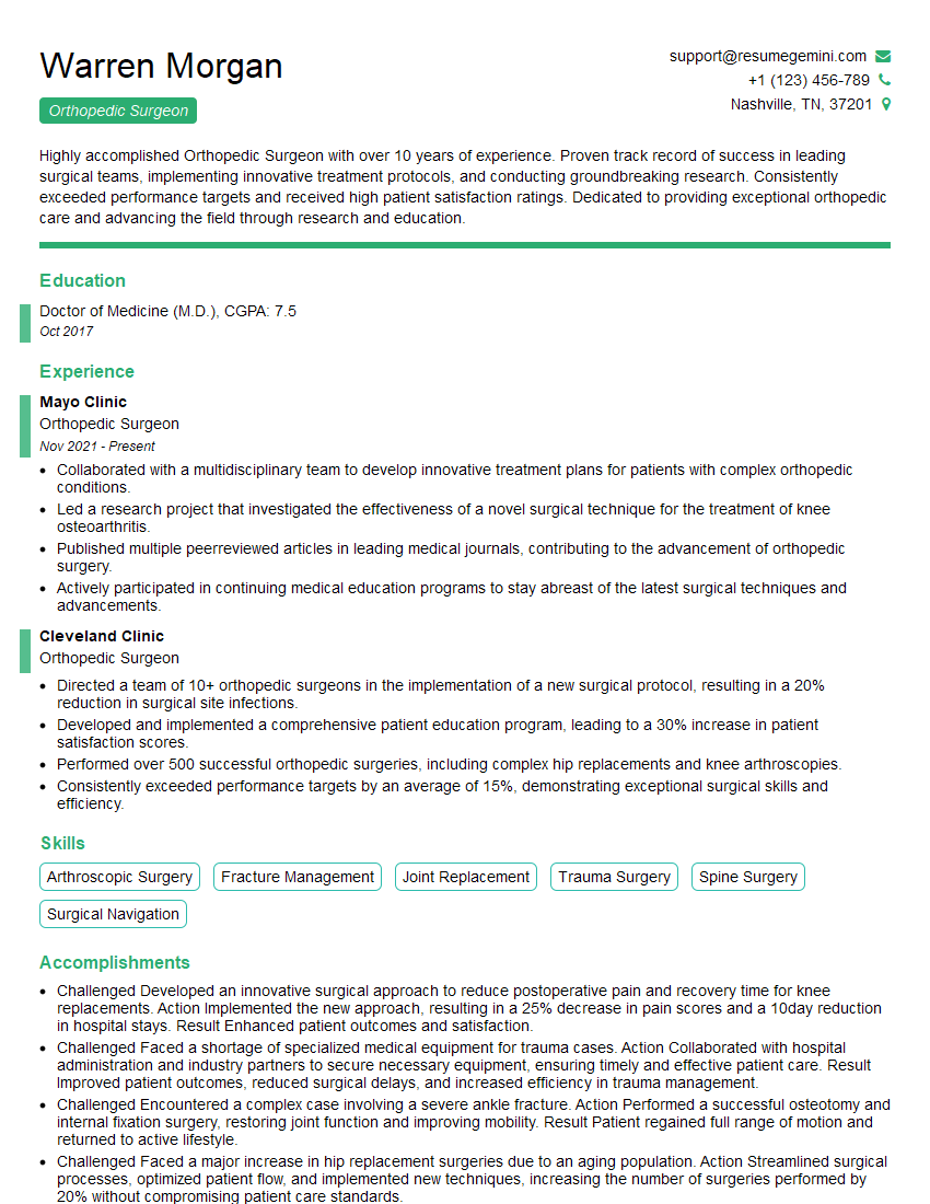
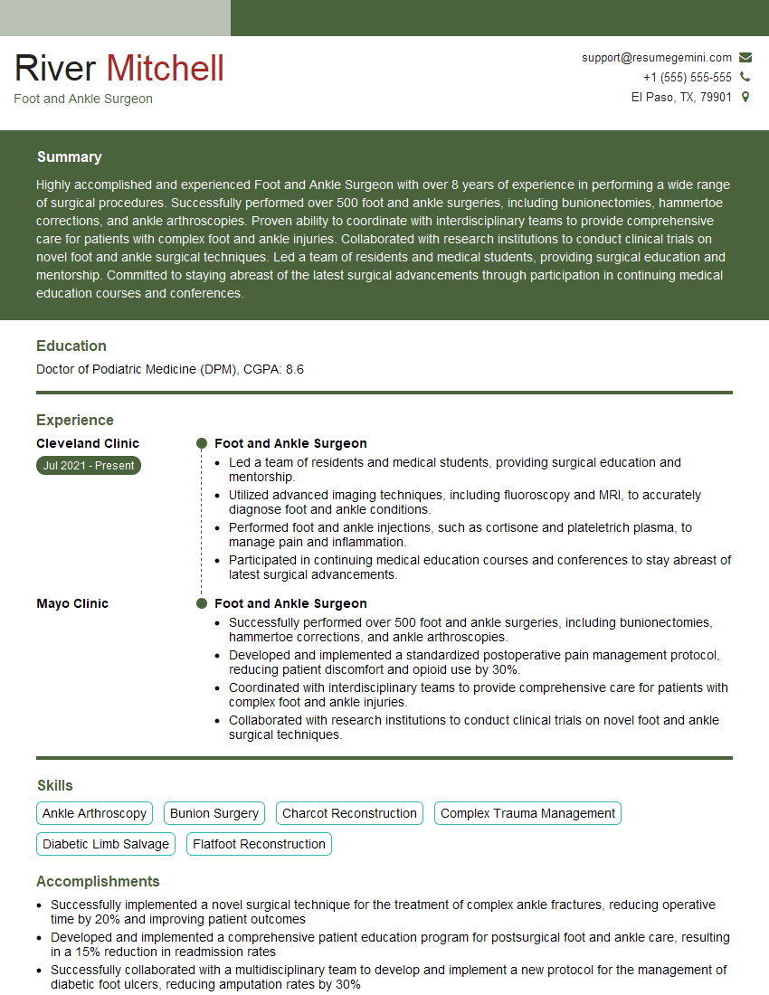
Mastering Hallux Valgus Repair significantly enhances your career prospects in orthopedics and podiatric surgery. Demonstrating a thorough understanding of this complex condition is crucial for securing a competitive advantage in the job market. To maximize your chances of landing your dream role, crafting a strong, ATS-friendly resume is essential. ResumeGemini is a trusted resource that can help you build a professional and impactful resume. They provide examples of resumes tailored to Hallux Valgus Repair, ensuring your qualifications are effectively highlighted for potential employers. Take the next step in your career journey and create a resume that showcases your expertise.
Explore more articles
Users Rating of Our Blogs
Share Your Experience
We value your feedback! Please rate our content and share your thoughts (optional).
What Readers Say About Our Blog
This was kind of a unique content I found around the specialized skills. Very helpful questions and good detailed answers.
Very Helpful blog, thank you Interviewgemini team.

