Preparation is the key to success in any interview. In this post, we’ll explore crucial Heel Pain Treatment interview questions and equip you with strategies to craft impactful answers. Whether you’re a beginner or a pro, these tips will elevate your preparation.
Questions Asked in Heel Pain Treatment Interview
Q 1. Describe the differential diagnosis for heel pain.
Heel pain can stem from various sources, making a precise diagnosis crucial. The differential diagnosis involves systematically considering several possibilities to arrive at the most likely cause. This process often begins with a thorough history and physical examination, focusing on the location, nature, and aggravating factors of the pain.
- Plantar Fasciitis: This is the most common cause, characterized by pain in the heel and arch, often worse in the morning or after periods of rest. It’s caused by inflammation of the plantar fascia, a thick band of tissue on the bottom of the foot.
- Heel Spur: A bony growth on the heel bone (calcaneus) can cause heel pain, often in conjunction with plantar fasciitis. However, a heel spur itself isn’t always painful; the pain is usually related to the associated inflammation.
- Achilles Tendinitis: Inflammation of the Achilles tendon, located at the back of the heel, presents with pain and stiffness in the back of the heel and may extend upwards along the tendon.
- Stress Fracture: A tiny crack in the heel bone, often caused by repetitive stress or overuse, can lead to localized heel pain, typically worse with activity.
- Tarsal Tunnel Syndrome: Compression of the tibial nerve as it passes through the tarsal tunnel on the inside of the ankle can cause heel pain and numbness extending to the sole of the foot.
- Sever’s Disease: This is a common cause of heel pain in children, resulting from inflammation of the growth plate at the back of the heel.
- Other conditions: Less common causes include nerve entrapment, arthritis (e.g., rheumatoid arthritis), and inflammatory conditions like reactive arthritis.
A comprehensive approach involving a detailed history, physical examination, and, in some cases, imaging studies is vital for accurate diagnosis and effective treatment.
Q 2. What are the common causes of plantar fasciitis?
Plantar fasciitis, the leading cause of heel pain, arises from repetitive stress and strain on the plantar fascia. Several factors contribute to its development:
- Overpronation: This is the excessive inward rolling of the foot during walking or running, placing extra stress on the plantar fascia.
- High-impact activities: Sports involving jumping, running, or prolonged standing can significantly increase the load on the plantar fascia.
- Improper footwear: Flat shoes or shoes lacking adequate arch support can worsen the condition.
- Tight calf muscles: Tightness in the calf muscles can pull on the heel and plantar fascia, increasing tension and inflammation.
- Obesity: Excess weight increases the stress placed on the plantar fascia.
- Age: The plantar fascia loses elasticity with age, making it more susceptible to injury.
- Underlying conditions: Conditions like diabetes and rheumatoid arthritis can increase susceptibility to plantar fasciitis.
Imagine the plantar fascia like a rubber band constantly stretched; over time, the repeated stress can lead to inflammation and pain. Addressing these contributing factors is key to effective management.
Q 3. Explain the role of imaging (X-ray, MRI, Ultrasound) in diagnosing heel pain.
Imaging plays a supportive role in diagnosing heel pain, helping to rule out certain conditions and confirm others. While not always necessary, it can be invaluable in complex cases.
- X-ray: Primarily used to identify bony abnormalities such as heel spurs or stress fractures. An X-ray clearly shows bone structure, providing a definitive answer regarding fractures or bone spurs. However, it won’t show soft tissue issues like plantar fasciitis.
- MRI (Magnetic Resonance Imaging): MRI provides detailed images of both bone and soft tissue, making it useful for visualizing the plantar fascia itself and identifying inflammation or tears. It’s particularly helpful in complex cases where other causes are suspected.
- Ultrasound: Ultrasound uses sound waves to create images of soft tissues. It’s less expensive than MRI but can also show plantar fascia thickening and inflammation. It’s also useful in guiding injections.
The choice of imaging modality depends on the clinical presentation and the suspicion of specific conditions. For example, if a stress fracture is suspected, an X-ray is sufficient. But if the diagnosis remains unclear after a physical exam, an MRI might be the next step.
Q 4. Outline your approach to managing acute plantar fasciitis.
Managing acute plantar fasciitis focuses on reducing pain and inflammation while promoting healing. My approach involves a combination of strategies:
- Rest and activity modification: Avoiding activities that aggravate the pain is crucial. This might involve reducing running or standing for long periods. Switching to lower-impact activities like swimming can be beneficial.
- Ice: Applying ice packs to the affected area for 15-20 minutes several times a day can help reduce inflammation.
- Elevation: Elevating the foot can help reduce swelling.
- Over-the-counter pain relief: Nonsteroidal anti-inflammatory drugs (NSAIDs) like ibuprofen or naproxen can help manage pain and inflammation.
- Night splints: These devices gently stretch the plantar fascia overnight, helping to reduce morning stiffness.
- Initial physical therapy: Gentle stretching exercises targeting the plantar fascia and calf muscles are critical. A physical therapist can guide you through appropriate exercises and help to determine if other soft tissue issues are contributing to the pain.
The goal is to quickly control pain and inflammation, allowing the patient to return to their normal activities as tolerated. Regular follow-up is important to monitor progress and adjust treatment as needed.
Q 5. Discuss conservative treatment options for heel pain (e.g., stretching, orthotics, medication).
Conservative treatment is the cornerstone of managing heel pain, and often successful for most people. A multi-faceted approach typically yields the best outcomes.
- Stretching exercises: Regular stretching of the plantar fascia and calf muscles is crucial. Examples include towel stretches (pulling the toes towards the shin with a towel), gastrocnemius and soleus stretches (stretching the calf muscles). These should be done multiple times throughout the day.
- Orthotics: Custom or over-the-counter orthotics can provide arch support and cushioning, helping to reduce stress on the plantar fascia. These act like shock absorbers, reducing the load on the injured tissue.
- Medication: NSAIDs are commonly used to relieve pain and inflammation. In some cases, your doctor might recommend corticosteroid injections directly into the plantar fascia to provide more targeted relief; however, this isn’t a long-term solution and repeated injections can weaken the plantar fascia.
- Physical Therapy: A physical therapist can provide a personalized exercise program, teach proper stretching techniques, and guide you through strengthening exercises for the muscles supporting the foot and ankle.
- Extracorporeal shockwave therapy (ESWT): This non-invasive treatment uses sound waves to stimulate healing in the plantar fascia. It’s an option for those who haven’t responded to other conservative treatments.
Conservative treatment requires patience and consistency. Results aren’t always immediate, and it may take several weeks or months to see significant improvement.
Q 6. When is surgical intervention indicated for heel pain?
Surgical intervention for heel pain is rarely necessary and is typically considered only after conservative treatment has failed for at least 6-12 months. This is a case-by-case decision made in consultation with the patient and based on several factors:
- Severe, persistent pain despite conservative management: If the pain significantly impairs daily life and doesn’t improve with conservative measures, surgery may be considered.
- Plantar fascia rupture: A complete tear of the plantar fascia often requires surgical repair to restore function.
- Failed conservative treatment for plantar fasciitis: After trying various conservative approaches without success, surgery can be an option, although less common.
- Other underlying conditions: In some cases, surgery might be necessary to address other structural issues causing heel pain, such as severe bone spurs or nerve entrapment.
It’s vital to exhaust all conservative options before considering surgery, as surgery carries its own risks and recovery time.
Q 7. Describe different surgical techniques used for heel pain.
Several surgical techniques are available for heel pain, each tailored to the specific cause and severity. The choice of procedure depends on the individual patient’s needs and the surgeon’s expertise.
- Plantar Fascia Release: This involves surgically releasing the tension in the plantar fascia, relieving pressure and reducing inflammation. It can be performed through a minimally invasive technique or through open surgery. A small incision is made, and the plantar fascia is released, allowing it to heal in a more relaxed position.
- Heel Spur Removal: If a heel spur is contributing significantly to the pain, it can be surgically removed. This procedure is often combined with plantar fascia release.
- Achilles Tendon Repair: If the pain stems from an Achilles tendon problem, surgical repair might be needed to address a tear or other damage.
- Neurolysis: For nerve entrapment causing heel pain, neurolysis involves releasing the compressed nerve to alleviate symptoms.
Post-operative rehabilitation is essential after any heel pain surgery. It typically involves a period of immobilization followed by gradual weight-bearing and physical therapy to restore function and prevent recurrence.
Q 8. What are the potential complications of heel pain treatment?
Complications from heel pain treatment are thankfully uncommon, but it’s crucial to be aware of potential issues. These can range from minor inconveniences to more serious problems. For instance, some patients experience temporary skin irritation or discomfort from topical medications or injections. Improperly fitted orthotics can lead to further foot problems, such as calluses or blisters. In rare cases, steroid injections, while often effective, can cause local tissue atrophy or even infection if proper sterile techniques aren’t followed. Surgical intervention, while a last resort, carries inherent risks like infection, nerve damage, or delayed healing. Therefore, a thorough assessment and careful treatment plan are paramount to minimize any potential adverse effects.
Example: A patient receiving corticosteroid injections might experience temporary skin thinning at the injection site. This typically resolves, but it highlights the need for monitoring and adherence to post-injection care instructions.
Q 9. How do you assess the effectiveness of heel pain treatment?
Assessing the effectiveness of heel pain treatment involves a multi-faceted approach, going beyond simply asking, ‘Does it hurt less?’ We utilize several methods. Firstly, we track the patient’s pain levels using validated pain scales like the Visual Analog Scale (VAS) or Numeric Rating Scale (NRS) at regular intervals. Secondly, we observe improvements in functional abilities. Can the patient walk, run, or stand for longer periods without pain? Thirdly, we evaluate the range of motion in the ankle and foot. Are there any lingering stiffness or limitations? Finally, we examine the physical examination findings – is there a reduction in tenderness, inflammation, or swelling? Combining these assessments provides a comprehensive picture of treatment success. If progress is unsatisfactory, we reassess the diagnosis and adjust the treatment strategy.
Example: A patient initially reported a pain level of 8/10 on the VAS. After four weeks of physical therapy and orthotic use, their pain level is 2/10, they can walk a mile without significant discomfort, and their ankle range of motion has improved. This indicates successful treatment.
Q 10. Explain the role of patient education in managing heel pain.
Patient education is the cornerstone of successful heel pain management. It empowers patients to actively participate in their recovery. We explain the underlying causes of their heel pain, the proposed treatment plan, and the importance of adherence. We discuss potential lifestyle modifications, including appropriate footwear, weight management strategies if obesity is a contributing factor, and the importance of stretching and strengthening exercises. We also educate them on recognizing warning signs, such as increased pain, persistent swelling, or signs of infection, and when to seek immediate medical attention. The goal is to foster a collaborative relationship built on mutual understanding and shared responsibility for their recovery. Providing written materials, including exercise diagrams and self-care instructions, complements verbal explanations.
Example: A patient with plantar fasciitis is educated on the importance of wearing supportive footwear, performing regular calf stretches, and using ice to manage inflammation. They’re also given a handout illustrating the correct stretching techniques.
Q 11. How do you manage heel pain in patients with comorbidities (e.g., diabetes, arthritis)?
Managing heel pain in patients with comorbidities requires a more nuanced approach. For example, in patients with diabetes, meticulous foot care is crucial to prevent complications like infection or ulceration. We carefully assess for signs of neuropathy and vascular disease, which can affect healing and increase the risk of complications. Treatment might involve conservative methods like custom orthotics, modified footwear, and meticulous foot hygiene, but we closely monitor their progress due to potentially slower healing times. Similar caution is needed for patients with arthritis. We carefully consider the effects of medication, potential joint instability, and the patient’s overall functional status when developing a treatment plan. A multidisciplinary approach, involving podiatrists, endocrinologists (for diabetes), and rheumatologists (for arthritis), may be necessary to provide optimal care.
Example: A diabetic patient with heel pain might require regular foot exams to check for signs of infection or ulceration, along with modified footwear and meticulous wound care if a minor lesion develops. Treatment must balance pain relief with the preservation of foot health.
Q 12. Describe your experience with different types of orthotics for heel pain.
My experience encompasses a wide range of orthotics, each suited for specific needs. I frequently prescribe custom-molded orthotics, which are created from plaster casts of the patient’s feet to provide highly individualized support. These are particularly useful for patients with complex foot deformities or severe plantar fasciitis. Pre-fabricated orthotics, while less expensive, are also effective for many individuals, offering readily available options for various foot types and levels of support. I often utilize heel cups or heel lifts to provide targeted cushioning and shock absorption in the heel region. For patients with significant arch problems, arch supports are essential for proper weight distribution. The choice depends on the individual’s needs, the severity of their condition, and their budget. Patient feedback on comfort and effectiveness plays a key role in determining the right orthotic solution.
Example: A patient with a high arch and significant plantar fasciitis would benefit most from a custom orthotic with increased arch support and cushioning in the heel. A patient with mild heel pain might find sufficient relief from a simple heel cup.
Q 13. What are the common causes of heel spur?
Heel spurs, bony projections on the heel bone, often develop as a result of repetitive strain and micro-tears in the plantar fascia. These micro-tears cause inflammation and eventually calcium deposits to form, leading to a spur. Conditions like plantar fasciitis frequently accompany heel spurs. Other contributing factors include biomechanical abnormalities like overpronation (the inward rolling of the foot during gait), tight calf muscles, excessive weight, and certain footwear. Essentially, anything that increases stress on the plantar fascia can contribute to heel spur formation. Importantly, while heel spurs might be visible on imaging, they don’t always cause pain. Pain is often related to the associated inflammation, not the spur itself.
Example: A runner who consistently trains on hard surfaces and wears inadequate footwear is at higher risk of developing heel spurs and plantar fasciitis due to the repetitive stress on the plantar fascia.
Q 14. How do you differentiate between plantar fasciitis and Achilles tendinitis?
Differentiating between plantar fasciitis and Achilles tendinitis involves careful examination and consideration of symptoms. Plantar fasciitis, inflammation of the plantar fascia, typically causes pain in the heel, often worse in the morning or after periods of rest. The pain is typically located at the bottom of the heel and may radiate along the arch. Achilles tendinitis, on the other hand, involves inflammation of the Achilles tendon, resulting in pain located in the back of the heel and sometimes higher up the calf. The pain is often worse after exercise or activity and may be accompanied by stiffness. Palpation (physical examination) can help pinpoint the area of tenderness – on the bottom of the heel for plantar fasciitis and the back of the heel for Achilles tendinitis. Imaging, like ultrasound or MRI, might be used to confirm the diagnosis in unclear cases. Both conditions can co-exist, making accurate diagnosis even more important for effective treatment.
Example: A patient experiencing sharp pain in their heel first thing in the morning, relieved by activity, with tenderness along the bottom of their heel and arch, is more likely to have plantar fasciitis. In contrast, a patient experiencing pain in their back heel after running, with stiffness, and tenderness along the Achilles tendon, is more likely to have Achilles tendinitis.
Q 15. What are the key elements of a comprehensive physical examination for heel pain?
A comprehensive physical examination for heel pain starts with a thorough history taking, understanding the patient’s symptoms, onset, and any aggravating or relieving factors. Then, a meticulous physical exam follows. This involves:
Visual Inspection: Assessing the heel for swelling, redness, deformity, and any visible signs of injury.
Palpation: Gently feeling the heel and surrounding tissues to identify areas of tenderness, particularly the plantar fascia, heel bone (calcaneus), and surrounding muscles.
Range of Motion (ROM): Evaluating the flexibility of the ankle and foot, checking for limitations in dorsiflexion (bringing toes towards shin) and plantarflexion (pointing toes down). Restricted ROM can indicate plantar fasciitis or Achilles tendon issues.
Neurological Examination: Checking for any nerve involvement causing numbness, tingling, or weakness in the foot. This is important to rule out conditions like nerve entrapment.
Gait Analysis: Observing the patient’s walking pattern to identify any compensatory movements or limping that could indicate underlying problems.
Special Tests: Specific tests may be performed, such as the Thompson test for Achilles tendon rupture (squeezing the calf muscle to see if the foot plantarflexes) or a windlass test to assess plantar fascia tension.
For example, a patient presenting with sharp heel pain upon first steps in the morning, that gradually improves throughout the day, suggests plantar fasciitis. A physical exam would likely reveal tenderness along the plantar fascia and limited ankle dorsiflexion.
Career Expert Tips:
- Ace those interviews! Prepare effectively by reviewing the Top 50 Most Common Interview Questions on ResumeGemini.
- Navigate your job search with confidence! Explore a wide range of Career Tips on ResumeGemini. Learn about common challenges and recommendations to overcome them.
- Craft the perfect resume! Master the Art of Resume Writing with ResumeGemini’s guide. Showcase your unique qualifications and achievements effectively.
- Don’t miss out on holiday savings! Build your dream resume with ResumeGemini’s ATS optimized templates.
Q 16. Discuss the role of physical therapy in the management of heel pain.
Physical therapy plays a crucial role in heel pain management, often forming the cornerstone of conservative treatment. It focuses on restoring function, reducing pain, and preventing recurrence. Key components include:
Stretching Exercises: Specifically targeting the plantar fascia, Achilles tendon, calf muscles, and surrounding tissues. These help improve flexibility and reduce muscle tightness, which are major contributors to heel pain. Examples include calf stretches, plantar fascia stretches (using a towel or rolling a ball under the foot), and toe curls.
Strengthening Exercises: Building strength in the muscles supporting the foot and ankle, such as the calf muscles and intrinsic foot muscles. This helps provide better support and stability to the heel.
Manual Therapy: Techniques like soft tissue mobilization and joint mobilization can help to improve tissue mobility and reduce pain and inflammation.
Orthotics and Footwear Advice: Custom or over-the-counter orthotics can help to provide better support to the arch and heel, reducing strain on the plantar fascia. Advice on appropriate footwear is crucial to minimize stress and irritation.
Modalities: Therapeutic modalities like ultrasound, ice, and heat can be used to reduce inflammation and pain.
For instance, a patient with plantar fasciitis would benefit from a program including plantar fascia stretches, calf raises, and the use of supportive orthotics. Progress is closely monitored, and exercises are adjusted as needed.
Q 17. Describe different types of injections used in managing heel pain.
Various injections can be used to manage heel pain, primarily targeting inflammation and pain relief. These include:
Corticosteroid Injections: These are the most common type of injection, effectively reducing inflammation in conditions like plantar fasciitis. They offer temporary but significant pain relief. However, repeated injections can weaken the tissues over time.
Platelet-Rich Plasma (PRP) Injections: PRP is prepared from the patient’s own blood, concentrating platelets that contain growth factors to promote healing and tissue regeneration. It’s used for chronic heel pain conditions that haven’t responded to conservative treatment.
Hyaluronic Acid Injections: This viscosupplementation therapy is sometimes used for heel pain associated with osteoarthritis, improving joint lubrication and reducing pain.
Local Anesthetic Injections: These provide temporary pain relief, often used diagnostically to pinpoint the source of pain or in conjunction with other injections.
The choice of injection depends on the underlying diagnosis and the patient’s response to conservative treatment. For example, a patient with recalcitrant plantar fasciitis might be a candidate for a PRP injection after failing to respond adequately to physical therapy and corticosteroid injections.
Q 18. Explain the use of extracorporeal shockwave therapy (ESWT) for heel pain.
Extracorporeal shockwave therapy (ESWT) is a non-invasive treatment that uses sound waves to stimulate healing in the tissues. For heel pain, it’s often used for chronic plantar fasciitis and other conditions that haven’t responded to other conservative therapies. The shockwaves are delivered through the skin to the affected area, promoting tissue regeneration, reducing inflammation, and pain relief. The exact mechanism of action is still under research, but it’s thought to enhance blood flow and stimulate the body’s natural healing processes.
Typically, patients receive several ESWT sessions over a few weeks. The procedure is usually well-tolerated, although some patients may experience mild discomfort or bruising at the treatment site. ESWT is not suitable for all patients, and a thorough assessment is necessary to determine suitability. For example, it might not be appropriate for patients with active infections or bleeding disorders.
Q 19. What are the common causes of heel pain in athletes?
Athletes frequently experience heel pain due to the repetitive stress and impact forces on their feet. Common causes include:
Plantar Fasciitis: This is the most common cause, resulting from repetitive strain on the plantar fascia, often exacerbated by activities like running, jumping, and high-impact sports.
Achilles Tendinitis: Inflammation of the Achilles tendon, often related to overuse, improper footwear, or inadequate warm-up.
Stress Fractures: Tiny cracks in the heel bone, often caused by repetitive impact forces from running or jumping.
Heel Spurs: Bony growths on the heel bone that can irritate the surrounding tissues, often asymptomatic but can cause pain if they irritate the plantar fascia.
Sever’s Disease: A common heel pain condition in young, growing athletes, involving inflammation of the growth plate at the back of the heel.
For example, a long-distance runner may develop plantar fasciitis due to repetitive impact on their feet, while a basketball player might experience Achilles tendinitis from frequent jumping.
Q 20. How do you manage heel pain during pregnancy?
Heel pain during pregnancy is relatively common, often attributed to hormonal changes, weight gain, and altered posture. Management focuses on reducing stress on the feet and providing pain relief. Strategies include:
Supportive Footwear: Wearing comfortable shoes with good arch support and cushioning helps distribute weight evenly, reducing stress on the heel.
Weight Management: Maintaining a healthy weight reduces stress on the feet and joints.
Rest and Ice: Resting the feet and applying ice packs can reduce inflammation and pain.
Gentle Stretching and Exercises: Specific stretches for the plantar fascia and calf muscles can help improve flexibility and reduce pain.
Orthotics: Custom or over-the-counter orthotics can provide additional arch support and cushioning.
Medication: Over-the-counter pain relievers like acetaminophen or ibuprofen can be used to manage pain, but should be used cautiously and under the guidance of a physician.
For instance, a pregnant woman experiencing heel pain could benefit from wearing supportive shoes, avoiding prolonged standing, performing gentle calf stretches, and applying ice to her heel as needed. It’s important to discuss any heel pain with a healthcare provider to rule out more serious conditions.
Q 21. Discuss the role of NSAIDs and other medications in heel pain management.
Nonsteroidal anti-inflammatory drugs (NSAIDs) like ibuprofen and naproxen are often used to manage heel pain by reducing inflammation and pain. They are generally well-tolerated but can have side effects such as stomach upset and increased risk of bleeding. Acetaminophen, a non-NSAID pain reliever, can also be used for pain management but doesn’t reduce inflammation.
Other medications may be considered in specific cases. For instance, muscle relaxants might be helpful if muscle spasms contribute to the pain, while nerve pain might require the use of neuropathic pain medications. It’s crucial to remember that medication should be used as part of a broader treatment plan, usually in combination with physical therapy, rest, and other conservative measures. A doctor should always be consulted before starting any new medication, particularly during pregnancy or if there are pre-existing health conditions.
Q 22. What are the long-term prognosis for different types of heel pain?
The long-term prognosis for heel pain varies significantly depending on the underlying cause. Many cases resolve completely with conservative management within a few months. However, some conditions can lead to persistent symptoms. Let’s look at a few examples:
- Plantar Fasciitis: In the majority of cases, plantar fasciitis responds well to conservative treatments like stretching, orthotics, and physical therapy. Most patients experience significant improvement within 6-12 months. However, a small percentage might experience persistent pain, requiring more aggressive interventions.
- Heel Spurs: Heel spurs themselves don’t always cause pain. The pain is usually related to the inflammation of the surrounding tissues. With appropriate treatment, pain typically reduces, but the spur may remain visible on imaging studies. Long-term prognosis is generally good with proper management.
- Stress Fractures: These require immobilization and rest. The prognosis is generally excellent with proper healing, but recurrence is possible without addressing the underlying cause (e.g., excessive training, improper footwear).
- Sever’s Disease (in children): This typically resolves with rest, stretching, and supportive footwear as the child grows. The prognosis is excellent, with complete resolution expected.
- Other conditions: Conditions like rheumatoid arthritis, nerve entrapment (tarsal tunnel syndrome), or inflammatory conditions can cause heel pain with varying prognoses depending on the underlying disease management.
It’s crucial to accurately diagnose the cause of heel pain to provide an accurate prognosis and tailored treatment plan.
Q 23. How do you counsel patients regarding the expected recovery time from heel pain treatment?
Counseling patients about recovery time is a crucial part of managing expectations and ensuring adherence to the treatment plan. I always emphasize that recovery is a process, not a quick fix. I typically break down the conversation into stages:
- Initial Phase (first few weeks): I explain that initial pain reduction can be seen in the first few weeks with rest, ice, and over-the-counter pain relievers. Significant improvement often takes longer.
- Intermediate Phase (weeks 4-12): This phase focuses on active treatment like physical therapy and orthotics. I stress the importance of consistent exercise and adherence to the home care program. Improvement should be noticeable, but complete pain resolution might not occur yet.
- Long-term Management (months 3-6 and beyond): For most conditions, this phase involves maintaining an exercise program, wearing supportive footwear, and making lifestyle modifications to prevent recurrence. I emphasize that complete resolution can take several months, and occasional flare-ups are possible.
I always provide realistic expectations based on the individual’s condition, age, activity level, and response to treatment. I also encourage open communication, assuring them that we will adjust the treatment plan if needed.
Q 24. Describe your experience with using diagnostic ultrasound in evaluating heel pain.
Diagnostic ultrasound has been invaluable in my practice for evaluating heel pain. It allows for real-time visualization of soft tissues, which is crucial for differentiating between various causes of heel pain. For example:
- Plantar Fasciitis: Ultrasound can visualize thickening and inflammation of the plantar fascia, confirming the diagnosis. It can also help identify tears or other abnormalities.
- Achilles Tendonitis: Ultrasound clearly shows the tendon, allowing assessment of thickness, inflammation, and the presence of partial or full-thickness tears.
- Bursitis: Ultrasound can identify fluid collection in the bursa, a key feature of bursitis.
- Stress Fractures: While less sensitive than X-rays for detecting stress fractures initially, ultrasound can help assess the extent of injury and monitor healing.
I find that ultrasound complements other imaging modalities such as X-rays and MRIs, providing a more complete picture of the patient’s condition. It’s particularly helpful in guiding interventions such as injections.
Q 25. Explain the use of night splints in managing heel pain.
Night splints are a valuable tool in the conservative management of heel pain, especially plantar fasciitis. They work by gently stretching the plantar fascia overnight, reducing morning stiffness and improving flexibility. The splint holds the foot in a position of dorsiflexion (toes pointing upwards), thus reducing tension on the plantar fascia.
Proper use of a night splint is crucial. The splint should be comfortable and not cause any pain. The degree of stretch needs to be gradually increased to avoid injuring the tissue. I often instruct patients to start with shorter periods of use (e.g., 30 minutes) and gradually increase the duration as tolerated.
The effectiveness of night splints varies from patient to patient. I consider them most beneficial for patients who experience significant morning stiffness and pain. They are often used in combination with other treatments like stretching exercises, orthotics, and physical therapy for optimal results.
Q 26. How do you assess patient compliance with treatment recommendations?
Assessing patient compliance is vital for successful treatment outcomes. I use a multi-faceted approach:
- Regular Follow-up Appointments: Scheduled appointments allow me to monitor progress, address any questions or concerns, and ensure the patient is adhering to the treatment plan.
- Detailed Questioning: I directly ask about adherence to exercises, usage of orthotics, and compliance with medication regimens. I try to understand the barriers they might face.
- Visual Examination: I physically examine the patient’s foot and ankle to assess for improvements and identify any issues related to non-compliance.
- Activity Diaries/Logs: Some patients are asked to maintain a diary or log tracking their daily activities, pain levels, and adherence to the treatment program. This allows for a better understanding of pain patterns and compliance.
- Open Communication: Creating a safe space for open discussion about the challenges they face in following the treatment plan is key to improving compliance.
By addressing the barriers to compliance and offering support, I aim to improve patient outcomes and promote long-term health.
Q 27. Describe your approach to managing a patient who is not responding to conservative treatment for heel pain.
When a patient doesn’t respond to conservative management, a thorough reassessment is necessary. This might involve:
- Re-evaluation of the Diagnosis: I re-examine the patient, review imaging studies (X-rays, ultrasound, MRI), and consider other potential diagnoses.
- Referral to Specialists: If needed, I might refer the patient to a podiatrist, orthopedist, or rheumatologist for further evaluation and management.
- Consideration of Injections: Corticosteroid injections can provide temporary pain relief, but they aren’t a long-term solution and should be used judiciously.
- Extracorporeal Shock Wave Therapy (ESWT): This non-invasive treatment may help stimulate healing in cases of chronic plantar fasciitis that haven’t responded to other therapies.
- Surgical Intervention: In rare cases, surgery might be considered as a last resort for conditions like severe plantar fasciitis or other structural abnormalities.
The decision on the next steps always involves shared decision-making, ensuring the patient understands the risks and benefits of each option.
Q 28. What are your professional development activities related to heel pain management?
My professional development in heel pain management is an ongoing process. I actively participate in:
- Continuing Medical Education (CME) Courses and Conferences: I regularly attend conferences and webinars focused on musculoskeletal medicine and podiatry to stay updated on the latest research and treatment techniques.
- Reading Peer-Reviewed Journals and Literature: I regularly review current research publications to deepen my understanding of the pathophysiology, diagnosis, and treatment of heel pain.
- Collaboration with Colleagues: I actively engage with colleagues in my field, sharing experiences and discussing complex cases. This allows for a broader perspective and the exchange of best practices.
- Mentorship and Teaching: I mentor younger colleagues and participate in teaching activities, sharing my expertise and fostering future professionals in this area.
This commitment to lifelong learning helps me provide the best possible care to my patients and improve their outcomes.
Key Topics to Learn for Heel Pain Treatment Interview
- Anatomy and Biomechanics of the Foot and Ankle: Understanding the structures involved in heel pain (plantar fascia, Achilles tendon, bones, etc.) and how their function contributes to or causes pain.
- Differential Diagnosis of Heel Pain: Distinguishing between various causes of heel pain (plantar fasciitis, Achilles tendinitis, heel spurs, stress fractures, nerve entrapment) through thorough patient history and physical examination.
- Conservative Treatment Modalities: Familiarity with non-surgical treatment options, including rest, ice, compression, elevation (RICE), stretching exercises, orthotics, physical therapy, and medication management (NSAIDs, etc.).
- Advanced Imaging Techniques: Understanding the role and interpretation of X-rays, ultrasound, and MRI in diagnosing heel pain and guiding treatment decisions.
- Injections: Knowledge of different types of injections used in heel pain treatment (cortisone, platelet-rich plasma (PRP), etc.) and their indications and limitations.
- Surgical Treatment Options: Familiarity with surgical procedures for recalcitrant heel pain and their appropriate application.
- Rehabilitation and Post-Operative Care: Understanding the process of rehabilitation after surgical or conservative treatment, including physical therapy protocols and patient education.
- Patient Communication and Education: The importance of clear and empathetic communication with patients, explaining diagnoses, treatment options, and expectations effectively.
- Evidence-Based Practice: Ability to interpret and apply the latest research findings in the diagnosis and treatment of heel pain.
- Case Management and Problem-Solving: Developing effective treatment plans based on individual patient needs and adapting strategies based on treatment response.
Next Steps
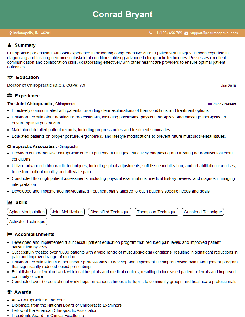
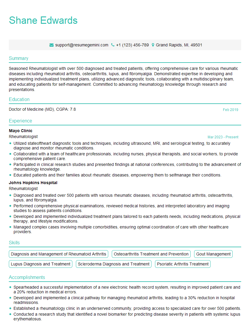
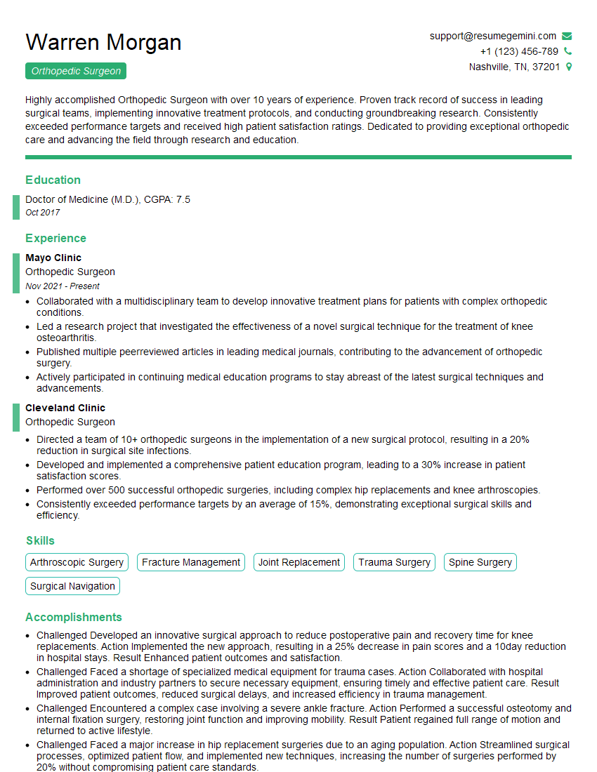
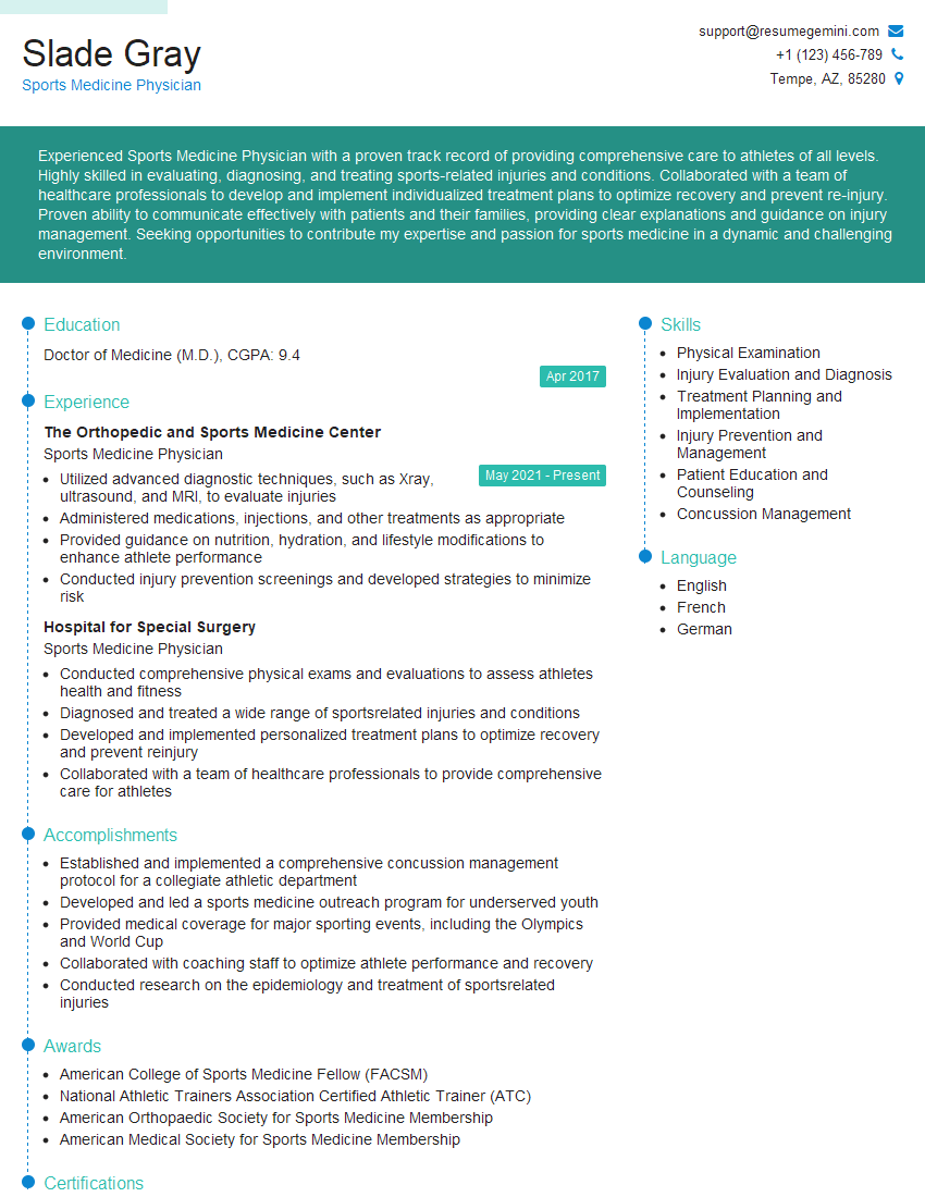
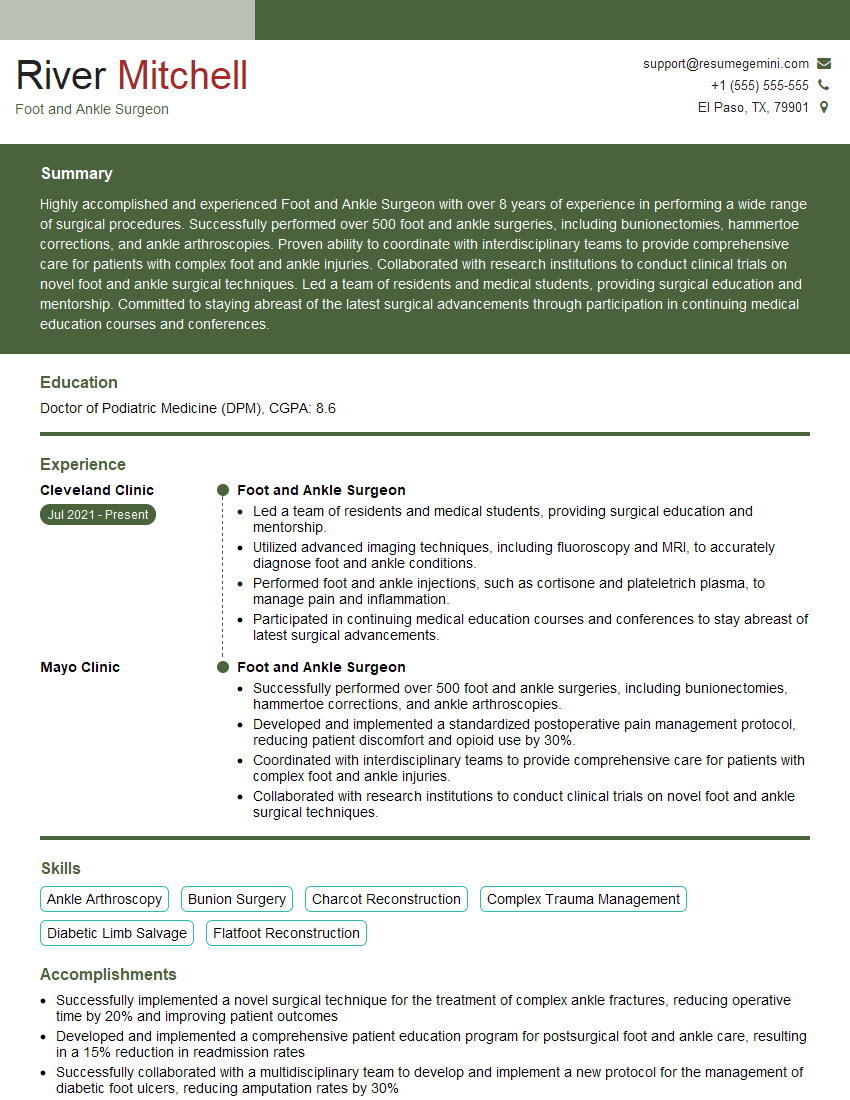
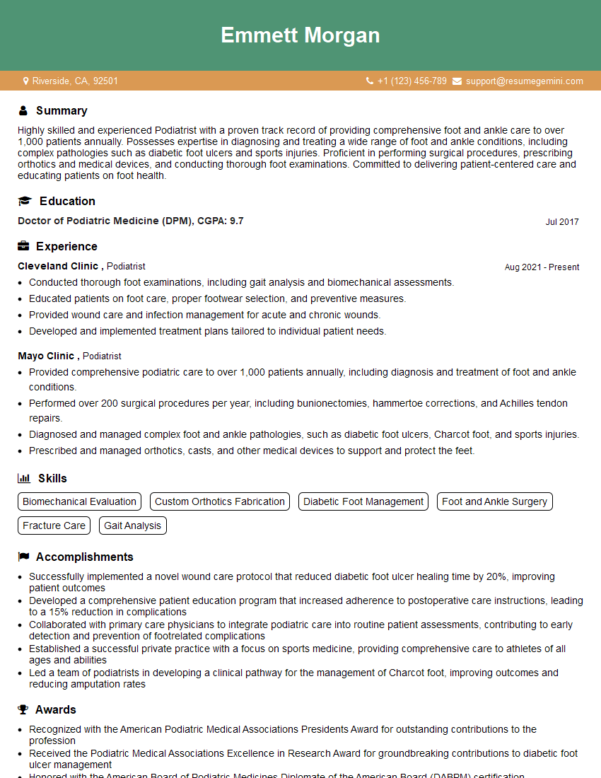
Mastering heel pain treatment is crucial for a successful and rewarding career in healthcare. A deep understanding of this area positions you for advanced roles and allows you to make a significant impact on patients’ lives. To maximize your job prospects, crafting an ATS-friendly resume is essential. ResumeGemini can help you build a professional and effective resume that showcases your skills and experience. ResumeGemini provides examples of resumes tailored to the Heel Pain Treatment field, giving you a head start in creating a compelling application.
Explore more articles
Users Rating of Our Blogs
Share Your Experience
We value your feedback! Please rate our content and share your thoughts (optional).
What Readers Say About Our Blog
This was kind of a unique content I found around the specialized skills. Very helpful questions and good detailed answers.
Very Helpful blog, thank you Interviewgemini team.
