Interviews are opportunities to demonstrate your expertise, and this guide is here to help you shine. Explore the essential Immunodeficiency Evaluation and Management interview questions that employers frequently ask, paired with strategies for crafting responses that set you apart from the competition.
Questions Asked in Immunodeficiency Evaluation and Management Interview
Q 1. Describe the diagnostic approach to suspected primary immunodeficiency.
Diagnosing primary immunodeficiency (PI) requires a systematic approach, starting with a thorough clinical history focusing on recurrent infections. The type of infections (bacterial, viral, fungal, opportunistic), their severity, age of onset, and response to treatment are crucial. Family history of immunodeficiency is also vital.
Initial investigations often include a complete blood count (CBC) with differential, immunoglobulin levels (IgG, IgA, IgM), and assessment of lymphocyte subsets. Abnormal findings trigger further testing, which may include:
- Specific antibody responses: Assessing the antibody response to specific vaccines (e.g., pneumococcal, tetanus) or antigens.
- Functional assays: These evaluate the ability of immune cells to perform their functions, such as phagocytosis (ability of cells to engulf and destroy pathogens) or lymphocyte proliferation (the ability of lymphocytes to multiply in response to stimulation).
- Genetic testing: This is crucial for identifying genetic defects underlying PI, especially in cases of severe disease or strong family history. Targeted gene sequencing or whole exome sequencing might be employed depending on the clinical suspicion.
- Flow cytometry: This technique quantifies and characterizes different immune cell populations, allowing for the identification of specific immune cell deficiencies.
The diagnostic pathway is iterative. Initial tests may identify broad defects, guiding further targeted investigations. Ultimately, the goal is to pinpoint the specific immune defect and guide appropriate management.
Q 2. What are the key differences between primary and secondary immunodeficiencies?
The key difference lies in the origin of the immune deficiency. Primary immunodeficiencies (PIs) are congenital; they are caused by genetic defects affecting the development or function of the immune system. These are present from birth, although symptoms may not appear until later in life. Examples include severe combined immunodeficiency (SCID) and common variable immunodeficiency (CVID).
Secondary immunodeficiencies, on the other hand, are acquired. They develop later in life due to various factors like malnutrition, viral infections (like HIV), certain medications (e.g., immunosuppressants), or malignancies (e.g., lymphoma). These deficiencies are a consequence of an underlying condition rather than an inherent genetic defect. For example, someone with poorly controlled diabetes might develop recurrent infections due to impaired immune function.
Q 3. Explain the role of flow cytometry in immunodeficiency evaluation.
Flow cytometry is an indispensable tool in immunodeficiency evaluation. It allows for the precise quantification and characterization of different immune cell populations in the blood. This includes lymphocytes (T cells, B cells, NK cells), as well as other immune cells like monocytes and neutrophils.
By using specific antibodies labeled with fluorescent dyes, flow cytometry can identify different cell subsets based on the expression of surface markers. This helps pinpoint defects in lymphocytic populations, for instance, low numbers of CD4+ T cells or B cells, or abnormal expression of activation markers. This detailed analysis provides crucial information for classifying PI and guiding further investigation. For example, a significant reduction in CD3+ T cells may suggest SCID, while reduced B cells with decreased antibody production could indicate CVID.
Q 4. Discuss the interpretation of a complete blood count (CBC) with differential in a patient with suspected immunodeficiency.
Interpreting a CBC with differential in a patient with suspected immunodeficiency requires careful attention to several parameters. A low white blood cell count (leukopenia) may suggest bone marrow dysfunction. A decrease in neutrophils (neutropenia) increases susceptibility to bacterial infections. Lymphopenia (low lymphocyte count) is common in many PIs, reflecting a deficit in T cells, B cells, or NK cells. The differential counts help to determine if the lymphopenia is due to T cell, B cell or NK cell deficiency.
However, it’s important to note that normal CBC findings do not rule out PI. Some individuals with PIs may have normal total lymphocyte counts but have abnormal proportions of different lymphocyte subsets. For example, they may have a normal total lymphocyte count, but the numbers of CD4+ or CD8+ T cells may be markedly reduced. This emphasizes the need for further specialized immunological tests beyond the CBC.
Q 5. How would you manage a patient with recurrent bacterial infections suggestive of antibody deficiency?
Management of recurrent bacterial infections suggestive of antibody deficiency involves several key strategies: identifying and treating the infection aggressively, providing prophylactic antibiotics, and replacing missing antibodies. A thorough workup to confirm the diagnosis is essential, including measurement of serum immunoglobulin levels and antibody responses to specific antigens.
Treatment typically includes:
- Intravenous immunoglobulin (IVIG): This provides passive immunity, replacing the missing antibodies and reducing the frequency and severity of infections.
- Prophylactic antibiotics: These are often prescribed to prevent recurrent infections, especially in those with recurrent sinopulmonary infections. Choice depends on the pathogens causing infections.
- Vaccination: While antibody responses may be poor, vaccinations (with appropriate precautions) can still provide some level of protection. It is important to monitor antibody levels after vaccinations.
- Supportive care: This involves managing symptoms, such as chronic sinusitis or bronchitis.
Regular monitoring of infections, immunoglobulin levels, and overall health status are vital components of long-term management.
Q 6. Outline the management of a patient with severe combined immunodeficiency (SCID).
SCID is a life-threatening condition requiring immediate and aggressive intervention. Management focuses on preventing infections and restoring immune function. The cornerstone of treatment is hematopoietic stem cell transplantation (HSCT), ideally from a matched sibling donor. This procedure replaces the defective immune system with a healthy one.
Prior to HSCT, supportive care includes:
- Strict infection control: Protecting the patient from infectious agents through isolation and prophylactic antibiotics and antivirals.
- Intravenous immunoglobulin (IVIG): provides passive immunity while waiting for the transplant.
- Gene therapy: In specific cases, gene therapy is an alternative to HSCT, aiming to correct the genetic defect in the patient’s own cells.
Post-HSCT, ongoing monitoring for graft rejection, infection, and other complications is critical. Careful management of potential side effects from the transplant is also essential. Early diagnosis and prompt treatment are crucial for survival in SCID.
Q 7. Describe the indications for intravenous immunoglobulin (IVIG) therapy.
Intravenous immunoglobulin (IVIG) therapy is used to replace missing antibodies in individuals with antibody deficiencies. Its indications are broad and include several primary and secondary immunodeficiency conditions.
Common indications for IVIG include:
- Primary antibody deficiencies: Conditions like CVID, X-linked agammaglobulinemia, and common variable immunodeficiency.
- Recurrent bacterial infections: Frequent infections despite appropriate antibiotic treatment, often sinopulmonary infections.
- Secondary antibody deficiencies: Immunodeficiencies resulting from conditions like HIV, malnutrition, or certain medications.
- Autoimmune diseases: In some cases, IVIG can modulate the immune response in autoimmune disorders.
- Inflammatory conditions: IVIG can be used to treat certain inflammatory conditions where antibody deficiencies play a role.
The decision to use IVIG is made based on the patient’s clinical presentation, immunological workup, and the severity of the immunodeficiency. Careful monitoring of infection rates, immunoglobulin levels, and adverse effects of the treatment are important.
Q 8. What are the potential complications of IVIG therapy?
Intravenous immunoglobulin (IVIG) therapy, while highly effective in treating various immunodeficiencies, carries potential complications. These can range from mild to severe and are often related to the infusion process itself or the components of the IVIG preparation.
- Infusion Reactions: These are the most common complications and can manifest as fever, chills, headache, nausea, vomiting, and back pain. Pre-medication with antihistamines and/or acetaminophen can often mitigate these reactions. Severe reactions, such as anaphylaxis, are rare but require immediate medical attention.
- Aseptic Meningitis: A sterile inflammation of the meninges, sometimes associated with IVIG infusion. Symptoms include headache, fever, and neck stiffness. This usually resolves spontaneously but warrants careful monitoring.
- Renal Dysfunction: IVIG can, rarely, cause acute kidney injury. This is more common in patients with pre-existing renal impairment. Careful monitoring of renal function is crucial, especially in high-risk individuals.
- Thromboembolic Events: Although uncommon, IVIG has been associated with an increased risk of blood clots. Patients with a history of thrombosis are particularly vulnerable.
- Transmission of Infectious Agents: While rigorous screening and processing minimize this risk, there’s always a theoretical possibility of transmitting infectious agents through IVIG, though this is exceptionally rare.
Managing these potential complications involves careful patient selection, pre-infusion assessment, slow infusion rates, appropriate pre-medication, and vigilant monitoring during and after the infusion. A detailed discussion of risks and benefits is essential before initiating IVIG therapy.
Q 9. Explain the role of genetic testing in diagnosing primary immunodeficiencies.
Genetic testing plays a crucial role in diagnosing primary immunodeficiencies (PIDs), which are inherited disorders affecting the immune system. These disorders are often heterogeneous, meaning they can result from mutations in a wide variety of genes. Genetic testing helps pinpoint the specific genetic defect responsible for the immune dysfunction.
The approach typically involves:
- Next-Generation Sequencing (NGS): This powerful technology allows for simultaneous analysis of numerous genes associated with PIDs, significantly increasing the diagnostic yield. It’s often the first-line approach due to its comprehensive nature.
- Targeted Gene Sequencing: If NGS doesn’t identify a causative mutation, or if a specific gene is suspected based on the clinical presentation, targeted sequencing of that gene may be performed.
- Chromosomal Microarray Analysis: This technique identifies large-scale genomic changes, such as deletions or duplications, that might be responsible for a PID.
- Quantitative PCR (qPCR): Used to measure the expression levels of specific genes, which can provide clues about the underlying genetic abnormality.
Genetic testing not only confirms the diagnosis but also allows for accurate genetic counseling, informing family members about their risk of carrying or inheriting the condition. It’s vital for early intervention and personalized management strategies.
For example, identifying a specific mutation in the IL7R gene confirms a diagnosis of severe combined immunodeficiency (SCID), paving the way for appropriate bone marrow transplantation or gene therapy.
Q 10. How do you counsel patients and families regarding the diagnosis and management of an immunodeficiency?
Counseling patients and families facing an immunodeficiency diagnosis is a critical aspect of care. It’s a multi-faceted process requiring empathy, patience, and a clear, straightforward communication style.
The counseling process usually involves:
- Explaining the diagnosis: Using clear, non-technical language to explain the specific type of immunodeficiency, its impact on the immune system, and the associated risks.
- Discussing treatment options: Presenting various treatment options, including IVIG, antibiotics, antifungals, antiviral agents, and possibly hematopoietic stem cell transplantation or gene therapy, while carefully explaining their benefits, risks, and limitations.
- Addressing emotional concerns: Creating a safe space for patients and families to express their emotions—fear, anxiety, grief—and providing emotional support.
- Developing a management plan: Collaboratively developing an individualized plan that encompasses regular monitoring, infection prophylaxis strategies, and lifestyle adjustments.
- Providing ongoing support: Ensuring access to resources such as support groups, educational materials, and specialists. Regular follow-up appointments are crucial.
Example: Explaining to a family that their child has X-linked agammaglobulinemia (XLA) requires carefully explaining the absence of antibodies, increased susceptibility to infections, and the need for lifelong immunoglobulin replacement therapy, while simultaneously offering reassurance and hope for a positive outcome with proper management.
Q 11. Describe the different types of antibody deficiencies.
Antibody deficiencies, also known as humoral immunodeficiencies, are characterized by impaired production or function of antibodies (immunoglobulins). They encompass a spectrum of disorders, differing in severity and underlying mechanisms.
- Common Variable Immunodeficiency (CVID): Characterized by low levels of IgG, IgA, and/or IgM antibodies in adulthood. The underlying cause varies, often involving B cell dysfunction.
- X-linked Agammaglobulinemia (XLA): A severe primary immunodeficiency caused by a mutation in the Bruton tyrosine kinase (BTK) gene, leading to a complete absence of B cells and consequently, antibodies.
- Selective IgA Deficiency: The most common primary antibody deficiency, involving a deficiency in IgA, the major antibody found in mucosal tissues. Individuals may be asymptomatic or experience recurrent infections.
- IgG Subclass Deficiencies: Involve low levels of one or more IgG subclasses (IgG1, IgG2, IgG3, IgG4), leading to varying degrees of immune compromise.
- Transient Hypogammaglobulinemia of Infancy (THI): A temporary condition characterized by low antibody levels during the first few years of life, usually resolving spontaneously.
These deficiencies can result in recurrent sinopulmonary infections, gastrointestinal problems, and an increased risk of autoimmune disorders.
Q 12. What are the common clinical presentations of cellular immunodeficiencies?
Cellular immunodeficiencies primarily affect T cells, the key players in cell-mediated immunity. These deficiencies result in impaired immune responses against intracellular pathogens, viruses, fungi, and some cancers.
Common clinical presentations include:
- Recurrent or severe viral infections: Such as herpes simplex virus, varicella-zoster virus, cytomegalovirus infections.
- Opportunistic infections: Infections with pathogens that typically do not cause disease in individuals with normal immune systems, such as Pneumocystis jirovecii pneumonia (PCP).
- Persistent thrush (oral candidiasis): A fungal infection of the mouth.
- Failure to thrive: Poor growth and development in infants and young children.
- Autoimmune diseases: An overactive immune system attacking the body’s own tissues.
- Increased susceptibility to malignancies: Some cellular immunodeficiencies increase the risk of certain types of cancers.
Specific examples include Severe Combined Immunodeficiency (SCID) characterized by profound defects in both B and T cells, leading to life-threatening infections, and DiGeorge syndrome, characterized by defects in the development of the thymus gland resulting in severely reduced T cell numbers.
Q 13. Discuss the use of live attenuated vaccines in immunodeficient patients.
Live attenuated vaccines contain weakened forms of the pathogen, inducing a strong and lasting immune response. However, their use in immunodeficient patients is generally contraindicated due to the risk of vaccine-derived infection. The weakened virus might cause severe or disseminated disease in individuals with compromised immune systems.
Therefore, live attenuated vaccines such as the measles, mumps, rubella (MMR) vaccine, varicella vaccine, and rotavirus vaccine are generally avoided in patients with significant immunodeficiencies, particularly those with severe T cell defects or those receiving immunosuppressive therapy. Instead, inactivated or subunit vaccines (containing only specific parts of the pathogen) are preferred when available.
In certain cases, the use of live vaccines might be considered for some patients with less severe immunodeficiencies, after careful assessment of individual risks and benefits by a specialist. This might involve measuring the patient’s antibody response after vaccination or using a lower dose of the vaccine.
The decision to use a live attenuated vaccine is made on a case-by-case basis, balancing the risk of vaccine-related disease with the benefits of the protection it could provide.
Q 14. What are the principles of infection prophylaxis in immunodeficient patients?
Infection prophylaxis in immunodeficient patients is crucial due to their increased susceptibility to infections. The strategy is multifaceted and tailored to the specific immunodeficiency and the patient’s risk factors.
Key principles include:
- Vaccination: Administering inactivated or subunit vaccines, whenever possible and appropriate, to protect against common infections.
- Antimicrobial Prophylaxis: This involves administering antibiotics, antifungals, or antivirals to prevent specific infections based on the patient’s risk profile. For example, patients with recurrent respiratory infections might receive prophylactic antibiotics, while those at risk of PCP might receive PCP prophylaxis.
- Infection Control Measures: Hygiene measures such as handwashing, avoidance of sick contacts, and prompt treatment of minor infections are essential.
- Environmental Modification: Changes to the environment may be necessary to minimize exposure to pathogens. This could include using air purifiers, regular cleaning and disinfection of surfaces, and avoiding certain types of pets.
- Prompt Treatment of Infections: Early diagnosis and appropriate treatment of infections are critical to prevent complications.
- Regular Monitoring: Close monitoring of the patient’s health through regular blood tests, physical examinations, and imaging studies are important.
For instance, a patient with CVID might receive regular IVIG infusions, prophylactic antibiotics for recurrent sinus infections, and influenza and pneumococcal vaccines. A patient with SCID might require strict isolation and broad-spectrum antimicrobial prophylaxis.
Q 15. How would you approach the management of a patient with recurrent viral infections?
Recurrent viral infections are a significant indicator of potential immunodeficiency. Managing such a patient requires a systematic approach beginning with a thorough history and physical examination. We need to ascertain the frequency, severity, and types of infections, along with any family history of immunodeficiency.
Diagnostic Evaluation: This involves a comprehensive immunological workup, including complete blood counts (CBC) with differential, serum immunoglobulin levels (IgG, IgA, IgM, IgE), and assessment of T-cell function (e.g., lymphocyte subsets, delayed-type hypersensitivity skin testing). Further investigations may include flow cytometry for specific lymphocyte populations, and if indicated, genetic testing to identify potential primary immunodeficiencies.
Treatment Strategy: Treatment is tailored to the underlying cause. If a specific immunodeficiency is identified (e.g., common variable immunodeficiency – CVID), targeted therapies like immunoglobulin replacement therapy (IVIG or subcutaneous immunoglobulin) may be employed. In other cases, supportive care with antiviral medications, vaccinations (where appropriate, considering the immune status), and meticulous infection control measures might be necessary. Regular monitoring of the patient’s clinical status and immune parameters is crucial. For example, a patient presenting with recurrent respiratory infections might need regular pulmonary function testing and close observation for potential lung damage.
Career Expert Tips:
- Ace those interviews! Prepare effectively by reviewing the Top 50 Most Common Interview Questions on ResumeGemini.
- Navigate your job search with confidence! Explore a wide range of Career Tips on ResumeGemini. Learn about common challenges and recommendations to overcome them.
- Craft the perfect resume! Master the Art of Resume Writing with ResumeGemini’s guide. Showcase your unique qualifications and achievements effectively.
- Don’t miss out on holiday savings! Build your dream resume with ResumeGemini’s ATS optimized templates.
Q 16. Describe the role of bone marrow transplantation in the management of certain immunodeficiencies.
Bone marrow transplantation (BMT), also known as hematopoietic stem cell transplantation (HSCT), is a powerful therapeutic modality for certain severe, life-threatening immunodeficiencies, particularly those with defects in hematopoietic stem cells or their progeny. In these cases, the patient’s faulty bone marrow is replaced with healthy donor marrow containing normal immune cells.
Mechanism: The transplanted stem cells engraft in the patient’s bone marrow and reconstitute the immune system, correcting the underlying defect. This leads to a sustained improvement in immune function and a reduction in infections.
Examples: Severe Combined Immunodeficiency (SCID), Wiskott-Aldrich syndrome, and certain types of chronic granulomatous disease are among the conditions where BMT is a potentially curative treatment.
Challenges: BMT is a complex procedure with inherent risks, including graft-versus-host disease (GVHD), infection, and the need for lifelong immunosuppressive therapy to prevent rejection. Careful patient selection, meticulous donor matching, and rigorous post-transplant management are crucial for successful outcomes.
Q 17. Explain the mechanism of action of different classes of immunosuppressants.
Immunosuppressants are drugs used to dampen the activity of the immune system. They are crucial in managing autoimmune diseases, preventing organ rejection after transplantation, and controlling inflammation. Different classes target distinct aspects of the immune response.
- Calcineurin Inhibitors (e.g., Cyclosporine, Tacrolimus): These block calcineurin, an enzyme crucial for T-cell activation, thus suppressing the production of interleukin-2 (IL-2), a key cytokine in immune responses.
- mTOR Inhibitors (e.g., Sirolimus, Everolimus): They target the mammalian target of rapamycin (mTOR), a protein kinase involved in T-cell proliferation and activation. These drugs are more effective against T-cell responses.
- Corticosteroids (e.g., Prednisone): These are broad-spectrum immunosuppressants that have multiple mechanisms of action, including suppressing inflammation, inhibiting cytokine production, and altering lymphocyte trafficking.
- Antimetabolites (e.g., Azathioprine, Mycophenolate mofetil): They interfere with DNA synthesis and inhibit the proliferation of lymphocytes. Azathioprine blocks purine synthesis, while mycophenolate mofetil inhibits inosine monophosphate dehydrogenase (IMPDH), an enzyme involved in guanine nucleotide synthesis.
- Monoclonal Antibodies (e.g., Basiliximab, Daclizumab): These target specific components of the immune system, such as T-cell activation molecules (e.g., IL-2 receptor).
Q 18. What are the adverse effects of commonly used immunosuppressants?
Immunosuppressants, while crucial for managing immune disorders, are associated with several adverse effects that vary depending on the drug and individual patient factors. Some common side effects are:
- Infections: Increased susceptibility to infections (bacterial, viral, fungal) due to immune suppression.
- Renal toxicity: Calcineurin inhibitors can cause kidney damage, manifested by reduced kidney function.
- Gastrointestinal problems: Nausea, vomiting, diarrhea, and abdominal pain are common, especially with some antimetabolites.
- Neurotoxicity: Some drugs can cause tremors, headaches, seizures, and cognitive impairment.
- Hepatotoxicity: Liver damage can occur with certain immunosuppressants.
- Hyperlipidemia: Elevated cholesterol and triglyceride levels are a frequent side effect.
- Hypertension: Increased blood pressure.
- Diabetes Mellitus: Increased risk of developing diabetes.
- Bone Marrow Suppression: Decreased production of blood cells.
The specific adverse effects and their severity can vary significantly between individuals, necessitating close monitoring and management.
Q 19. How do you monitor the efficacy and toxicity of immunosuppressive therapy?
Monitoring the efficacy and toxicity of immunosuppressive therapy is critical to ensure optimal patient outcomes. A multi-faceted approach is required.
- Clinical assessment: Regular evaluation of the patient’s clinical status, including monitoring for infection, organ function (kidney, liver), blood pressure, and other potential side effects.
- Laboratory tests: Periodic blood tests are vital. This includes complete blood counts (CBC), serum creatinine (kidney function), liver function tests (LFTs), lipid profile, and blood glucose levels. Therapeutic drug monitoring (TDM) is crucial for many immunosuppressants, ensuring optimal drug levels while minimizing toxicity.
- Imaging studies: Depending on the underlying condition, imaging modalities like ultrasound, CT scans, or MRI might be utilized to assess organ function or detect complications.
- Immunological monitoring: In certain cases, periodic monitoring of immune parameters may be essential, such as lymphocyte counts, immunoglobulin levels, or specific immune function assays. This is crucial for assessing the impact of immunosuppressive therapy on the immune response. For example, in transplantation, regular monitoring of the recipient’s immune response to the graft is important.
The frequency of monitoring depends on the type and dose of immunosuppressant, the patient’s clinical condition, and the presence of any adverse effects. This requires a collaborative effort between the physician, nurse, and other healthcare professionals involved in the patient’s care.
Q 20. What are the common causes of secondary immunodeficiency?
Secondary immunodeficiencies result from an underlying condition rather than an intrinsic defect in the immune system itself. Numerous factors can contribute.
- Infections: HIV, measles, and other infections can severely compromise immune function.
- Malignancies: Cancers like leukemia and lymphoma directly affect immune cells or disrupt their production.
- Autoimmune diseases: Conditions like lupus and rheumatoid arthritis can impair the immune system’s function.
- Nutritional deficiencies: Protein-energy malnutrition, deficiencies of vitamins (e.g., Vitamin A, Vitamin D), and minerals (e.g., zinc) can cause significant immune dysfunction.
- Medications: Many medications, including corticosteroids, chemotherapy drugs, and certain immunosuppressants, can have immunosuppressive effects.
- Chronic diseases: Renal failure, liver disease, and diabetes can weaken the immune system.
- Stress: Severe or prolonged stress can suppress immune responses.
- Aging: The immune system naturally declines with age, increasing susceptibility to infections.
- Medical treatments: Radiation therapy, chemotherapy, and splenectomy (surgical removal of the spleen) can all result in significant immunosuppression.
Q 21. How do you distinguish between primary and secondary immunodeficiency in clinical practice?
Differentiating between primary and secondary immunodeficiencies is crucial for appropriate diagnosis and management. The key distinction lies in the underlying cause: primary immunodeficiencies are congenital (present at birth) due to genetic defects, while secondary immunodeficiencies are acquired later in life due to external factors.
Clinical presentation: While both can manifest with recurrent or severe infections, the pattern and age of onset might provide clues. Primary immunodeficiencies often present in early childhood with severe or unusual infections, while secondary immunodeficiencies often manifest later in life and are frequently associated with an identifiable underlying condition or risk factor.
Immunological workup: This is the cornerstone of diagnosis. A comprehensive assessment of immune parameters (e.g., CBC, immunoglobulin levels, lymphocyte subsets, T cell function tests) helps identify specific immune defects suggestive of primary immunodeficiencies. Further investigations, such as genetic testing, can be crucial in confirming the diagnosis. In contrast, in secondary immunodeficiencies, the immunological findings may reflect the impact of the underlying condition, such as lymphopenia in HIV or decreased immunoglobulin levels in chronic liver disease.
Patient History: A detailed family history, along with a thorough review of systems and exploration of potential environmental or medical risk factors, helps determine whether the immunodeficiency is likely to be primary or secondary.
Example: A young child with recurrent severe infections since birth and a family history of immunodeficiency would raise suspicion for a primary immunodeficiency like SCID, whereas an older adult with recurrent infections following chemotherapy for cancer would be suggestive of a secondary immunodeficiency.
Q 22. Describe the evaluation of a patient with suspected phagocytic defect.
Evaluating a patient with suspected phagocytic defects requires a multi-pronged approach focusing on identifying impairments in the ability of phagocytes (neutrophils, macrophages) to engulf and kill pathogens. This begins with a thorough clinical history, paying close attention to recurrent and severe bacterial or fungal infections, often involving unusual organisms. For example, a child presenting with multiple Staphylococcus aureus skin infections or Aspergillus pneumonia should raise suspicion.
Next, we perform a complete blood count (CBC) with differential to assess neutrophil count and morphology. Low neutrophil counts may indicate a separate issue, but normal or high counts do not rule out a phagocytic defect. Functional assays are crucial. These include:
- Nitroblue tetrazolium (NBT) test: This assesses the ability of neutrophils to generate reactive oxygen species (ROS), essential for killing ingested microbes. A negative NBT test is highly suggestive of Chronic Granulomatous Disease (CGD).
- Dihydrorhodamine (DHR) flow cytometry: A more sensitive and quantitative method for assessing ROS production than the NBT test.
- Chemiluminescence assay: Measures the light emitted during phagocytosis, reflecting oxidative burst activity.
- Phagocytosis assay: Directly assesses the ability of phagocytes to engulf and ingest bacteria or other particles.
Genetic testing might be necessary to pinpoint the specific genetic defect causing the phagocytic dysfunction, particularly if functional assays are abnormal. This helps confirm the diagnosis and allows for genetic counseling.
Q 23. Discuss the management of chronic granulomatous disease (CGD).
Managing Chronic Granulomatous Disease (CGD) involves a multifaceted approach centered around infection prevention and treatment. This is a lifelong commitment as CGD is incurable.
- Prophylactic antibiotics: Trimethoprim-sulfamethoxazole (TMP-SMX) is a cornerstone of prophylaxis, reducing the incidence of bacterial infections. Other antibiotics may be added depending on the individual’s infection history.
- Inflammatory bowel disease (IBD) management: Patients with CGD frequently develop granulomas in the gastrointestinal tract, leading to IBD. This requires close monitoring and management with medications like anti-inflammatory drugs, immunomodulators, or even surgery if necessary.
- Fungal prophylaxis: Itraconazole or posaconazole can be considered for patients with recurrent fungal infections or those at high risk.
- Intravenous immunoglobulin (IVIG): May be used for severe or recurrent infections to provide passive immunity.
- Hematopoietic stem cell transplantation (HSCT): Considered for patients with severe disease and life-threatening infections. It offers a potential cure but comes with significant risks.
- Gene therapy: This is an emerging therapeutic approach holding promise for a cure in some CGD patients.
Regular monitoring for infections, particularly through blood cultures and imaging studies as needed, is essential. Patient education regarding infection prevention – hand hygiene, avoiding exposure to sick individuals – is crucial for successful management. Close collaboration between the immunologist, infectious disease specialist, and gastroenterologist is vital for optimal care.
Q 24. What is the role of complement testing in immunodeficiency evaluation?
Complement testing plays a vital role in immunodeficiency evaluation because the complement system is a crucial part of the innate immune system, mediating the lysis of pathogens, inflammation, and opsonization (enhancing phagocytosis). Testing can identify deficiencies or dysfunctions within this system, helping diagnose a range of conditions.
Complement testing typically involves several assays:
- CH50 (classical pathway): Measures the overall function of the classical complement pathway.
- AH50 (alternative pathway): Measures the overall function of the alternative complement pathway.
- Individual complement component levels: Quantifies the levels of specific complement proteins (e.g., C3, C4, factor B, factor D) to identify deficiencies in particular components.
Abnormal results can indicate various complement deficiencies, ranging from deficiencies in specific components to dysregulation of the system’s function. This guides further investigation and treatment.
Q 25. Explain the various types of complement deficiencies and their clinical manifestations.
Complement deficiencies can affect various components of the complement cascade, leading to diverse clinical manifestations. The severity and types of infections vary depending on the deficient component.
- C3 deficiency: This is the most common and severe deficiency, leading to recurrent and severe bacterial infections (pyogenic).
- C1 inhibitor deficiency: Causes hereditary angioedema, characterized by recurrent episodes of swelling in various parts of the body (e.g., face, throat). This is a distinct clinical picture not primarily focused on increased susceptibility to infections.
- Factor I deficiency: Leads to increased susceptibility to infections, often with severe consequences.
- Properdin deficiency: Similar to factor I deficiency, with a higher risk of severe infections.
- Deficiencies in other components (C4, C2, C5-C9): These deficiencies can cause increased susceptibility to infections, but with less severity and frequency compared to C3 deficiency.
It’s crucial to note that the clinical presentation can overlap with other immunodeficiencies, highlighting the importance of a thorough evaluation.
Q 26. Describe the diagnostic approach to suspected complement deficiencies.
Suspected complement deficiencies are investigated based on clinical suspicion and laboratory findings. The initial approach involves:
- Thorough clinical history: Focusing on recurrent and severe infections, particularly bacterial, their type, and response to treatment. Family history is also important.
- CH50 and AH50 assays: These screening tests assess the overall function of the classical and alternative complement pathways. Low values suggest a deficiency.
- Individual complement component levels: If the screening tests are abnormal, measurement of individual complement components helps pinpoint the specific deficiency.
- Genetic testing: May be necessary to confirm the diagnosis and identify the underlying genetic defect.
Additional investigations might be needed depending on the clinical picture, such as assessing autoantibodies that could be interfering with complement function.
Q 27. How do you manage a patient with a complement deficiency?
Managing a patient with a complement deficiency depends on the specific deficiency and its severity. The overall goal is to prevent infections and minimize their impact.
- Prophylactic antibiotics: Especially important for patients with C3 or other severe deficiencies to prevent life-threatening bacterial infections.
- Intravenous immunoglobulin (IVIG): Can provide passive immunity and help combat infections in cases where the body’s own complement system is deficient.
- Treatment for specific manifestations: For conditions like hereditary angioedema (associated with C1 inhibitor deficiency), specific treatments like C1 esterase inhibitor concentrate or other medications may be necessary to manage acute attacks.
- Supportive care: Includes prompt treatment of infections, supportive care measures like hydration and oxygen therapy when needed.
Regular monitoring is critical, including regular blood work to track complement levels and assess for infections. A multidisciplinary approach involving immunologists, infectious disease specialists, and other specialists may be needed, especially for individuals with severe deficiencies.
Q 28. Discuss the challenges in managing patients with rare immunodeficiencies.
Managing patients with rare immunodeficiencies poses unique challenges. The rarity itself creates hurdles:
- Limited diagnostic experience: Physicians may encounter these conditions infrequently, leading to delayed or inaccurate diagnoses.
- Lack of established treatment guidelines: The scarcity of data makes it difficult to establish evidence-based treatment protocols.
- Challenges in finding appropriate specialists: Patients may need to travel long distances or rely on telehealth consultations to access specialists with expertise in rare immunodeficiencies.
- Difficulty participating in clinical trials: Small patient populations may hinder the development of new therapies.
- High cost of treatments: Specialized treatments and monitoring can be incredibly expensive, placing a significant burden on patients and their families.
Addressing these challenges requires collaborative efforts: building national and international registries to gather more data on rare conditions, fostering collaboration between specialists, encouraging research into novel treatments, and advocating for policies that improve access to care and reduce financial barriers.
For example, a patient with a newly identified complement deficiency might require extensive genetic testing and consultations with multiple specialists, which is a major burden without access to comprehensive healthcare coverage and experienced medical teams.
Key Topics to Learn for Immunodeficiency Evaluation and Management Interview
- Primary Immunodeficiencies: Understanding the classifications, diagnostic criteria, and clinical presentations of various primary immunodeficiencies (e.g., SCID, CVID, IgA deficiency).
- Immunodeficiency Workup: Mastering the interpretation of laboratory data, including flow cytometry, immunoglobulins, and specific antibody responses. Know how to strategically select appropriate diagnostic tests based on clinical suspicion.
- Secondary Immunodeficiencies: Familiarize yourself with the causes and impact of secondary immunodeficiencies, such as those related to HIV, malnutrition, medications, and malignancy. Understand how to differentiate between primary and secondary causes.
- Treatment Strategies: Develop a strong understanding of various treatment approaches, including immunoglobulin replacement therapy, hematopoietic stem cell transplantation, and other targeted therapies. Be prepared to discuss the benefits and risks of each.
- Infection Prevention and Management: Know the strategies for infection prevention in immunocompromised patients and how to manage common opportunistic infections. This includes vaccination strategies and prophylactic antibiotics.
- Patient Case Management: Practice approaching complex patient scenarios and developing comprehensive management plans. Focus on integrating diagnostic findings with clinical presentation to guide decision-making.
- Ethical and Legal Considerations: Be aware of the ethical implications of genetic testing, informed consent, and patient confidentiality in the context of immunodeficiency management.
Next Steps
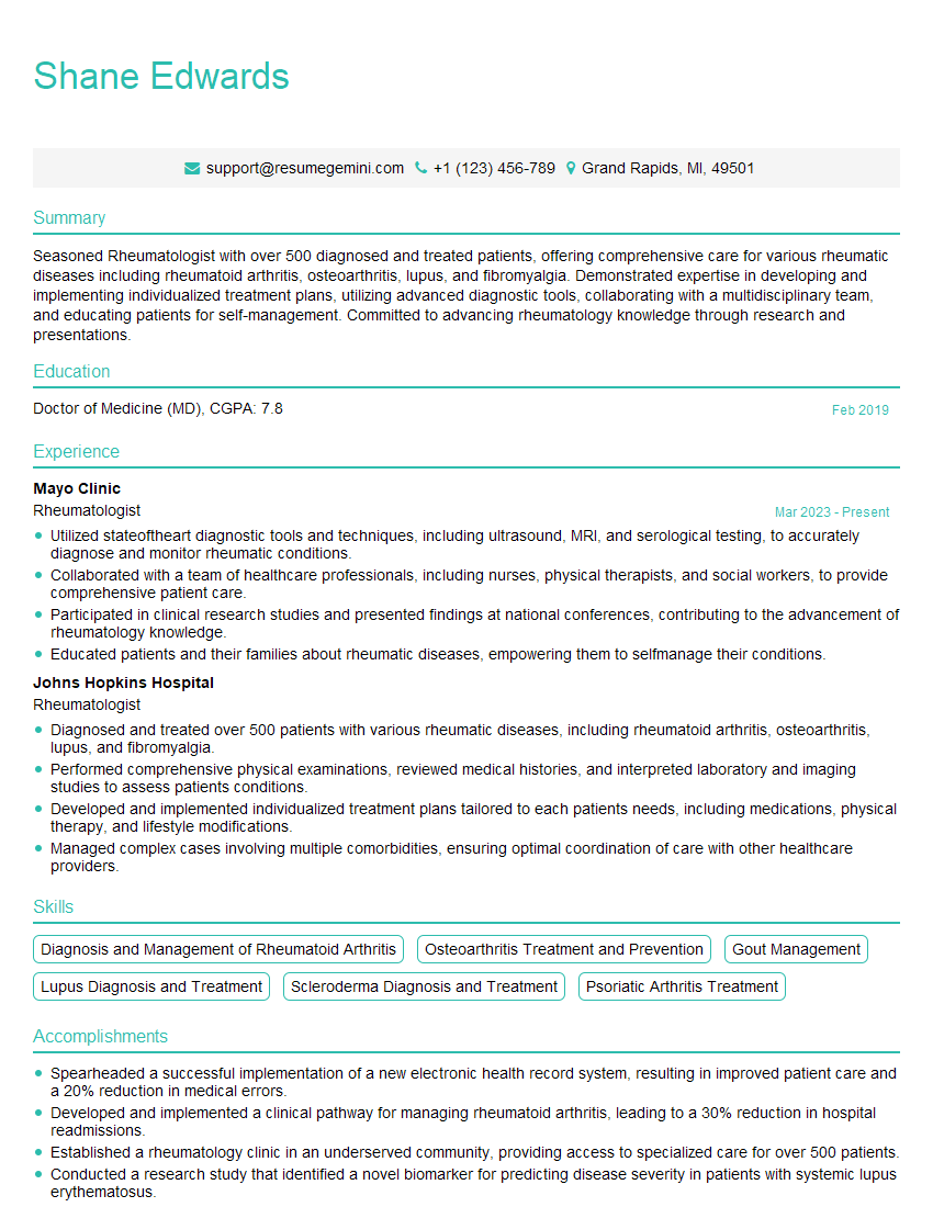
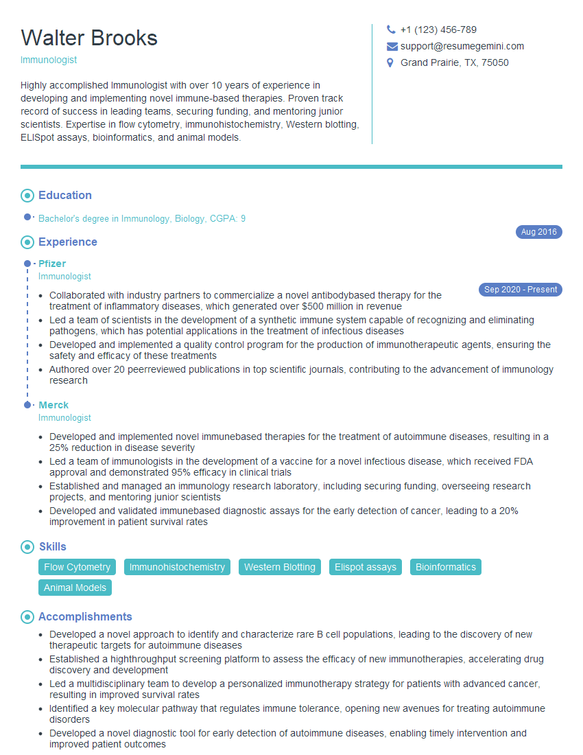
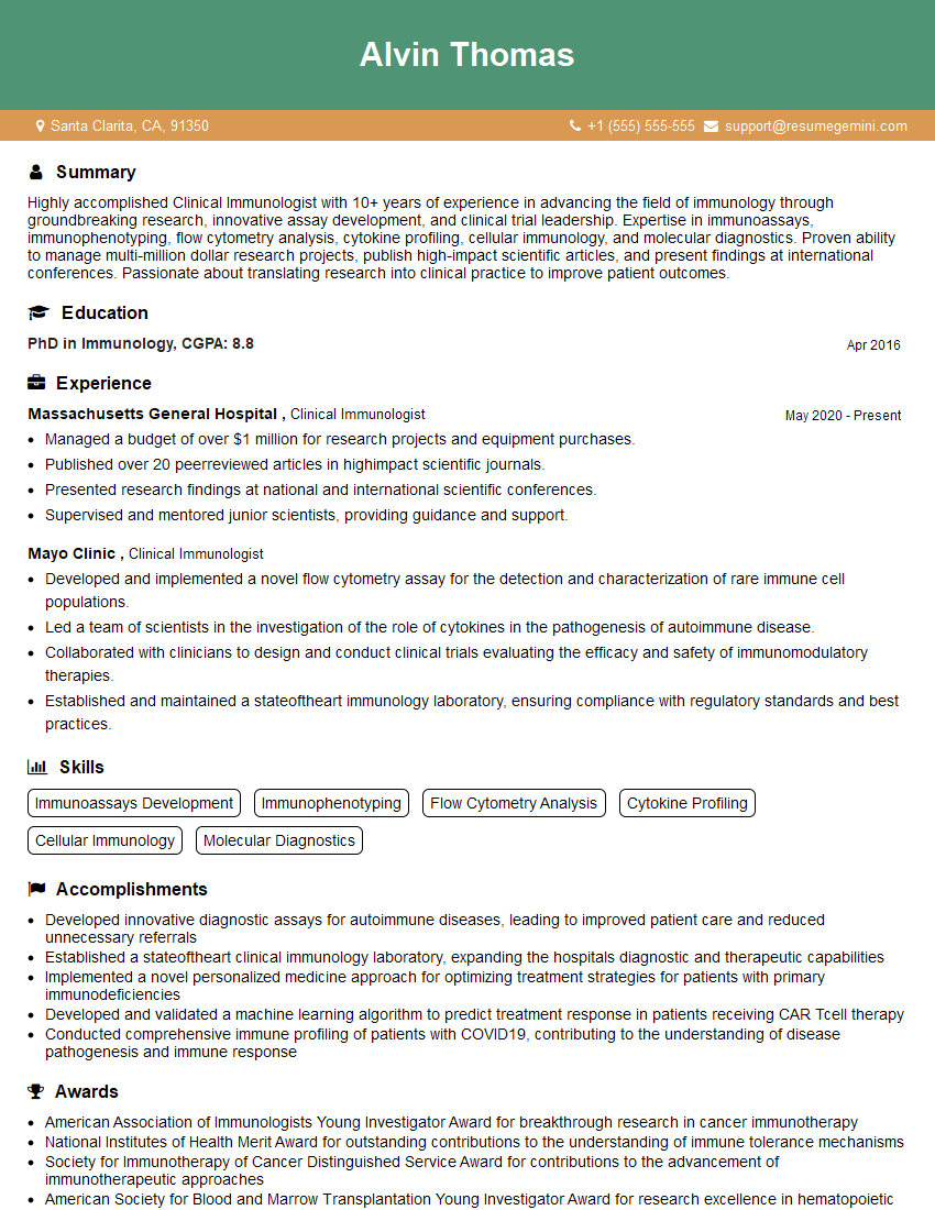
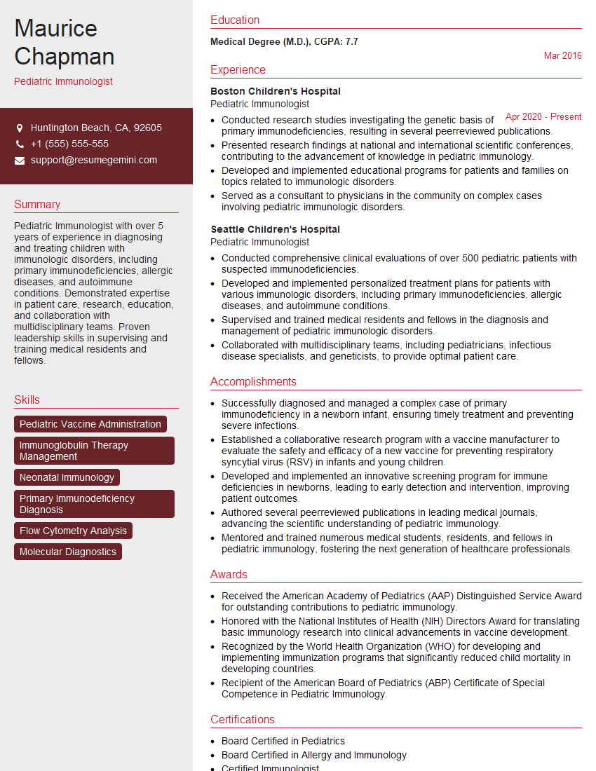
Mastering Immunodeficiency Evaluation and Management opens doors to rewarding and impactful careers in immunology, infectious disease, and hematology-oncology. A strong foundation in this area positions you for leadership roles and advanced research opportunities. To maximize your job prospects, it’s crucial to present your skills and experience effectively. Creating an ATS-friendly resume is key to getting noticed by recruiters. We highly recommend using ResumeGemini, a trusted resource, to build a professional and impactful resume that showcases your expertise. ResumeGemini provides examples of resumes tailored to Immunodeficiency Evaluation and Management to guide you in this process.
Explore more articles
Users Rating of Our Blogs
Share Your Experience
We value your feedback! Please rate our content and share your thoughts (optional).
What Readers Say About Our Blog
Hi, I have something for you and recorded a quick Loom video to show the kind of value I can bring to you.
Even if we don’t work together, I’m confident you’ll take away something valuable and learn a few new ideas.
Here’s the link: https://bit.ly/loom-video-daniel
Would love your thoughts after watching!
– Daniel
This was kind of a unique content I found around the specialized skills. Very helpful questions and good detailed answers.
Very Helpful blog, thank you Interviewgemini team.


