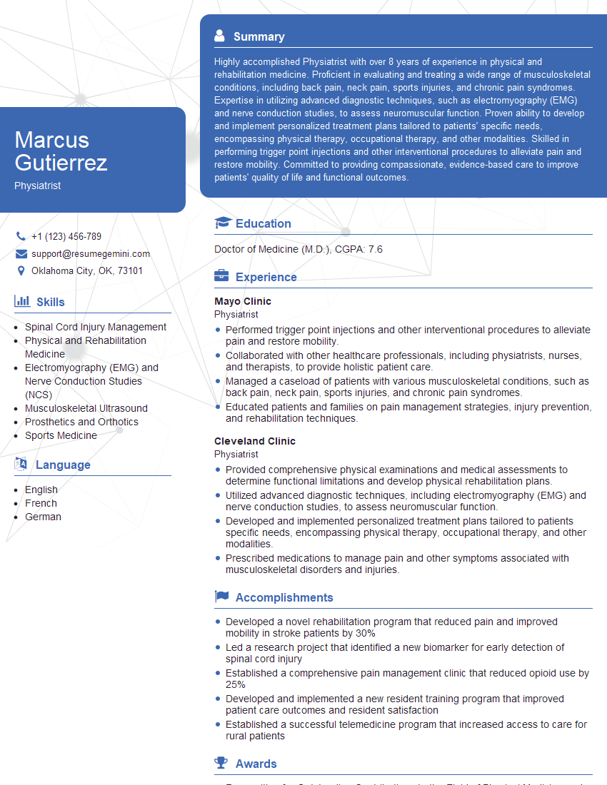
Preparation is the key to success in any interview. In this post, we’ll explore crucial Inflammatory Muscle Diseases Management interview questions and equip you with strategies to craft impactful answers. Whether you’re a beginner or a pro, these tips will elevate your preparation.
Questions Asked in Inflammatory Muscle Diseases Management Interview
Q 1. Describe the diagnostic criteria for Polymyositis.
Diagnosing Polymyositis, a form of inflammatory myopathy, requires a multi-faceted approach. There isn’t one single definitive test. Instead, diagnosis relies on a combination of clinical features, laboratory findings, and imaging studies. The key is to establish evidence of proximal muscle weakness, meaning weakness in muscles closer to the body’s core, like the shoulders and hips. This weakness is typically symmetrical, affecting both sides of the body equally.
- Clinical Presentation: Patients present with progressive weakness in proximal muscles, often leading to difficulty climbing stairs, rising from a chair (the ‘getting up from the floor’ test is often used), or lifting objects. Fatigue is also a very common symptom.
- Laboratory Findings: Elevated muscle enzymes, such as creatine kinase (CK), aldolase, and lactate dehydrogenase (LDH), are often present, indicating muscle damage. However, these enzymes can be elevated in other conditions, so they are not diagnostic on their own.
- Electromyography (EMG): This test assesses the electrical activity of muscles. In Polymyositis, EMG typically shows myopathic changes, characteristic patterns indicative of muscle damage and dysfunction.
- Muscle Biopsy: This is a crucial diagnostic tool. A small sample of muscle tissue is examined under a microscope, revealing characteristic inflammatory changes, such as infiltration of inflammatory cells and muscle fiber necrosis (death).
It’s important to note that a definitive diagnosis requires integration of all these findings. No single element guarantees the diagnosis. For example, elevated CK alone doesn’t confirm Polymyositis; it could be due to other muscle problems.
Q 2. Differentiate between Polymyositis and Dermatomyositis.
Polymyositis and Dermatomyositis are both inflammatory myopathies characterized by muscle weakness, but they differ significantly in their clinical presentation. The key difference lies in the skin involvement.
- Polymyositis: Primarily involves muscle weakness. Skin manifestations are absent or minimal. The weakness is usually symmetrical and affects proximal muscles (shoulders, hips, thighs).
- Dermatomyositis: Characterized by both muscle weakness and distinctive skin rashes. These rashes can be quite varied but often involve a characteristic heliotrope rash (a purplish discoloration) around the eyelids, Gottron’s papules (raised scaly lesions on the knuckles), and shawl sign (a V-shaped rash on the chest and back).
Think of it this way: Polymyositis affects the muscles ‘under the hood,’ while Dermatomyositis shows its effects both ‘under the hood’ and on the ‘body’s exterior’. Both conditions share similar laboratory findings like elevated muscle enzymes and myopathic changes on EMG, but the presence of skin rash strongly points towards Dermatomyositis.
Q 3. Explain the role of muscle biopsies in diagnosing Inflammatory Myopathies.
Muscle biopsies play a pivotal role in diagnosing inflammatory myopathies, offering a direct look at the underlying muscle pathology. While blood tests and EMG provide valuable clues, the biopsy provides the definitive histological evidence.
During a muscle biopsy, a small sample of muscle tissue is extracted (usually from the vastus lateralis muscle in the thigh) and examined under a microscope. Pathologists look for several key features:
- Inflammatory cell infiltration: The presence of increased numbers of inflammatory cells (lymphocytes and macrophages) surrounding and within muscle fibers is a hallmark of inflammatory myopathies.
- Muscle fiber necrosis: Damage and death of muscle fibers is another key feature, indicated by changes in fiber size, shape, and staining characteristics.
- Specific patterns of muscle fiber damage: Different inflammatory myopathies can show distinct patterns of muscle fiber damage, helping differentiate between conditions like Polymyositis, Dermatomyositis, and inclusion body myositis.
For instance, a biopsy might reveal the characteristic perifascicular atrophy in dermatomyositis, where muscle fibers at the edge of muscle bundles are more affected than those in the center.
The results of a muscle biopsy, combined with clinical features and laboratory findings, allow for an accurate diagnosis and guide treatment decisions.
Q 4. What are the common laboratory findings in patients with Inflammatory Muscle Diseases?
Common laboratory findings in patients with inflammatory muscle diseases often point towards muscle damage and inflammation. However, it’s important to remember that these findings aren’t specific to these diseases and can be seen in other muscle disorders.
- Elevated muscle enzymes: Creatine kinase (CK), aldolase, and lactate dehydrogenase (LDH) are frequently elevated, indicating muscle breakdown and release of intracellular contents into the bloodstream. The degree of elevation doesn’t always correlate with disease severity.
- Elevated inflammatory markers: Erythrocyte sedimentation rate (ESR) and C-reactive protein (CRP) may be elevated, reflecting systemic inflammation. However, these are non-specific markers of inflammation, seen in many conditions.
- Autoantibodies: The presence of specific autoantibodies, like anti-Jo-1, anti-Mi-2, and anti-SRP antibodies, can be helpful in diagnosing specific types of inflammatory myopathies and predicting disease course. For example, anti-Jo-1 antibodies are associated with interstitial lung disease, a common complication.
- Low serum levels of Vitamin D Hypovitaminosis D is increasingly recognized as being associated with inflammatory myopathies.
It’s crucial to interpret these laboratory findings in the context of the clinical presentation, EMG results, and muscle biopsy findings for an accurate diagnosis.
Q 5. Discuss the treatment strategies for Polymyositis, including corticosteroids and immunosuppressants.
Treatment of Polymyositis aims to reduce inflammation, improve muscle strength, and enhance the patient’s quality of life. The cornerstone of therapy is often immunosuppression.
- Corticosteroids: Prednisone is usually the first-line treatment. It quickly reduces inflammation and improves muscle strength. However, corticosteroids are associated with significant side effects (discussed below), so the goal is to use the lowest effective dose for the shortest possible duration.
- Immunosuppressants: If corticosteroids alone are insufficient or cause unacceptable side effects, immunosuppressants are added. Common choices include methotrexate, azathioprine, mycophenolate mofetil, and rituximab. These medications work by suppressing the immune system’s attack on the muscles. The choice of immunosuppressant depends on several factors, including patient characteristics, response to corticosteroids, and presence of other organ involvement.
- Intravenous Immunoglobulin (IVIG): In severe cases or those unresponsive to other treatments, IVIG may be used to dampen the immune response.
- Physical and Occupational Therapy: These are essential components of management, aiming to maintain muscle function, improve range of motion, and prevent contractures. Regular exercise helps prevent muscle atrophy and promotes strength.
Treatment plans are individualized, tailored to the patient’s specific needs and response to therapy. Regular monitoring of muscle strength, laboratory markers, and potential side effects is crucial.
Q 6. What are the potential side effects of corticosteroids in the treatment of Inflammatory Myopathies?
Corticosteroids, while highly effective in reducing inflammation in inflammatory myopathies, carry a substantial risk of side effects. These side effects can be significant and often necessitate careful monitoring and management.
- Metabolic effects: Weight gain, hyperglycemia (high blood sugar), hyperlipidemia (high cholesterol), and osteoporosis (weakening of bones) are common. These can increase the risk of cardiovascular disease and fractures.
- Gastrointestinal effects: Peptic ulcers, gastritis (inflammation of the stomach lining), and pancreatitis (inflammation of the pancreas) can occur.
- Musculoskeletal effects: Although used to treat muscle inflammation, high-dose corticosteroids can lead to muscle weakness and atrophy over time, a paradoxical effect.
- Endocrine effects: Corticosteroids can suppress the hypothalamic-pituitary-adrenal (HPA) axis, leading to adrenal insufficiency (the body’s inability to produce enough cortisol). This can be life-threatening if corticosteroids are abruptly stopped.
- Infections: Immunosuppression associated with corticosteroids increases susceptibility to infections.
- Ophthalmologic effects: Cataracts and glaucoma are potential long-term complications.
- Psychiatric effects: Mood changes, including depression and psychosis, are possible.
Careful monitoring and proactive strategies to mitigate side effects, such as bone density monitoring, blood sugar control, and lifestyle modifications, are vital during corticosteroid therapy.
Q 7. How do you manage the complications of Inflammatory Muscle Diseases, such as dysphagia or respiratory compromise?
Complications of inflammatory muscle diseases, such as dysphagia (difficulty swallowing) and respiratory compromise, require prompt and multidisciplinary management.
- Dysphagia: This is a common complication, particularly in Dermatomyositis, potentially leading to aspiration pneumonia (lung infection due to food or saliva entering the lungs). Management involves a speech-language pathologist (SLP) assessment to evaluate swallowing function and recommend strategies, such as dietary modifications, swallowing exercises, or feeding tubes if necessary.
- Respiratory compromise: Weakness of respiratory muscles can lead to reduced lung capacity and increased risk of respiratory infections. Pulmonary function tests are crucial to assess lung function. Non-invasive ventilation (NIV), such as CPAP or BiPAP, may be required to support breathing, especially at night. Respiratory physiotherapy can help maintain respiratory muscle strength and prevent complications.
- Cardiac involvement: Myocarditis (inflammation of the heart muscle) can occur in inflammatory myopathies. Echocardiography and electrocardiography are used to assess cardiac function.
- Interstitial Lung Disease: Certain autoantibodies, particularly anti-Jo-1, are associated with an increased risk of interstitial lung disease. This requires close monitoring of lung function with pulmonary function tests and high resolution CT scans. Treatment might include immunosuppressants and supportive care.
Managing these complications requires a collaborative approach involving rheumatologists, pulmonologists, gastroenterologists, SLPs, and physiotherapists. Regular monitoring and proactive intervention are crucial to prevent further deterioration and improve the patient’s overall well-being.
Q 8. What are the different types of Inflammatory Myopathies?
Inflammatory myopathies are a group of muscle diseases characterized by muscle inflammation and weakness. They’re often autoimmune in nature, meaning the body’s immune system mistakenly attacks its own muscle tissue. The major types include:
- Polymyositis (PM): This affects multiple muscles symmetrically (both sides of the body equally), typically proximal muscles (those closer to the body’s core, like the shoulders and hips).
- Dermatomyositis (DM): Similar to PM in muscle involvement, but also features characteristic skin rashes. These rashes can manifest in various ways, including a heliotrope rash (purple discoloration around the eyelids) and Gottron’s papules (raised, scaly lesions on the knuckles).
- Inclusion Body Myositis (IBM): A more severe and less responsive to treatment form characterized by the presence of distinctive inclusions within muscle cells. It primarily affects proximal muscles but can also involve distal muscles (farther from the core).
- Overlap syndromes: These encompass cases with features of more than one inflammatory myopathy, often combining aspects of PM, DM, and other connective tissue diseases.
Understanding these distinctions is critical for appropriate diagnosis and treatment planning, as each type might respond differently to therapy.
Q 9. Describe the role of genetic testing in the diagnosis of Inflammatory Muscle Diseases.
Genetic testing plays an increasingly important, albeit sometimes limited, role in diagnosing inflammatory muscle diseases. While many cases are sporadic (without a clear genetic cause), genetic testing can help identify specific genes associated with some forms and subtypes. This can aid in:
- Confirming a diagnosis: Certain gene mutations are strongly linked to specific inflammatory myopathies, providing supporting evidence for a clinical diagnosis.
- Identifying subtypes: Genetic testing may distinguish between different forms of inflammatory myopathies, especially when clinical presentation is ambiguous.
- Predicting disease course and response to treatment: Some genetic variations might correlate with disease severity or treatment response, allowing for personalized management strategies.
- Facilitating family screening: If a genetic cause is identified, family members can be screened for the same mutation to assess their risk.
However, it’s crucial to understand that negative results don’t rule out an inflammatory myopathy; many cases remain idiopathic (of unknown cause). Genetic testing should be considered as one piece of the puzzle alongside clinical examination, muscle biopsy, and other laboratory tests.
Q 10. Explain the pathogenesis of Inclusion Body Myositis (IBM).
The pathogenesis of IBM is complex and not fully understood. It’s considered a sporadic disease, typically affecting older adults. The key features include:
- Immune dysregulation: While not as clearly autoimmune as PM or DM, there’s evidence suggesting an immune component. Abnormal immune cell activity and inflammatory processes within the muscle contribute to muscle fiber damage.
- Protein aggregation: The hallmark of IBM is the accumulation of abnormal proteins (such as amyloid-beta and TDP-43) within muscle fibers, forming characteristic inclusions visible under a microscope. These inclusions disrupt muscle cell function and lead to progressive muscle damage.
- Oxidative stress: Increased oxidative stress, an imbalance between free radicals and antioxidants, plays a crucial role. This stress contributes to muscle fiber degeneration and inflammation.
- Muscle fiber degeneration and regeneration: Muscle fibers undergo degeneration, and attempts at regeneration are ineffective, leading to progressive weakness and atrophy.
Research continues to unravel the precise mechanisms driving protein aggregation, oxidative stress, and immune dysregulation in IBM. This knowledge is essential for developing effective therapeutic strategies for this challenging condition.
Q 11. What are the long-term management considerations for patients with Inflammatory Muscle Diseases?
Long-term management of inflammatory muscle diseases requires a multidisciplinary approach focusing on symptom control, disease progression mitigation, and improvement of quality of life. Key considerations include:
- Regular monitoring: Frequent clinical assessments, muscle strength testing, and laboratory tests (including muscle enzymes) are needed to track disease activity and response to treatment.
- Medication management: This often includes immunosuppressants (e.g., corticosteroids, methotrexate, azathioprine) to control inflammation. Biological therapies targeting specific immune pathways are also used in some cases.
- Physical and occupational therapy: These are crucial for maintaining muscle strength, improving functional abilities, and preventing contractures (muscle shortening).
- Respiratory support: If respiratory muscles are affected, respiratory support (including non-invasive ventilation) might be necessary.
- Nutritional support: Maintaining adequate nutrition is important for muscle health and overall well-being.
- Psychosocial support: These diseases can significantly impact quality of life; access to psychological counseling and support groups is vital.
Long-term management necessitates a collaborative relationship between the patient, physician, physical therapist, and other healthcare professionals to tailor the care plan to individual needs.
Q 12. How do you monitor the response to treatment in patients with Inflammatory Myopathies?
Monitoring response to treatment in inflammatory myopathies is essential to ensure effectiveness and adjust the therapy as needed. Several methods are used:
- Clinical assessment: Regular evaluations of muscle strength (using manual muscle testing or quantitative methods), functional capacity, and symptom severity provide crucial information about treatment efficacy.
- Laboratory tests: Monitoring muscle enzyme levels (such as creatine kinase) helps track muscle damage and inflammation. Other blood tests may assess immune markers.
- Imaging studies: Magnetic resonance imaging (MRI) can visualize muscle inflammation and atrophy, providing objective evidence of disease activity.
- Electromyography (EMG) and nerve conduction studies (NCS): These studies help evaluate muscle and nerve function, detecting abnormalities consistent with inflammatory myopathy.
- Quality of life questionnaires: Measuring patients’ quality of life helps assess the impact of the disease and treatment on daily living.
Combining these methods provides a comprehensive picture of treatment response, allowing for timely adjustments in the therapeutic strategy. A lack of response might necessitate a change in medication, addition of other therapies, or referral to a specialist.
Q 13. Discuss the role of physical therapy and occupational therapy in managing Inflammatory Muscle Diseases.
Physical therapy and occupational therapy play a vital role in managing inflammatory muscle diseases. They focus on:
- Improving muscle strength and endurance: Tailored exercise programs, ranging from low-impact activities to resistance training, are designed to strengthen muscles without causing further damage.
- Enhancing functional abilities: Therapists help patients perform daily activities, such as dressing, bathing, and mobility tasks, with improved ease and independence.
- Preventing contractures: Stretching and range-of-motion exercises help prevent muscle shortening and stiffness, maintaining joint flexibility.
- Improving respiratory function: Breathing exercises and techniques can improve respiratory muscle strength and capacity.
- Ergonomic advice: Occupational therapists can advise on modifications to workspaces and daily tasks to minimize muscle strain and fatigue.
- Assistive devices: They may recommend or provide assistive devices, such as walkers or adaptive equipment, to enhance independence.
These therapies are not just supportive but actively contribute to improved muscle function, increased independence, and enhanced quality of life for patients with inflammatory myopathies.
Q 14. What are the challenges in diagnosing Inflammatory Muscle Diseases?
Diagnosing inflammatory muscle diseases can be challenging due to several factors:
- Overlapping symptoms: Symptoms like muscle weakness and fatigue can mimic other conditions, leading to diagnostic delays.
- Variable clinical presentation: The symptoms and their severity vary significantly between individuals and among different types of inflammatory myopathies.
- Absence of specific diagnostic markers: There isn’t a single blood test or imaging technique that definitively diagnoses inflammatory myopathy. Diagnosis often relies on a combination of clinical findings, laboratory tests, and muscle biopsy.
- Need for muscle biopsy: Muscle biopsy, though invasive, is often required to confirm the diagnosis and distinguish between different types of myopathies by visualizing characteristic histopathological features.
- Subtle early symptoms: In the early stages, symptoms may be subtle and nonspecific, delaying diagnosis until the disease has progressed further.
The diagnostic process requires a high index of suspicion and careful evaluation by experienced clinicians to differentiate between inflammatory myopathies and other muscle disorders. A comprehensive approach, combining clinical assessment, laboratory tests, and imaging, is crucial to reach an accurate and timely diagnosis.
Q 15. Describe the different types of immunosuppressants used in Inflammatory Myopathies and their mechanisms of action.
Immunosuppressants are cornerstone treatments in Inflammatory Myopathies (IM), aiming to dampen the overactive immune response causing muscle inflammation and damage. Several classes are used, each with a unique mechanism.
Corticosteroids (e.g., Prednisone): These are often the first-line treatment. They act broadly by suppressing inflammation throughout the body, reducing the production of inflammatory cytokines. However, long-term use carries significant side effects like osteoporosis, weight gain, and diabetes. We carefully monitor patients on corticosteroids and strive to taper dosages as soon as clinically feasible.
Methotrexate: This is a disease-modifying antirheumatic drug (DMARD) that inhibits dihydrofolate reductase, interfering with DNA synthesis and cell proliferation, thus reducing inflammation. It’s frequently used in combination with corticosteroids to minimize steroid dosage.
Azathioprine: Another DMARD, azathioprine inhibits the synthesis of purines, essential for immune cell function. It’s a slower-acting immunosuppressant often used as a steroid-sparing agent or in cases where methotrexate is ineffective.
Mycophenolate mofetil: This drug inhibits inosine monophosphate dehydrogenase (IMPDH), crucial for lymphocyte proliferation, thus selectively suppressing the immune response. It’s increasingly used as a steroid-sparing agent, especially in severe cases.
Rituximab: A monoclonal antibody targeting CD20 on B lymphocytes, rituximab depletes B cells which are key players in the autoimmune process. It’s reserved for more severe cases resistant to other treatments and requires careful monitoring for side effects.
Cyclosporine: A calcineurin inhibitor, cyclosporine inhibits T-cell activation and cytokine production. It’s less commonly used for IM due to its significant nephrotoxic potential.
The choice of immunosuppressant and the combination regimen depend on the severity of the disease, patient response, and the presence of comorbidities. Careful monitoring of blood counts, liver function, and kidney function is crucial throughout treatment.
Career Expert Tips:
- Ace those interviews! Prepare effectively by reviewing the Top 50 Most Common Interview Questions on ResumeGemini.
- Navigate your job search with confidence! Explore a wide range of Career Tips on ResumeGemini. Learn about common challenges and recommendations to overcome them.
- Craft the perfect resume! Master the Art of Resume Writing with ResumeGemini’s guide. Showcase your unique qualifications and achievements effectively.
- Don’t miss out on holiday savings! Build your dream resume with ResumeGemini’s ATS optimized templates.
Q 16. How do you differentiate Inflammatory Muscle Diseases from other neuromuscular disorders?
Differentiating Inflammatory Muscle Diseases from other neuromuscular disorders requires a thorough clinical evaluation, incorporating detailed history, physical examination, and laboratory investigations. Key distinctions lie in the pattern of weakness, associated symptoms, and diagnostic tests.
Inflammatory Myopathies typically present with proximal muscle weakness (difficulty with lifting arms or climbing stairs), often symmetrically affecting both sides of the body. Elevated muscle enzymes (creatine kinase) are a common finding. The clinical course can be variable, with periods of exacerbation and remission. Biopsy showing muscle inflammation is often definitive.
Other neuromuscular disorders like muscular dystrophies (genetic, progressive weakness), motor neuron diseases (ALS), myasthenia gravis (autoimmune, fluctuating weakness), and metabolic myopathies (enzyme deficiencies) have distinct clinical characteristics and diagnostic features. For example, muscular dystrophies usually present with progressive weakness starting distally, often with family history. Myasthenia gravis involves fatigable muscle weakness worsened by exertion, and diagnostic testing includes EMG and antibody studies. A detailed neurological examination and appropriate investigations are crucial for accurate diagnosis.
Imaging techniques such as MRI can help visualize muscle inflammation in IM, but this is not always diagnostic. Electromyography (EMG) can identify myopathic changes (e.g., short duration, low amplitude potentials). Muscle biopsy remains the gold standard for definitive diagnosis of IM, revealing inflammatory cell infiltrates within the muscle tissue.
Q 17. What are the prognostic factors for Inflammatory Muscle Diseases?
Prognosis in Inflammatory Muscle Diseases varies greatly depending on several factors. Some key prognostic indicators include:
Disease subtype: Some subtypes, like dermatomyositis, often have a better prognosis than others, like inclusion body myositis.
Severity at presentation: Patients presenting with severe muscle weakness and significant organ involvement tend to have a more challenging course.
Response to treatment: A good response to initial immunosuppressive therapy is generally a positive prognostic sign.
Presence of extramuscular manifestations: Involvement of other organs, such as the lungs or skin, can complicate management and impact prognosis.
Age at diagnosis: Younger patients often have a more favorable outcome compared to older individuals.
Presence of interstitial lung disease: This serious complication is associated with poorer outcomes.
It’s important to note that even with seemingly poor prognostic factors, advancements in treatment are improving long-term outcomes for many patients. Regular monitoring and proactive management of complications are vital in optimizing prognosis.
Q 18. Describe your experience with managing patients with Inflammatory Muscle Diseases.
Throughout my career, I have managed a significant number of patients with Inflammatory Muscle Diseases. This has involved a multidisciplinary approach, working closely with other specialists including rheumatologists, pulmonologists, and physical therapists. My experience encompasses all stages of the disease, from initial diagnosis and treatment selection to long-term monitoring and management of complications.
One memorable case involved a young woman with rapidly progressive dermatomyositis, presenting with significant muscle weakness and severe skin rashes. We initiated high-dose corticosteroids followed by methotrexate and close monitoring for side effects. With careful treatment optimization and rehabilitation, she achieved significant improvement in her muscle strength and quality of life. This case highlighted the importance of aggressive early intervention and the collaborative nature of care.
In another case, an elderly gentleman with inclusion body myositis initially responded well to treatment, but subsequently experienced a decline. This underscores the challenges inherent in managing chronic inflammatory myopathies and the need for ongoing assessment and adjustment of treatment strategies. We focused on managing his symptoms, optimizing his mobility with assistive devices, and providing support to address his quality-of-life concerns.
My experience emphasizes the importance of personalized management tailored to individual patient characteristics and response to treatment.
Q 19. How would you approach a patient presenting with proximal muscle weakness and elevated muscle enzymes?
A patient presenting with proximal muscle weakness and elevated muscle enzymes (CK) necessitates a thorough investigation to consider Inflammatory Muscle Diseases (IM) and other potential causes. My approach would be:
Detailed history: This includes the onset, duration, and progression of symptoms; presence of rash (especially Gottron’s papules or heliotrope rash suggestive of dermatomyositis), dysphagia (difficulty swallowing), Raynaud’s phenomenon, or other systemic symptoms.
Thorough physical examination: Focus on assessing muscle strength, particularly proximal muscles (shoulder, hip girdle). A complete skin and neurological examination is critical to identify other systemic manifestations.
Laboratory investigations: This includes repeat CK measurement to confirm elevation, complete blood count, inflammatory markers (ESR, CRP), autoantibody testing (anti-Jo-1, anti-Mi-2, anti-SRP, etc.), and liver and kidney function tests. Autoantibody profiles often help guide subtype diagnosis.
Electrodiagnostic studies (EMG/NCS): EMG can reveal myopathic changes confirming muscle damage. Nerve conduction studies rule out other neurological causes.
Muscle biopsy: This is often the gold standard for diagnosis, revealing inflammatory cell infiltrates within muscle tissue. Histological findings are pivotal in confirming the diagnosis and determining the specific type of inflammatory myopathy.
Imaging studies (MRI): MRI scans can help evaluate muscle involvement, but this is supplementary to the other investigations.
Based on the results, a differential diagnosis will be established. If IM is suspected, treatment would be initiated with a multidisciplinary approach involving rheumatology, physical therapy, and potentially other specialists depending on extramuscular manifestations.
Q 20. What are the ethical considerations in managing patients with Inflammatory Muscle Diseases?
Managing patients with Inflammatory Muscle Diseases presents several ethical considerations.
Informed consent: Patients must fully understand the risks and benefits of various treatments, including immunosuppressants with their potential side effects. Shared decision-making is crucial.
Balancing benefits and harms: Immunosuppressants can effectively reduce inflammation, but they also carry risks of infections, malignancies, and other adverse effects. The potential benefits must outweigh the risks, and close monitoring is essential.
Resource allocation: The cost of long-term immunosuppression and supportive care can be significant. Clinicians must consider resource allocation and access to appropriate therapies.
Quality of life: The goal is not only to control disease activity but also to maintain or improve the patient’s quality of life. This includes addressing functional limitations, psychological impact, and social support.
Autonomy and decision-making capacity: Respecting patient autonomy is paramount. In cases where patients lack decision-making capacity, careful consideration of advance directives and surrogate decision-makers is needed.
End-of-life care: For patients with severe, progressive disease, ethical considerations surrounding end-of-life care, such as palliative care and advance care planning, become crucial.
Navigating these ethical complexities requires open communication with patients, families, and other healthcare providers, ensuring patient-centered care and adherence to ethical guidelines.
Q 21. What are the latest advancements in the treatment of Inflammatory Myopathies?
Recent advancements in the treatment of Inflammatory Myopathies are focused on refining existing therapies and developing novel approaches:
Biologics: Studies are exploring the use of targeted biologics, such as those inhibiting specific cytokines or immune pathways, to better address the underlying autoimmune mechanisms with potentially fewer side effects compared to traditional immunosuppressants. These are still under investigation and not yet widely used in routine clinical practice.
Improved monitoring techniques: Developments in imaging (MRI) and blood biomarkers improve disease monitoring, allowing for earlier detection of relapses and better tailoring of treatment.
Personalized medicine: Research is focused on understanding the genetic and immunological factors that contribute to disease variability. This has the potential for targeted therapies based on individual patient characteristics and disease subtypes.
Combination therapies: More research is exploring optimized combination therapies to minimize side effects and maximize efficacy while tailoring therapy based on the individual patient’s unique response profile.
Rehabilitation and supportive care: Advancements in physical and occupational therapy, respiratory care, and assistive devices improve functional outcomes and quality of life.
Clinical trials are constantly evaluating new treatments and strategies to improve outcomes for patients with Inflammatory Myopathies. Staying updated on the latest research and clinical guidelines is crucial for providing optimal care.
Q 22. Discuss your understanding of clinical trials related to Inflammatory Muscle Diseases.
Clinical trials for Inflammatory Muscle Diseases (IMDs) are crucial for developing and evaluating new treatments. These trials follow rigorous scientific methodologies to assess the safety and efficacy of interventions, ranging from novel drugs to innovative therapies. They involve carefully selected patient populations meeting specific inclusion and exclusion criteria, ensuring the results are reliable and generalizable.
For example, a Phase III clinical trial might compare a new drug for dermatomyositis against a placebo, measuring improvements in muscle strength, fatigue, and inflammatory markers. The trial’s design would involve randomization, blinding (where neither the patient nor the researcher knows who receives the drug vs. the placebo), and robust data collection to minimize bias. Data analysis would employ statistical methods to determine if the new drug significantly improves outcomes compared to the placebo. The results then undergo peer review before publication, contributing to the body of evidence guiding IMD treatment.
Different trial designs exist, such as randomized controlled trials (RCTs), observational studies, and cohort studies. Each has its strengths and limitations, influencing the strength of evidence generated. Understanding these methodologies is paramount in critically evaluating research findings and informing treatment decisions.
Q 23. Explain your experience with patient education and counseling in the context of Inflammatory Muscle Diseases.
Patient education and counseling are cornerstones of effective IMD management. It’s not just about explaining diagnoses and treatments; it’s about empowering patients to actively participate in their care. I approach this using a patient-centered, empathetic style, adapting my communication based on individual needs and learning styles.
I begin by ensuring patients understand their specific diagnosis, its implications, and the potential course of the disease. I explain treatment options clearly, addressing any concerns or misunderstandings. This might involve using visual aids, simplifying medical jargon, or providing written materials in easy-to-understand language. I also emphasize the importance of lifestyle modifications, such as regular exercise (adapted to their capabilities), a healthy diet, stress management, and avoiding triggers (like excessive sun exposure in dermatomyositis).
For example, when counseling a patient newly diagnosed with polymyositis, I’d not only explain the disease’s inflammatory nature and impact on muscle function, but also provide practical strategies for managing daily tasks, accessing resources like physical therapy, and recognizing warning signs of complications. Follow-up sessions are crucial to assess progress, adjust treatment strategies as needed, and offer ongoing support.
Q 24. How do you incorporate shared decision-making in the management of Inflammatory Muscle Diseases?
Shared decision-making is central to my approach. It’s not about me telling the patient what to do; it’s about collaboratively developing a treatment plan that aligns with their values, preferences, and lifestyle. I present patients with the evidence-based options, discuss the potential benefits and risks of each, and then help them weigh these factors in light of their individual circumstances.
For example, when considering immunosuppressants for a patient with inclusion body myositis, we’d discuss the potential for improved muscle strength, the risks of side effects (like infections), and the patient’s personal tolerance for medication. We’d also consider the patient’s employment, social support system, and overall health status. This collaborative process leads to a treatment plan that is not only medically sound but also personally acceptable and sustainable for the patient.
I use tools and techniques like decision aids, shared decision-making guides, and open-ended questions to facilitate this process. The goal is to empower the patient to become an active participant in their own healthcare journey, leading to better adherence and improved outcomes.
Q 25. Describe your experience collaborating with other healthcare professionals in managing complex cases of Inflammatory Muscle Diseases.
Managing complex IMD cases often requires a multidisciplinary approach. I routinely collaborate with rheumatologists, neurologists, pulmonologists, physical therapists, occupational therapists, and other specialists, as needed. Effective communication and coordinated care are paramount.
For instance, a patient with overlap syndrome (features of several IMDs) might require the expertise of a rheumatologist to manage the inflammatory aspects, a neurologist to assess neurological involvement, and a pulmonologist to address respiratory complications. Regular team meetings, shared electronic health records, and clear communication channels are vital in ensuring everyone is informed and contributing to the patient’s overall care plan. This collaborative approach helps to provide holistic care addressing the myriad of potential challenges a complex case presents.
I also value open communication with the patient’s family or caregivers, ensuring they understand the treatment plan and can provide support. This holistic approach improves outcomes and addresses the often profound impact of IMDs on patients and their loved ones.
Q 26. What are the key challenges in the research of Inflammatory Muscle Diseases?
Researching IMDs faces several key challenges. One is the heterogeneity of these diseases. IMDs encompass a wide range of conditions with varying clinical presentations, genetic backgrounds, and responses to treatment. This complexity makes it difficult to establish clear diagnostic criteria and develop targeted therapies. Furthermore, many IMDs are rare diseases, limiting the size of patient populations available for clinical trials, making it harder to collect sufficient data for robust statistical analysis and to develop clinically meaningful endpoints.
Another challenge is the lack of objective biomarkers. Diagnosing IMDs currently relies heavily on clinical symptoms and muscle biopsies, which are subjective and invasive. Identifying reliable biomarkers would improve early diagnosis and facilitate the evaluation of new treatments in clinical trials. Finally, the underlying pathophysiology of many IMDs is not fully understood, hindering the development of truly targeted therapies. More basic research is needed to fully understand the mechanisms driving inflammation and muscle damage.
Q 27. What are the potential future directions in the treatment of Inflammatory Muscle Diseases?
Future directions in IMD treatment are promising. Research focuses on developing targeted therapies based on a deeper understanding of disease mechanisms. This includes therapies aimed at specific inflammatory pathways, genetic abnormalities, or immune cells involved in disease pathogenesis. For example, research into the role of specific cytokines and immune checkpoints has led to the development of biologics targeting these pathways. Personalized medicine approaches, tailoring treatments based on an individual patient’s genetic profile and disease characteristics, also hold great potential.
Advances in gene therapy and regenerative medicine offer exciting possibilities, but are still in early stages of development. Improved diagnostic tools, including non-invasive biomarkers, will enhance early detection and facilitate more timely interventions. Finally, further research into lifestyle interventions and supportive care will improve patients’ quality of life and overall health outcomes.
Q 28. How do you stay up-to-date on the latest advancements in Inflammatory Muscle Diseases management?
Staying current in the rapidly evolving field of IMD management requires a multifaceted approach. I regularly attend professional conferences and workshops, participate in continuing medical education (CME) activities, and actively engage with leading experts in the field. I meticulously review peer-reviewed journals, including publications in leading rheumatology and neurology journals, for updates on research findings and new treatment guidelines.
I also participate in professional organizations, such as the American College of Rheumatology, and actively engage in online professional communities and forums to stay updated on the latest advancements and clinical trials. By combining these strategies, I ensure my knowledge and clinical practice remain current and aligned with the best available evidence, ensuring that I provide my patients with the most effective and up-to-date care possible.
Key Topics to Learn for Inflammatory Muscle Diseases Management Interview
- Disease Pathophysiology: Understanding the underlying mechanisms of inflammatory muscle diseases like polymyositis, dermatomyositis, and inclusion body myositis. This includes knowledge of immune system dysfunction and muscle fiber damage.
- Diagnostic Approaches: Familiarity with various diagnostic tools, including blood tests (e.g., muscle enzymes, autoantibodies), electromyography (EMG), muscle biopsy, and imaging techniques (e.g., MRI).
- Treatment Strategies: In-depth knowledge of current treatment modalities, encompassing immunosuppressants, corticosteroids, biologics, and supportive care. Consider the nuances of managing different disease severities and patient profiles.
- Clinical Presentation and Assessment: Ability to recognize and interpret the clinical manifestations of inflammatory muscle diseases, including muscle weakness, pain, fatigue, and extramuscular symptoms. Understanding patient history taking and physical examination techniques is crucial.
- Monitoring and Management of Side Effects: Expertise in recognizing and managing the potential side effects associated with various treatments, such as infections, immunosuppression, and metabolic disturbances. This includes developing strategies for patient safety and adherence.
- Disease Progression and Prognosis: Understanding the natural history of inflammatory muscle diseases, including factors influencing disease course and long-term outcomes. This includes discussion of potential complications and long-term management strategies.
- Interdisciplinary Collaboration: Awareness of the importance of collaborating with other healthcare professionals (e.g., physiatrists, pulmonologists, rheumatologists) in providing holistic patient care.
- Research and Evidence-Based Practice: Understanding the ongoing research in inflammatory muscle disease management and the ability to apply current evidence-based guidelines to clinical practice.
Next Steps
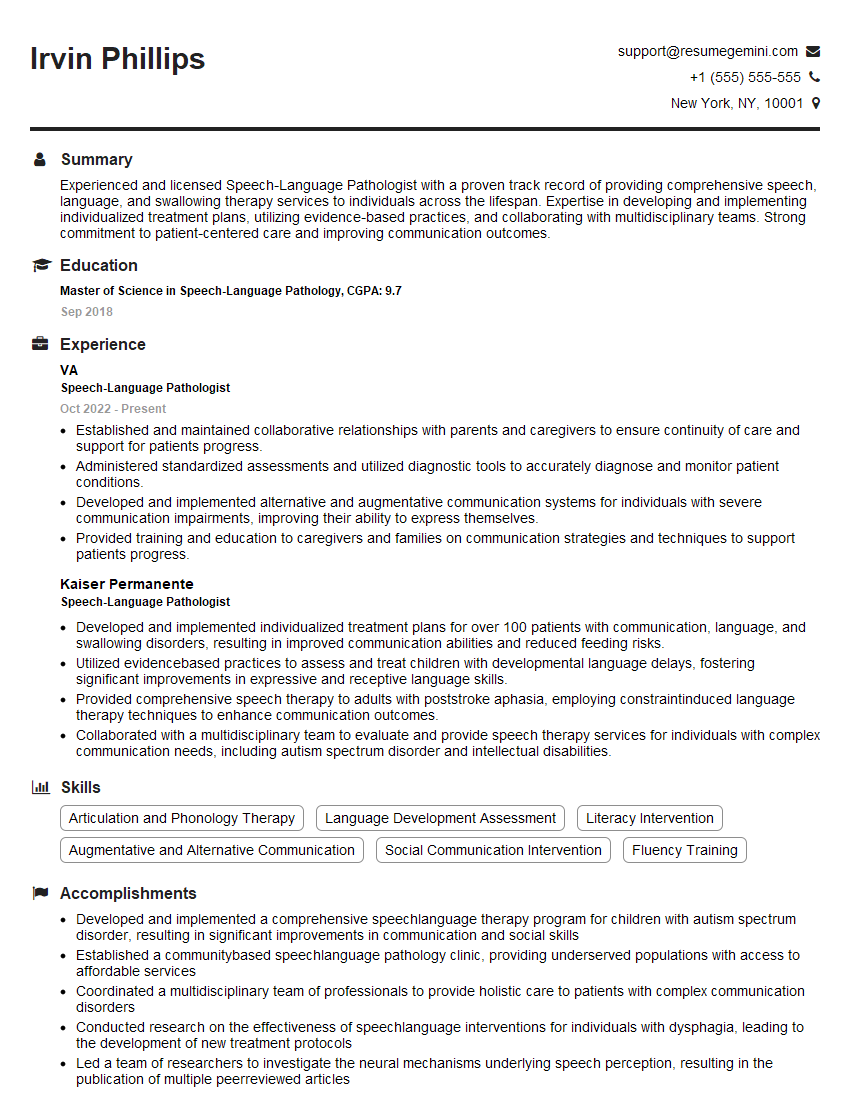
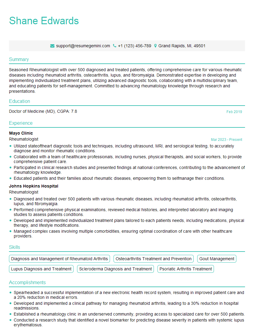
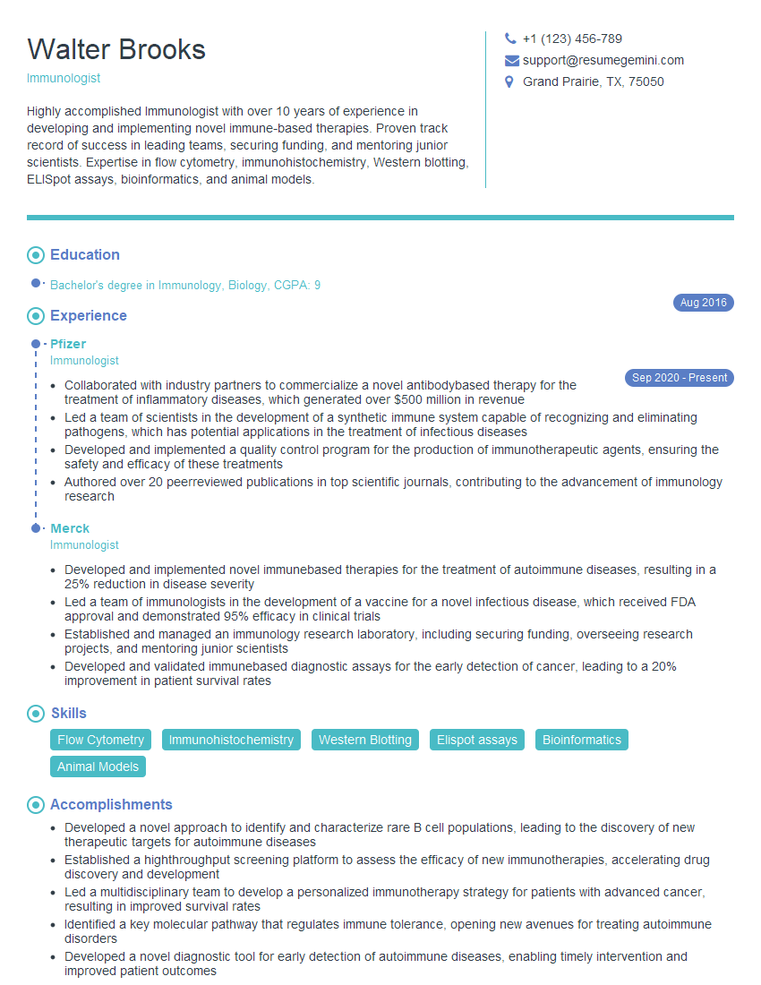
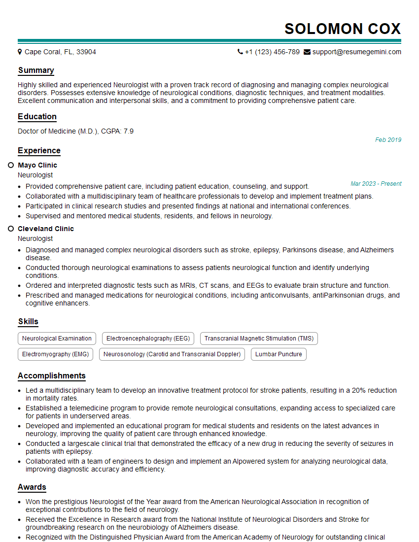
Mastering Inflammatory Muscle Diseases Management is key to advancing your career in a rapidly evolving field. A strong understanding of these complexities will significantly enhance your clinical skills and open doors to diverse opportunities. To showcase your expertise, creating an ATS-friendly resume is crucial for maximizing your job prospects. ResumeGemini is a trusted resource that can help you build a professional and impactful resume tailored to highlight your skills and experience in Inflammatory Muscle Diseases Management. Examples of resumes tailored specifically to this field are available to further assist you in creating a compelling application.
Explore more articles
Users Rating of Our Blogs
Share Your Experience
We value your feedback! Please rate our content and share your thoughts (optional).
What Readers Say About Our Blog
Hi, I have something for you and recorded a quick Loom video to show the kind of value I can bring to you.
Even if we don’t work together, I’m confident you’ll take away something valuable and learn a few new ideas.
Here’s the link: https://bit.ly/loom-video-daniel
Would love your thoughts after watching!
– Daniel
This was kind of a unique content I found around the specialized skills. Very helpful questions and good detailed answers.
Very Helpful blog, thank you Interviewgemini team.
