Feeling uncertain about what to expect in your upcoming interview? We’ve got you covered! This blog highlights the most important Middle Ear Analysis interview questions and provides actionable advice to help you stand out as the ideal candidate. Let’s pave the way for your success.
Questions Asked in Middle Ear Analysis Interview
Q 1. Describe the anatomy and physiology of the middle ear.
The middle ear is a small, air-filled cavity located within the temporal bone of the skull. It’s crucial for sound transmission from the outer to the inner ear. Its anatomy includes three tiny bones, the ossicles – the malleus (hammer), incus (anvil), and stapes (stirrup) – which form a chain articulating across the middle ear space. The malleus attaches to the tympanic membrane (eardrum), and the stapes footplate rests in the oval window of the inner ear. The middle ear also contains the Eustachian tube, connecting the middle ear to the nasopharynx, responsible for equalizing pressure across the eardrum. Physiologically, the middle ear acts as an impedance-matching transformer. Sound waves striking the tympanic membrane cause it to vibrate, these vibrations are amplified by the ossicular chain and transmitted to the inner ear’s fluid-filled structures. This amplification compensates for the significant impedance mismatch between air and fluid, ensuring efficient sound transmission.
Think of it like this: imagine trying to push a balloon into water – it’s difficult. The ossicles act like a lever system, reducing the force needed to move the fluid in the inner ear. This is essential for hearing faint sounds.
Q 2. Explain the acoustic reflex and its clinical significance.
The acoustic reflex, also known as the stapedius reflex, is an involuntary contraction of two middle ear muscles, the stapedius and tensor tympani, in response to loud sounds or intense vibrations. This contraction reduces the amount of sound energy transmitted to the inner ear by stiffening the ossicular chain. Clinically, this reflex is significant as its presence or absence helps in differentiating between conductive and sensorineural hearing loss. A normal acoustic reflex suggests the integrity of the middle ear and the neural pathways involved. Absence of the reflex, however, points toward a possible middle ear pathology, or a problem along the neural pathway affecting the reflex arc. Audiologists use acoustic reflex testing to identify conditions like otosclerosis or facial nerve dysfunction.
For instance, if someone has a conductive hearing loss (problem in the middle ear), the reflex might be absent or abnormal. But with sensorineural hearing loss (problem in the inner ear or auditory nerve), the reflex might be present, but the patient still experiences hearing difficulties. Therefore, acoustic reflex testing contributes valuable information to diagnose hearing disorders.
Q 3. What are the different types of tympanograms and what do they indicate?
Tympanometry is a procedure that measures the middle ear’s pressure and compliance. Different types of tympanograms reflect the status of the middle ear system. A Type A tympanogram is considered normal, showing good middle ear pressure and compliance. It indicates a healthy middle ear system. A Type B tympanogram indicates that the middle ear is filled with fluid (like in otitis media) and shows a flat peak or no peak at all. The compliance (ability to move easily) is severely reduced, so the system is very stiff. A Type C tympanogram displays a negative middle ear pressure, suggesting that the Eustachian tube is not functioning correctly to equalize pressure. This is seen in conditions such as Eustachian tube dysfunction. A Type As indicates reduced mobility of the eardrum, such as in otosclerosis, while Type Ad shows excessive mobility implying the system is too loose or compliant (maybe eardrum perforation) .
Each type of tympanogram provides valuable insights into the condition of the middle ear. A Type A suggests a normal functioning ear, while types B, C, As, and Ad indicate problems that may need further investigation.
Q 4. How is static admittance measured and what does it represent?
Static admittance, also known as compliance or admittance, is a measure of how easily the tympanic membrane and middle ear system can move in response to sound pressure. It’s measured using an impedance bridge or immittance meter during tympanometry. The probe tip delivers a series of sounds at various air pressures, and the device measures the amount of sound energy that is either reflected or admitted into the middle ear. The admittance is plotted against the applied pressure. Static admittance represents the flexibility of the eardrum and ossicles at a specific middle ear pressure. A high static admittance shows increased mobility (e.g., a flaccid eardrum), while a low static admittance indicates a stiff middle ear system (e.g., otosclerosis or otitis media with effusion).
Imagine a trampoline; high admittance is like a very bouncy, flexible trampoline, while low admittance is like a stiff, less responsive one. The measurement helps clinicians assess the mechanical function of the middle ear system.
Q 5. Explain the concept of middle ear impedance.
Middle ear impedance is the resistance to the flow of sound energy through the middle ear. It’s the opposite of admittance. It encompasses both resistance (energy loss due to friction) and reactance (energy storage due to stiffness and mass). High impedance means the sound energy has difficulty passing through, while low impedance indicates easy transmission. Impedance is influenced by the stiffness of the ossicles, the mass of the ossicles, the pressure in the middle ear cavity, and the condition of the eardrum. Measuring middle ear impedance is crucial in diagnosing conductive hearing loss and various middle ear pathologies.
Think of it as a water pipe: high impedance would be like a narrow, clogged pipe, whereas low impedance is like a wide, open pipe. The amount of water (sound energy) that can flow through depends on the impedance.
Q 6. Describe the different types of middle ear pathologies.
Various pathologies can affect the middle ear. These include:
- Otitis media: Inflammation or infection of the middle ear, often resulting in fluid buildup behind the eardrum.
- Otitis media with effusion (OME): Fluid accumulation in the middle ear without infection.
- Otosclerosis: Abnormal bone growth around the stapes footplate, reducing its movement.
- Tympanic membrane perforation: A hole or tear in the eardrum.
- Cholesteatoma: An abnormal skin growth in the middle ear, potentially leading to bone erosion.
- Middle ear tumors: Rare but potentially serious growths within the middle ear.
- Eustachian tube dysfunction: Impaired functioning of the Eustachian tube, preventing pressure equalization.
The specific symptoms and the diagnostic process vary depending on the type of pathology.
Q 7. What are the diagnostic tests used to assess middle ear function?
Several diagnostic tests are employed to assess middle ear function. These include:
- Otoscopy: Visual examination of the eardrum and ear canal using an otoscope.
- Tympanometry: Measurement of middle ear pressure and compliance, as discussed previously.
- Acoustic reflex testing: Assessment of the acoustic reflex, indicating the status of the middle ear and associated neural pathways.
- Auditory brainstem response (ABR): An electrophysiological test evaluating the electrical activity of the auditory nerve and brainstem, which can help differentiate conductive from sensorineural hearing loss.
- Computed tomography (CT) scan or Magnetic Resonance Imaging (MRI): Imaging techniques providing detailed images of the middle ear structures, useful in identifying anatomical abnormalities such as cholesteatoma or tumors.
The choice of tests depends on the suspected pathology and the information required for accurate diagnosis and management.
Q 8. How do you interpret results from a tympanometry test?
Tympanometry measures the middle ear’s ability to move in response to changes in air pressure. The results are displayed graphically as a tympanogram. We interpret the peak (highest point) of the curve and its shape to assess middle ear function.
- Type A: Normal middle ear pressure and compliance (movement). This is what we expect in a healthy ear. Think of it like a well-inflated balloon – it expands and contracts easily.
- Type As: Normal pressure but reduced compliance. This suggests stiffness in the middle ear system, possibly due to otosclerosis (hardening of the ossicles). Imagine a slightly less flexible balloon.
- Type Ad: Normal pressure but increased compliance. This often indicates a discontinuity in the ossicular chain or a perforation of the eardrum. Think of a balloon with a small hole – it moves easily but isn’t airtight.
- Type B: Flat tympanogram with no peak. This indicates fluid in the middle ear (effusion), completely obstructing movement. Imagine a balloon filled with water – it cannot expand or contract.
- Type C: Negative middle ear pressure with normal compliance. This suggests Eustachian tube dysfunction. The pressure in the middle ear is lower than atmospheric pressure, making it difficult for the eardrum to vibrate efficiently. It’s like a balloon that has been slightly squashed.
The interpretation isn’t always straightforward. We consider the tympanogram alongside other test results (like otoscopy and acoustic reflexes) and the patient’s clinical history to form a complete picture.
Q 9. What is otoscopy and how is it performed?
Otoscopy is a visual examination of the outer ear canal and eardrum (tympanic membrane) using an otoscope. It’s a fundamental procedure in audiology and ENT.
The procedure involves:
- Positioning the patient comfortably and appropriately.
- Selecting the correct sized speculum for the otoscope.
- Gently inserting the speculum into the external ear canal, pulling the auricle (outer ear) upwards and backwards for adults (downwards and backwards for children) to straighten the canal.
- Using the light source to visualize the ear canal for cerumen (earwax) or other obstructions.
- Observing the tympanic membrane for color, landmarks, and any perforations or inflammation.
- Documenting findings clearly and thoroughly.
It’s crucial to perform otoscopy gently to avoid injuring the delicate structures of the ear. For example, I once saw a patient with impacted cerumen that was causing significant hearing loss; otoscopy revealed this, allowing for safe removal and a resolution of the hearing impairment.
Q 10. Describe the procedure for performing acoustic reflex testing.
Acoustic reflex testing assesses the stapedius muscle’s response to intense sound. This muscle, located in the middle ear, contracts reflexively in response to loud sounds, stiffening the ossicular chain.
The procedure uses an acoustic immittance device to deliver a loud sound (usually 500, 1000, or 2000 Hz) to the ear while measuring changes in middle ear impedance. If the stapedius muscle contracts, we observe a change in impedance – this is the acoustic reflex.
The test is typically performed both ipsilaterally (sound and measurement in the same ear) and contralaterally (sound in one ear, measurement in the other). Absence of the acoustic reflex can indicate problems in the auditory pathway, such as a middle ear pathology, facial nerve paralysis, or sensorineural hearing loss.
For instance, a patient with a lesion on the facial nerve affecting the stapedius muscle might show an absent reflex even with normal hearing sensitivity.
Q 11. Explain the difference between conductive and sensorineural hearing loss.
Conductive hearing loss involves problems with the outer or middle ear that prevent sound from being conducted efficiently to the inner ear. Think of it like a blockage in a pipe preventing water from flowing. Sensorineural hearing loss, on the other hand, results from damage to the inner ear (cochlea) or auditory nerve, affecting the processing of sound signals. This is like a problem with the tap itself rather than the pipe.
- Conductive Hearing Loss: Causes include cerumen impaction, otitis media (middle ear infection), otosclerosis, and ossicular chain disruption. These problems impede the sound’s journey to the inner ear. The patient may have a normal hearing sensitivity if the sound is conducted directly to the inner ear (bone conduction).
- Sensorineural Hearing Loss: Causes include noise-induced hearing loss, age-related hearing loss (presbycusis), and certain medications or medical conditions. This type of loss affects the actual perception of sound. Bone conduction hearing is affected too.
A simple way to differentiate is to compare air conduction (testing through the outer and middle ear) and bone conduction (testing directly to the inner ear). A significant difference points towards conductive hearing loss.
Q 12. How does middle ear effusion affect hearing?
Middle ear effusion (fluid buildup in the middle ear) significantly impairs hearing because it reduces the efficient transmission of sound vibrations from the eardrum to the inner ear. The fluid acts as a barrier, damping the vibrations.
Imagine trying to hit a drum with a damp cloth instead of a drumstick; the sound will be muffled and less intense. Similarly, the fluid in the middle ear muffles sound waves, leading to conductive hearing loss. The extent of hearing loss depends on the volume and viscosity of the effusion.
Children with middle ear effusion often experience temporary hearing loss, impacting language development and academic performance. Adults may experience muffled hearing, difficulty understanding speech, especially in noisy environments.
Q 13. What are the treatment options for otitis media?
Treatment for otitis media (middle ear infection) depends on the severity and the patient’s age. Options include:
- Watchful waiting: For mild cases, particularly in young children, observing the infection’s progress without immediate intervention might be appropriate, given that many resolve spontaneously.
- Antibiotics: For moderate to severe infections, antibiotics, often administered orally, are prescribed to combat the infection. The choice of antibiotic depends on the causative organism.
- Pain relievers: Over-the-counter or prescription pain relievers can help manage discomfort.
- Myringotomy: If the infection persists, a myringotomy (small incision in the eardrum) may be necessary to drain the fluid and relieve pressure. Tubes may be placed in the eardrum (tympanostomy tubes) to facilitate drainage and prevent future fluid buildup.
The decision for a particular treatment approach depends on several factors including the age of the patient, severity of symptoms, and risk factors.
Q 14. What is the role of ossicular chain in sound transmission?
The ossicular chain – the malleus (hammer), incus (anvil), and stapes (stirrup) – plays a crucial role in transmitting sound vibrations from the eardrum to the inner ear. The sound waves hitting the eardrum cause it to vibrate. This vibration is mechanically amplified and transferred by the ossicles, which act like tiny levers, to the oval window of the inner ear. The increased pressure at the oval window efficiently stimulates the fluids within the cochlea, initiating the process of hearing.
Without the ossicular chain, the sound transmission would be extremely inefficient, resulting in significant conductive hearing loss. The chain’s unique mechanical advantage helps match the impedance between the air in the middle ear and the fluid in the inner ear, maximizing sound energy transfer. Any damage or disruption to the ossicular chain, such as from trauma or disease, can severely impair hearing.
Q 15. Explain the Eustachian tube function and its role in middle ear pressure.
The Eustachian tube is a vital canal connecting the middle ear to the nasopharynx (the upper part of the throat). Its primary function is to equalize pressure between the middle ear and the external environment. Think of it as a pressure valve. When we swallow or yawn, the tube opens briefly, allowing air to flow in or out of the middle ear, balancing the pressure. This pressure equalization is crucial for proper eardrum (tympanic membrane) movement and optimal hearing. Without this pressure equalization, the eardrum can become retracted or bulging, leading to hearing impairment and discomfort. For example, if you rapidly ascend in an airplane, the external air pressure drops, creating a pressure difference. The Eustachian tube opens to allow air to escape from the middle ear, equalizing the pressure and preventing pain and hearing changes.
Career Expert Tips:
- Ace those interviews! Prepare effectively by reviewing the Top 50 Most Common Interview Questions on ResumeGemini.
- Navigate your job search with confidence! Explore a wide range of Career Tips on ResumeGemini. Learn about common challenges and recommendations to overcome them.
- Craft the perfect resume! Master the Art of Resume Writing with ResumeGemini’s guide. Showcase your unique qualifications and achievements effectively.
- Don’t miss out on holiday savings! Build your dream resume with ResumeGemini’s ATS optimized templates.
Q 16. How do you differentiate between cholesteatoma and other middle ear pathologies?
Differentiating between cholesteatoma and other middle ear pathologies requires a careful clinical evaluation. Cholesteatoma is a destructive growth in the middle ear, characterized by a pearly white mass of keratinized squamous epithelium. It’s essentially abnormal skin growth in an area where it shouldn’t be. Unlike other middle ear infections or inflammations (like otitis media), cholesteatoma actively erodes bone structures, potentially damaging the ossicles (tiny bones of the middle ear) and leading to serious complications. Diagnostic tools include otoscopy (visual examination of the ear canal and eardrum), high-resolution computed tomography (HRCT) scans to visualize bone erosion, and sometimes surgical exploration. Other middle ear pathologies, like otitis media (middle ear infection) or otosclerosis (bone growth affecting the ossicles), present differently. Otitis media typically involves inflammation and fluid buildup, while otosclerosis presents with gradual hearing loss due to bone fixation. The key difference lies in the destructive nature and keratinous composition of a cholesteatoma which is not present in other conditions.
Q 17. What are the potential complications of untreated middle ear infections?
Untreated middle ear infections, particularly chronic ones, can lead to several serious complications. These include:
- Hearing loss: Persistent fluid buildup or damage to the ossicles can cause conductive hearing loss.
- Mastoiditis: Infection can spread to the mastoid air cells (bone behind the ear), causing severe pain, swelling, and potential intracranial complications.
- Cholesteatoma formation: Chronic inflammation can create an environment conducive to cholesteatoma growth, as mentioned earlier.
- Facial nerve paralysis: Inflammation or erosion of the facial nerve, which runs through the middle ear, can lead to facial weakness or paralysis.
- Meningitis: In rare but severe cases, infection can spread to the meninges (brain coverings), causing meningitis, a life-threatening condition.
- Brain abscess: Infection can spread to the brain, forming an abscess which can be fatal.
The severity of complications depends on the extent and duration of the infection, as well as the individual’s overall health. Early diagnosis and treatment are essential to prevent these serious outcomes.
Q 18. Describe the use of impedance audiometry in diagnosing middle ear disorders.
Impedance audiometry is a crucial objective test in diagnosing middle ear disorders. It measures the middle ear’s ability to conduct sound. It involves placing a probe tip in the ear canal, which delivers a sound stimulus and measures the amount of sound reflected back. The test provides several parameters, including tympanometry (measuring middle ear pressure and compliance) and acoustic reflexes (measuring the contraction of middle ear muscles in response to sound). Abnormal impedance results can indicate problems like middle ear fluid, ossicular chain discontinuity (broken tiny bones), or Eustachian tube dysfunction. For example, a flat tympanogram suggests middle ear fluid, while absent acoustic reflexes might point towards a problem in the middle ear or the pathways to the brainstem. This combined information provides a comprehensive picture of middle ear function and helps to differentiate various middle ear disorders.
Q 19. What are the limitations of tympanometry?
While tympanometry is a valuable tool, it has some limitations. It can’t distinguish between certain middle ear pathologies that present with similar tympanograms. For example, a flat tympanogram could be caused by otitis media with effusion (fluid in the middle ear), otosclerosis, or a perforated tympanic membrane. Furthermore, tympanometry relies on the patient’s cooperation, particularly in children. A child’s inability to remain still during the test can lead to inaccurate results. Additionally, tympanometry alone is not sufficient for a definitive diagnosis; it should be used in conjunction with other clinical findings and tests like otoscopy and pure-tone audiometry.
Q 20. Explain the role of the middle ear muscles in the acoustic reflex.
The middle ear contains two tiny muscles: the tensor tympani and the stapedius. These muscles play a significant role in the acoustic reflex, a protective mechanism that reduces the transmission of intense sounds to the inner ear. When exposed to loud sounds, these muscles contract, stiffening the ossicular chain and reducing sound transmission. This reflex is elicited by sounds above approximately 80 dB SPL (Sound Pressure Level). The stapedius muscle is the primary effector in this reflex, dampening the vibration of the stapes (stirrup) bone against the oval window of the inner ear. The tensor tympani muscle, which tenses the tympanic membrane, also plays a supporting role. This reflex protects the delicate inner ear structures from damage due to loud sounds. Testing of the acoustic reflex is an important component of impedance audiometry, allowing clinicians to assess the integrity of the middle ear and the neural pathways involved in the reflex.
Q 21. How does age affect middle ear function?
Age significantly affects middle ear function. Several age-related changes occur, impacting hearing and overall middle ear health. Eustachian tube function often declines with age, leading to reduced ability to equalize middle ear pressure. This can make individuals more susceptible to middle ear infections and hearing problems. The flexibility and compliance of the tympanic membrane decrease, reducing its ability to vibrate efficiently. Additionally, age-related changes in the ossicles can lead to decreased sound transmission and hearing loss. The ossicles can become more rigid or even fuse together. There’s also a gradual decrease in the acoustic reflex threshold with age, potentially impairing protection against loud noises. Overall, age-related changes contribute to a higher risk of hearing loss and middle ear dysfunction in older adults.
Q 22. Describe the different types of hearing aids and their suitability for different types of hearing loss.
Hearing aids are devices that amplify sound to compensate for hearing loss. The type best suited for an individual depends entirely on the type and severity of their hearing loss, as well as their lifestyle and preferences.
- Behind-the-ear (BTE) hearing aids: These are the most common type, fitting behind the ear and connecting to an earmold that sits in the ear canal. They are suitable for a wide range of hearing losses, including mild to profound sensorineural hearing loss and conductive hearing loss (when coupled with appropriate amplification strategies). They are particularly useful for individuals with significant hearing loss or those who need more powerful amplification.
- In-the-ear (ITE) hearing aids: These hearing aids fit completely inside the outer ear. They are a good option for mild to moderately severe hearing loss and are cosmetically appealing. However, they may be less powerful than BTE aids.
- In-the-canal (ITC) hearing aids: Smaller than ITE aids, these fit deeper within the ear canal. They are suitable for mild to moderate hearing loss and are less visible than ITE aids but can be more difficult to handle.
- Completely-in-canal (CIC) hearing aids: These are the smallest and least visible type, fitting entirely within the ear canal. They are best for mild to moderate hearing loss in individuals with relatively large ear canals. However, they may be more prone to earwax buildup and difficult to adjust.
- Invisible-in-canal (IIC) hearing aids: These are the smallest and most discreet, fitting deep within the ear canal. Suitable only for mild hearing loss.
For example, someone with primarily high-frequency sensorineural hearing loss might benefit from a BTE aid with directional microphones to improve speech understanding in noisy environments. Someone with mild conductive hearing loss might find an ITE or ITC device sufficient. A comprehensive hearing evaluation is crucial to determine the appropriate type of hearing aid.
Q 23. What are the advantages and disadvantages of different surgical techniques for middle ear reconstruction?
Surgical techniques for middle ear reconstruction aim to restore ossicular chain continuity and improve sound transmission to the inner ear. Several techniques exist, each with its own advantages and disadvantages:
- Tympanoplasty: This involves repairing a perforated eardrum and often includes ossicular chain reconstruction. Advantages include relatively low invasiveness and high success rates for certain types of hearing loss. Disadvantages may include potential for recurrence of perforation or incomplete ossicular chain repair.
- Ossicular chain reconstruction: This procedure aims to restore the continuity of the three tiny bones (malleus, incus, stapes) in the middle ear. Various materials like prostheses made of titanium or hydroxyapatite can be used. Advantages are improved hearing outcomes in cases of ossicular discontinuity, but disadvantages include potential complications such as prosthesis extrusion or infection.
- Stapedectomy/Stapedotomy: These procedures address otosclerosis, a condition causing the stapes to become fixed, impairing sound transmission. Stapedectomy involves removing the stapes and replacing it with a prosthesis; stapedotomy involves creating a small opening in the stapes footplate. Advantages include significant hearing improvement in otosclerosis. Disadvantages include risks of facial nerve injury, sensorineural hearing loss, and perilymph fistula.
The choice of surgical technique depends on the specific pathology, the extent of the damage, patient factors, and surgeon expertise. For instance, a patient with a simple perforation and intact ossicular chain might be a good candidate for tympanoplasty, while a patient with otosclerosis would require stapedectomy or stapedotomy. Pre-operative evaluation is crucial to weigh risks and benefits for each individual.
Q 24. How do you counsel patients about the results of middle ear tests?
Counseling patients about the results of middle ear tests requires sensitivity, clear communication, and a patient-centered approach. It’s crucial to explain the findings in simple terms, avoiding medical jargon.
My approach typically involves:
- Reviewing the test results: I explain each test (e.g., tympanometry, acoustic reflexes) and what the findings indicate about the function of the middle ear, without making a definitive diagnosis before confirming with other specialists as needed.
- Explaining the diagnosis (if any): If a problem is identified (e.g., otitis media, otosclerosis), I clearly explain the condition’s nature, cause, and potential complications in a way the patient understands.
- Discussing treatment options: I present various options (medical management, surgery, hearing aids) along with their benefits, risks, and limitations. This involves shared decision-making.
- Answering questions and addressing concerns: I allow ample time for the patient to ask questions and express concerns. I create a safe environment for open communication.
- Providing written summaries: I provide the patient with a written summary of the test results, diagnosis (if any), and treatment plan for future reference.
- Arranging follow-up appointments: I schedule appropriate follow-up visits to monitor progress and address any issues that may arise.
For example, if a patient’s tympanogram shows a type B pattern, indicative of middle ear fluid, I would explain that this suggests a possible infection or other obstruction in the middle ear. I’d discuss the implications, the need for further investigation (like an otoscopic examination) and possible treatments, rather than simply stating ‘your tympanogram is abnormal’. The goal is to empower the patient to make informed decisions about their care.
Q 25. Discuss the use of technology in Middle Ear Analysis (e.g., digital otoscopy, advanced tympanometry).
Technology has significantly advanced middle ear analysis, leading to more accurate and efficient diagnoses.
- Digital otoscopy: This technique uses a digital camera and computer to visualize the eardrum and external ear canal. It provides high-resolution images, allowing for better assessment of earwax, perforations, or other abnormalities. Images can be easily stored and shared with colleagues or other healthcare providers.
- Advanced tympanometry: This is an objective test that measures middle ear pressure and mobility. Advanced systems now incorporate features like wideband tympanometry, providing more detailed information about middle ear function and differentiating between various types of middle ear pathology.
- Acoustic reflexometry: Automated acoustic reflex testing is widely used to evaluate the integrity of the auditory pathway and identify the site of lesion.
- Computerized analysis: Software applications are widely used to analyze the data obtained from tympanometry and acoustic reflex testing, aiding in faster and more accurate diagnosis. They can also help predict potential treatment outcomes.
These technologies significantly improve the accuracy and efficiency of middle ear analysis, enabling early detection and improved management of middle ear disorders. For example, using digital otoscopy, a small perforation might be easily detected that would otherwise have been missed with a conventional otoscope. This allows for timely intervention, preventing further complications.
Q 26. Explain the concept of otoacoustic emissions and their relevance to middle ear function.
Otoacoustic emissions (OAEs) are faint sounds produced by the inner ear. They are thought to be generated by the outer hair cells within the cochlea. Their presence or absence provides valuable information about the function of the inner ear, which indirectly relates to middle ear function.
Relevance to Middle Ear Function:
OAEs are usually absent or significantly reduced when the sound transmission through the middle ear is impaired. This can be due to several conditions including:
- Middle ear effusion (fluid): Fluid in the middle ear acts as a barrier, reducing sound transmission and suppressing OAEs.
- Otosclerosis: The stiffening of the stapes bone can reduce sound transmission and OAEs.
- Otitis media (middle ear infection): Inflammation and fluid in the middle ear can suppress OAEs.
Therefore, OAE testing can be a valuable screening tool for identifying middle ear problems, particularly in infants and young children who may not be able to cooperate with other tests. Absence of OAEs does not always indicate a problem in the middle ear itself, and further investigation may be necessary. This requires considering the whole clinical picture and results from other tests such as tympanometry.
Q 27. What are the ethical considerations when diagnosing and treating middle ear disorders?
Ethical considerations in diagnosing and treating middle ear disorders are paramount. Several key aspects need careful consideration:
- Informed consent: Patients must be fully informed about the nature of their condition, the diagnostic tests to be performed, the treatment options available, the risks and benefits of each option, and the potential alternatives. Their consent must be obtained before any procedures are undertaken.
- Confidentiality: Patient information must be kept confidential and protected in accordance with relevant regulations and professional guidelines.
- Truthfulness and transparency: Physicians should be truthful and transparent with patients, avoiding unnecessary jargon or overly optimistic statements. They should manage expectations realistically.
- Justice and equity: All patients, regardless of their background, should have equal access to appropriate diagnosis and treatment. This includes addressing potential health disparities.
- Beneficence and non-maleficence: Physicians should always act in the best interests of their patients, minimizing risks and maximizing benefits. They must avoid unnecessary or harmful procedures.
- Competence: Physicians must possess the necessary skills and knowledge to provide appropriate care. Referral to specialists is warranted when needed.
For instance, a physician might need to consider the risks and benefits of surgery versus medical management, weighing factors such as age, overall health, and potential complications. Transparency in explaining the uncertainties associated with each choice is crucial.
Q 28. Describe a challenging case involving middle ear analysis and how you overcame it.
One particularly challenging case involved a young child presenting with significant conductive hearing loss and unusual tympanometric findings. Initial examinations revealed a normal-appearing eardrum, but tympanometry consistently showed a Type B pattern (suggesting middle ear fluid). However, the child did not exhibit symptoms of otitis media (such as fever, ear pain, or discharge).
The initial impression was incorrect. We proceeded with further investigations including high-resolution computed tomography (HRCT) of the temporal bones. This revealed a congenital ossicular malformation – the presence of an abnormal ossicle. This was not visible through conventional otoscopy. A surgical intervention involving ossicular chain reconstruction was performed, resulting in a significant improvement in hearing after surgery. The HRCT scan was crucial for proper diagnosis and treatment planning, leading to successful resolution of the previously unidentifiable conductive hearing loss.
This case highlighted the importance of comprehensive evaluation in cases of unusual or atypical findings. It demonstrates that reliance on standard procedures alone might not always provide a complete understanding of the problem. Integrating advanced imaging techniques with careful clinical observation is often key in resolving difficult cases.
Key Topics to Learn for Middle Ear Analysis Interview
- Anatomy and Physiology of the Middle Ear: Understanding the ossicular chain, tympanic membrane, and Eustachian tube’s function is fundamental. Be prepared to discuss their interrelationships and how dysfunction in one area impacts the others.
- Acoustic Impedance and Transfer Function: Grasp the concepts of impedance matching and how the middle ear efficiently transfers sound energy from air to fluid. Practice explaining this process and its clinical significance.
- Middle Ear Disorders and Diagnostics: Familiarize yourself with common pathologies like otitis media, cholesteatoma, and otosclerosis. Understand the diagnostic techniques used to identify these conditions, including tympanometry, acoustic reflexes, and imaging.
- Clinical Applications of Middle Ear Analysis: Explore how middle ear analysis informs treatment decisions, surgical planning, and patient management. Consider examples of how different test results guide your approach to a specific case.
- Signal Processing and Data Interpretation: Develop your ability to analyze and interpret data from various diagnostic tools. Understand the limitations of each technique and how to integrate findings for a comprehensive assessment.
- Hearing Loss and Middle Ear Contribution: Be able to discuss how middle ear pathology contributes to conductive, mixed, and sensorineural hearing loss. Understanding the differentiation between these types is crucial.
- Surgical Techniques and Post-Operative Care: For advanced roles, familiarity with common middle ear surgeries (e.g., myringoplasty, stapedectomy) and post-operative management is essential.
Next Steps
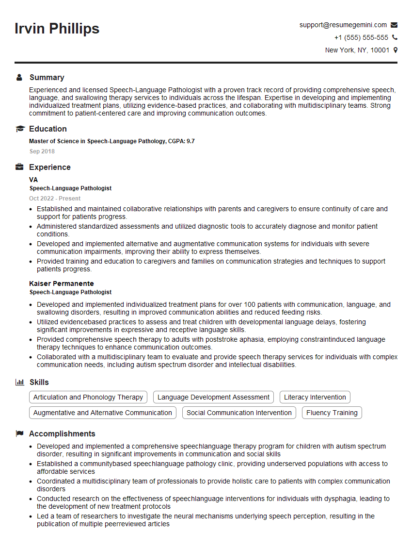
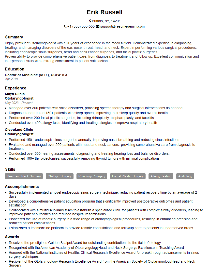
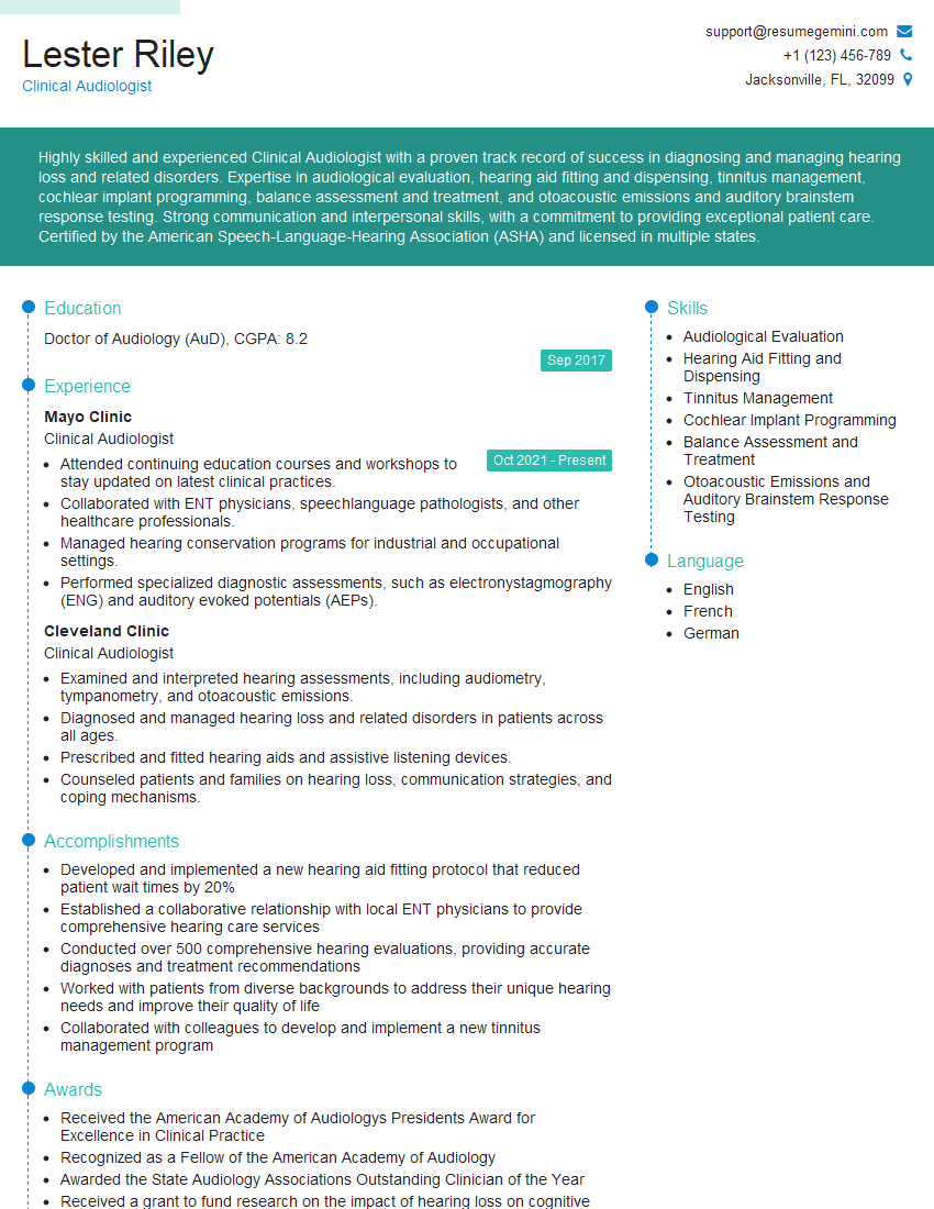
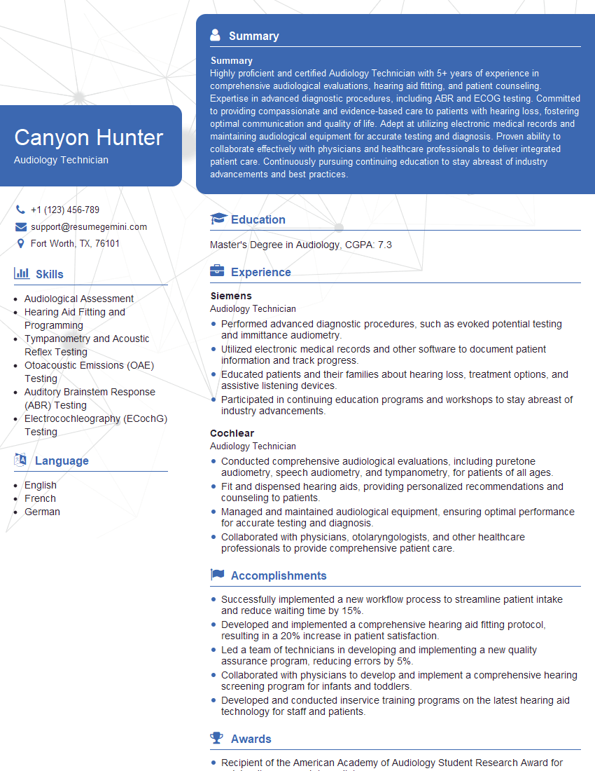
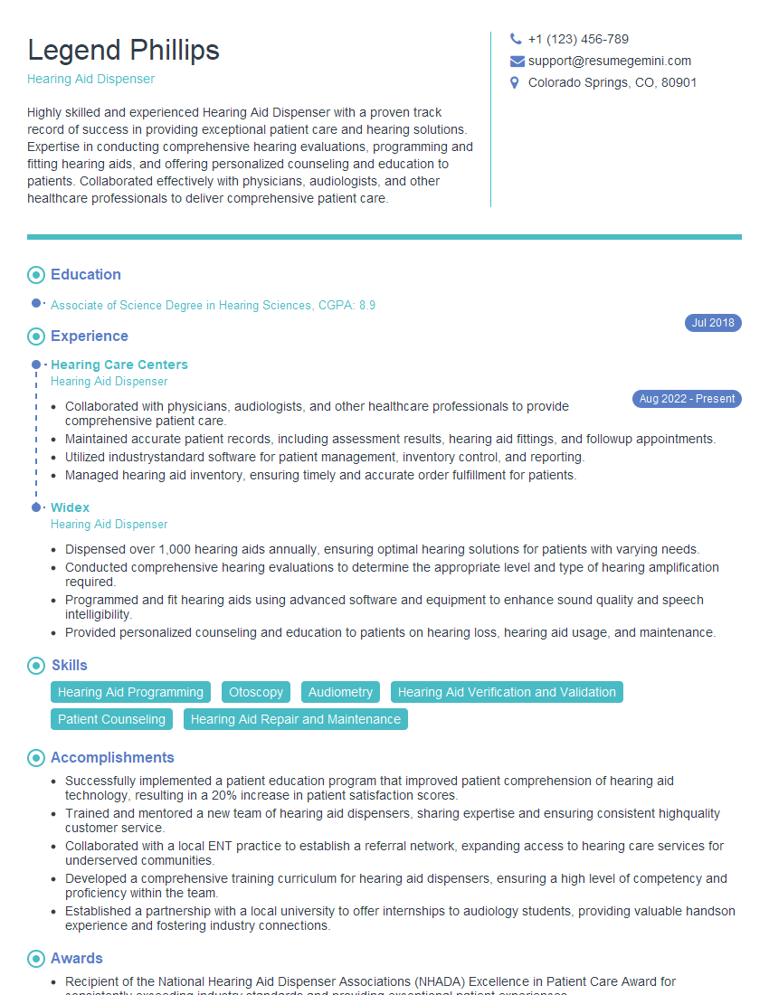
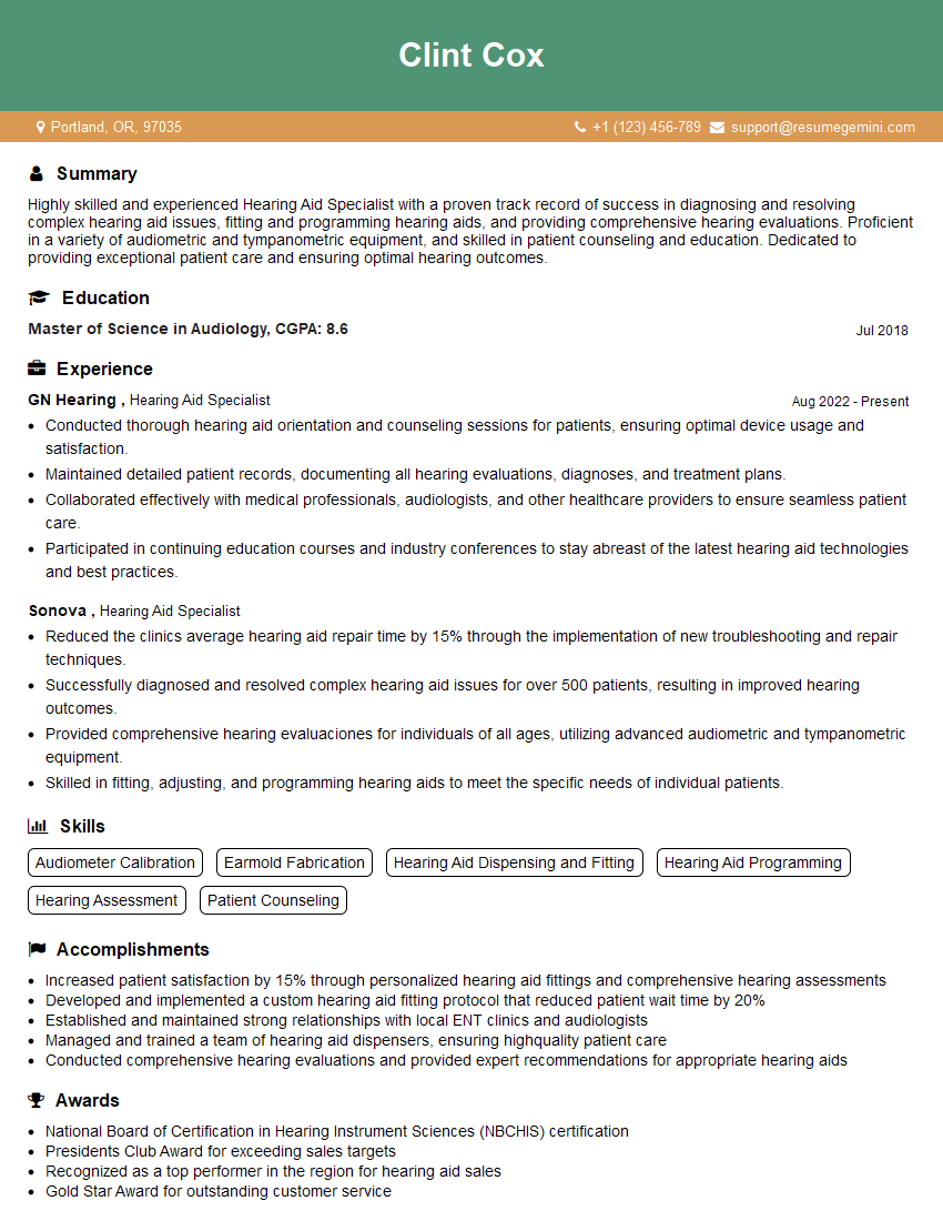
Mastering Middle Ear Analysis is crucial for career advancement in audiology, otolaryngology, and related fields. A strong understanding of these concepts will significantly enhance your interview performance and open doors to exciting opportunities. To maximize your job prospects, creating an ATS-friendly resume is essential. ResumeGemini is a trusted resource that can help you build a professional and impactful resume that gets noticed. We provide examples of resumes tailored to Middle Ear Analysis to help guide your creation process.
Explore more articles
Users Rating of Our Blogs
Share Your Experience
We value your feedback! Please rate our content and share your thoughts (optional).
What Readers Say About Our Blog
This was kind of a unique content I found around the specialized skills. Very helpful questions and good detailed answers.
Very Helpful blog, thank you Interviewgemini team.
