The thought of an interview can be nerve-wracking, but the right preparation can make all the difference. Explore this comprehensive guide to Otolaryngology Examination interview questions and gain the confidence you need to showcase your abilities and secure the role.
Questions Asked in Otolaryngology Examination Interview
Q 1. Describe the proper technique for performing an otoscopic examination.
Performing an otoscopic examination requires a gentle yet systematic approach. It’s crucial to ensure patient comfort and minimize any potential discomfort. First, you’ll need a good quality otoscope with a speculum of appropriate size. Then, follow these steps:
- Patient Positioning: Position the patient comfortably, either sitting or lying down, ensuring their head is tilted slightly away from you.
- External Ear Examination: Begin by visually inspecting the auricle (outer ear) for any deformities, lesions, redness, or discharge. Palpate for tenderness.
- Otoscopic Examination: Gently grasp the auricle and straighten the ear canal using the technique appropriate for the patient’s age (gentle upward and backward pull for adults; downward and backward for infants and young children). This straightens the ear canal, allowing better visualization. Insert the speculum slowly and carefully into the external auditory canal, avoiding forceful insertion which could damage the tympanic membrane. Observe the canal for cerumen (earwax), foreign bodies, erythema (redness), swelling, or discharge.
- Tympanic Membrane Examination: Once you have a clear view, examine the tympanic membrane (eardrum) for its color, light reflex (cone of light), landmarks, integrity (perforations), and any signs of inflammation or fluid.
- Documentation: Meticulously document your findings, including the presence of cerumen, any abnormalities, and the overall appearance of the tympanic membrane.
Example: A patient presents with ear pain. During the otoscopic examination, I observed a bulging, erythematous tympanic membrane with decreased mobility and absent light reflex, highly suggestive of acute otitis media (middle ear infection).
Q 2. How do you differentiate between conductive and sensorineural hearing loss during an examination?
Differentiating between conductive and sensorineural hearing loss involves a combination of history, physical examination, and audiometric testing. However, some clues can be gathered during the initial examination:
- Conductive Hearing Loss: This occurs when sound transmission is impaired in the outer or middle ear. Patients may report muffled hearing, and the physical examination might reveal cerumen impaction, otitis media, or ossicular chain dysfunction. Weber and Rinne tests can help differentiate. In a Weber test, sound lateralizes to the affected ear. In a Rinne test, bone conduction is greater than air conduction (BC > AC).
- Sensorineural Hearing Loss: This arises from damage to the inner ear or auditory nerve. Patients may report difficulty understanding speech, particularly in noisy environments. Physical exam might reveal nothing specific to the ear itself; the problem lies deeper. Weber test shows lateralization to the unaffected ear. Rinne test shows air conduction greater than bone conduction (AC > BC), though the overall hearing may be reduced.
It is important to remember that these bedside tests are screening tools and don’t replace formal audiometry. A complete audiological evaluation provides a precise diagnosis and identifies the nature and degree of hearing loss.
Q 3. Explain the steps involved in a comprehensive nasal examination.
A comprehensive nasal examination involves a systematic assessment of the external nose and the internal nasal cavity. The steps are:
- External Examination: Inspect the external nose for symmetry, deformities (e.g., deviation of the nasal septum), skin lesions, or any signs of trauma or inflammation.
- Rhinoscopy: Using a nasal speculum, gently insert it into each nasal cavity. Inspect the nasal mucosa for color, swelling, discharge (character, amount, color), and the presence of polyps, foreign bodies, or masses. Assess the patency of each nasal airway.
- Palpation: Gently palpate the nasal bones and sinuses for tenderness. This helps to assess for sinus inflammation.
- Examination of the nasal septum: Look for any deviation or perforation of the nasal septum. A deviated septum can obstruct airflow and contribute to sinusitis or nasal congestion.
- Assessment of the turbinates: Inspect the inferior, middle, and superior turbinates for size, swelling, and presence of inflammation.
Example: A patient with chronic nasal congestion reveals pale, boggy turbinates and clear rhinorrhea during rhinoscopy. This suggests allergic rhinitis.
Q 4. What are the key findings indicative of acute sinusitis during a physical examination?
Key findings indicative of acute sinusitis during a physical examination include:
- Facial Tenderness: Palpating over the sinuses (frontal and maxillary) may elicit tenderness, particularly in acute sinusitis. The patient might describe a deep, aching pain in the affected area.
- Nasal Congestion: Obstruction of the nasal passages is a common symptom. Rhinoscopy might reveal purulent or mucopurulent nasal discharge.
- Nasal Discharge: Thick, purulent (pus-like), colored nasal discharge is suggestive of infection.
- Fever: Patients with acute sinusitis often present with fever.
- Altered Sense of Smell (Hyposmia or Anosmia): Sinus inflammation can lead to reduced or loss of sense of smell.
- Facial Swelling: In severe cases, facial swelling may be present.
It’s essential to note that these findings are suggestive, but not diagnostic. Imaging studies like CT scans or X-rays are often needed for definitive diagnosis.
Q 5. How would you assess vocal cord function during a laryngeal examination?
Assessing vocal cord function during a laryngeal examination involves a combination of visual inspection and functional testing. The primary method is indirect laryngoscopy, which involves using a laryngeal mirror to visualize the larynx.
- Indirect Laryngoscopy: The patient is asked to phonate (make sounds) various vowels, and the examiner observes the vocal cord movement, symmetry, and closure during phonation. The examiner also assesses the color, texture, and presence of any lesions on the vocal cords.
- Stroboscopy: This advanced technique provides a magnified, slow-motion view of the vocal cords during phonation, allowing for a detailed assessment of vibratory patterns and identification of subtle abnormalities.
- Assessment of Voice Quality: The examiner assesses the voice quality (e.g., breathiness, hoarseness, roughness) in conversation to help identify underlying vocal cord dysfunction.
- Cough Assessment: Observing the patient’s cough can provide additional clues regarding vocal cord function and coordination.
Example: During indirect laryngoscopy, the patient’s vocal cords demonstrated incomplete closure during phonation, indicating vocal cord paralysis.
Q 6. Describe the examination techniques used to evaluate swallowing function.
Evaluating swallowing function, also known as dysphagia assessment, is a multi-faceted process that involves a thorough history, clinical examination, and often instrumental studies.
- History: Detailed information about the nature of the swallowing difficulty, including the onset, duration, consistency of foods affected, presence of choking or coughing episodes, and any associated symptoms.
- Clinical Examination: This involves assessing the patient’s oral cavity, oropharynx, and neck for any structural abnormalities that could contribute to dysphagia. This includes assessment of cranial nerves related to swallowing (CN V, VII, IX, X, XII).
- Bedside Swallowing Assessment: The patient is asked to swallow different consistencies of food and water (thin liquids, nectar-thick, pudding thick) while being observed for any signs of aspiration, coughing, or difficulty with swallowing. This also assesses the efficiency and coordination of the swallowing process.
- Instrumental Studies: Fiberoptic endoscopic evaluation of swallowing (FEES) or videofluoroscopic swallow study (VFSS) provides a detailed visual assessment of the swallowing process.
Example: A patient with stroke might present with difficulty swallowing liquids, and a bedside examination might reveal reduced oral sensation and weakness of the pharyngeal muscles. A VFSS can identify the specific stage of swallowing that’s impaired and help tailor management strategies.
Q 7. Explain your approach to examining a patient with suspected neck masses.
Examining a patient with suspected neck masses requires a systematic and thorough approach:
- History Taking: A detailed history including the duration, rate of growth, associated symptoms (pain, dysphagia, dyspnea, hoarseness), medical history, and family history is crucial. Understanding the patient’s symptoms helps guide the examination and narrow down potential diagnoses.
- Physical Examination: Begin with a general examination, looking for any signs of systemic illness. Next, focus on the neck:
- Inspection: Observe the size, location, shape, color, and mobility of the mass. Note any overlying skin changes (erythema, ulceration).
- Palpation: Gently palpate the mass to assess its size, consistency (hard, soft, fluctuant), mobility, tenderness, and any fixation to underlying structures.
- Auscultation: Listen for bruits (vascular sounds) over the mass, suggestive of a vascular lesion.
- Assessment of regional lymph nodes: Palpate the cervical lymph nodes to assess for enlargement, tenderness, and consistency, as these may be involved in metastatic disease.
- Further Investigations: Based on the history and physical examination, further investigations such as ultrasound, CT scan, or fine-needle aspiration biopsy may be necessary to establish the diagnosis.
Example: A patient presents with a painless, slowly growing, firm neck mass. Examination reveals a mobile, non-tender mass in the anterior neck, with no associated lymphadenopathy. Further imaging and biopsy could be indicated to differentiate between a benign thyroid nodule and other possibilities.
Q 8. What are the common signs and symptoms of a peritonsillar abscess, and how would you assess for it?
A peritonsillar abscess (PTA), also known as a Quinsy, is a collection of pus behind the tonsil. It’s a serious complication of tonsillitis.
Signs and Symptoms: Patients typically present with severe unilateral throat pain, often radiating to the ear. They may have difficulty swallowing (odynophagia), drooling, muffled voice (hot potato voice), and trismus (difficulty opening their mouth). Fever, chills, and swollen lymph nodes in the neck are also common. A characteristic finding is an asymmetrically swollen tonsil with a bulging, fluctuant mass visible on examination.
Assessment: The assessment begins with a thorough history taking, focusing on the onset, severity, and characteristics of the pain. A physical examination is crucial. I would inspect the oropharynx carefully, looking for asymmetry, swelling, and the presence of a yellowish-white exudate. Gentle palpation of the affected area might reveal a fluctuant mass, but this should be done cautiously to avoid rupturing the abscess. Sometimes, it’s difficult to visualize the abscess due to the patient’s inability to open their mouth fully (trismus). In such cases, imaging like a CT scan might be necessary to confirm the diagnosis. I would also check for signs of airway compromise like respiratory distress.
Q 9. How would you manage epistaxis encountered during an examination?
Managing epistaxis (nosebleed) during an examination requires a calm and systematic approach. The first priority is to ensure the patient’s airway is clear and the bleeding is controlled.
Immediate Steps: I would have the patient sit upright and lean forward, pinching the soft part of their nose (just below the bony bridge) firmly for 10-15 minutes. This helps to apply direct pressure to the bleeding vessels. I would then assess the patient’s vital signs (blood pressure, pulse, respiration) and address any signs of shock. If the bleeding persists despite this, I’d use a topical vasoconstrictor like phenylephrine nasal spray or cotton pledgets soaked in it to help constrict the blood vessels.
Further Management: If the bleeding continues, I would consider anterior nasal packing with either ribbon gauze or a commercial nasal tampon. Posterior epistaxis, which is bleeding from the back of the nose, is more dangerous and may require posterior nasal packing or cauterization (using electrocautery or silver nitrate). Intravenous fluids and blood transfusion may be necessary in cases of significant blood loss. In severe cases, referral to an ENT specialist for further evaluation and management, including angiography, is crucial. I’d also review the patient’s medication history, as certain medications like anticoagulants can increase the risk of bleeding.
Q 10. Describe your approach to evaluating a patient with vertigo.
Evaluating a patient with vertigo requires a systematic approach to differentiate between different causes. Vertigo is the sensation of spinning or whirling, often accompanied by nausea and vomiting.
History: I would start by obtaining a detailed history, focusing on the character of vertigo (rotational vs. non-rotational), its onset, duration, frequency, and associated symptoms such as hearing loss, tinnitus (ringing in the ears), imbalance, and neurological symptoms. I’d inquire about any precipitating factors (e.g., head position, certain activities) and any relevant medical history.
Physical Examination: A comprehensive neurological exam is critical, assessing cranial nerves (particularly those related to balance), cerebellar function, gait, and coordination. Otoscopic examination is crucial to assess for ear pathology. The Dix-Hallpike maneuver, a specific test for benign paroxysmal positional vertigo (BPPV), may be performed. I would also evaluate for other medical conditions potentially contributing to vertigo such as cardiovascular disease, neurological disorders, and inner ear infections.
Further Investigations: Depending on the clinical findings, further investigations may include audiometry, electronystagmography (ENG), or magnetic resonance imaging (MRI) of the brain and inner ear to identify the underlying cause. Differentiating between central (brain-related) and peripheral (inner ear-related) causes of vertigo is crucial for appropriate management.
Q 11. Explain the difference between otitis media with effusion and acute otitis media.
Both otitis media with effusion (OME) and acute otitis media (AOM) are middle ear infections, but they differ significantly in their presentation and management.
Otitis Media with Effusion (OME): This involves the presence of fluid in the middle ear without signs of acute infection. It’s often asymptomatic, though patients may experience a feeling of fullness or muffled hearing. The tympanic membrane (eardrum) typically appears retracted and dull, with the presence of fluid behind it visible on otoscopy. OME often follows an episode of AOM or an upper respiratory infection.
Acute Otitis Media (AOM): This is an acute infection of the middle ear characterized by inflammation and the accumulation of purulent (pus-containing) fluid. Patients typically experience ear pain (otalgia), fever, irritability, and hearing loss. Otoscopic examination reveals a bulging, erythematous (reddened) and opaque tympanic membrane.
Key Differences Summarized:
- OME: Fluid in the middle ear, no infection, often asymptomatic, retracted tympanic membrane.
- AOM: Infection, purulent fluid, ear pain, fever, bulging and red eardrum.
Management differs significantly. OME often resolves spontaneously, while AOM often requires antibiotic treatment. My approach would include a thorough history and examination, paying particular attention to the tympanic membrane.
Q 12. How do you differentiate between allergic rhinitis and a viral upper respiratory infection?
Differentiating between allergic rhinitis and a viral upper respiratory infection (URI) can be challenging as they share some symptoms, but key differences exist.
Allergic Rhinitis: This is an IgE-mediated hypersensitivity reaction to allergens (e.g., pollen, dust mites). Symptoms typically include itchy eyes, nose, and throat; sneezing; clear, watery rhinorrhea (runny nose); and nasal congestion. These symptoms tend to be seasonal or related to specific triggers.
Viral URI: This is caused by a viral infection. Symptoms can include sneezing, rhinorrhea (which may be clear initially but can become thicker and more colored), nasal congestion, cough, sore throat, and sometimes fever. These symptoms typically last for several days and resolve spontaneously.
Key Differentiating Factors:
- Itchiness: Significant itchiness of the eyes, nose, and throat is strongly suggestive of allergic rhinitis.
- Seasonal Pattern: Allergic rhinitis often follows a seasonal pattern, whereas viral URIs can occur at any time.
- Duration: Viral URIs typically resolve within a week or two, whereas allergic rhinitis symptoms can persist for longer periods if exposure to the allergen continues.
- Clear Rhinorrhea: While viral rhinorrhea can start clear, it usually becomes thicker and changes color.
A detailed history, focusing on the pattern and timing of symptoms, as well as the presence or absence of itchiness, is crucial for making the distinction. Further investigations, such as allergy testing, might be considered if the diagnosis remains unclear.
Q 13. What are the key components of a thorough head and neck examination?
A thorough head and neck examination is crucial in otolaryngology and involves a systematic assessment of several key areas.
Key Components:
- Inspection: This involves a visual examination of the entire head and neck region, looking for symmetry, swelling, masses, skin lesions, or scars. Note the patient’s facial expression and any signs of distress.
- Palpation: Palpable masses, lymph nodes (checking for size, consistency, tenderness), and the temporomandibular joint are assessed. Tenderness to palpation can suggest underlying infection or inflammation.
- Otoscopy: Examination of the ears using an otoscope to visualize the external auditory canal and tympanic membrane. This helps in detecting cerumen (earwax), foreign bodies, infections, or other abnormalities.
- Rhinoscopy: Inspection of the nasal cavity using a nasal speculum to assess the nasal mucosa, septum, and turbinates. This allows for detection of nasal polyps, deviations of the nasal septum, or signs of inflammation or infection.
- Oropharyngeal Examination: Inspection of the mouth, throat, and tonsils. This involves examining the tongue, teeth, gums, palate, uvula, and tonsils for lesions, inflammation, or any other abnormality. A tongue depressor is used to facilitate visualization.
- Neck Examination: Assessment of the neck for masses, lymph node enlargement, or thyroid abnormalities. The range of neck motion is also evaluated.
The order may vary based on the patient’s presentation and the suspected pathology. I would systematically proceed through each area to ensure no abnormalities are missed. Detailed documentation of findings is essential.
Q 14. Describe the use of a nasal speculum in ENT examination.
The nasal speculum is an essential instrument in ENT examination for visualizing the interior of the nasal cavity.
Use in ENT Examination: The speculum consists of two blades that can be gently spread apart to open the nasal passage, providing clear visualization of the nasal mucosa, septum, and turbinates. It allows for assessment of various conditions such as:
- Nasal Polyps: These are benign growths that can obstruct the nasal passages. The speculum allows for easy visualization of their size, location, and number.
- Nasal Septum Deviation: A deviated septum is a common cause of nasal obstruction. The speculum allows for evaluation of the extent of the deviation.
- Nasal Mucosal Inflammation: Signs of inflammation, such as swelling, redness, or bleeding, can indicate the presence of infections or allergic rhinitis.
- Foreign Bodies: The speculum can help identify and remove foreign bodies lodged in the nasal cavity.
- Nasal Tumors: While less common, the speculum plays a role in early detection of certain nasal tumors.
Proper technique involves gentle insertion and spreading of the speculum blades, avoiding undue force. The light source of the otoscope or head lamp is used for optimal illumination. The patient should be positioned comfortably and instructed to breathe through their mouth. This tool is simple but crucial for a thorough nasal assessment.
Q 15. How do you assess for cranial nerve palsies relevant to otolaryngology?
Assessing cranial nerves relevant to otolaryngology involves a systematic evaluation of the nerves impacting hearing, balance, facial movement, and swallowing. We meticulously examine cranial nerves VII (facial), VIII (vestibulocochlear), IX (glossopharyngeal), X (vagus), XI (accessory), and XII (hypoglossal).
Cranial Nerve VII (Facial): Assessed by observing facial symmetry at rest and during voluntary movements like smiling, frowning, and raising eyebrows. Weakness or asymmetry suggests a facial nerve palsy.
Cranial Nerve VIII (Vestibulocochlear): Evaluated through hearing tests (Rinne and Weber, audiometry) and balance assessments (Romberg test, observing gait).
Cranial Nerves IX (Glossopharyngeal) and X (Vagus): Assessed together by examining the palate for symmetry during phonation (saying ‘ah’), checking gag reflex, and observing swallowing. Asymmetry or absent gag reflex may indicate a lesion.
Cranial Nerve XI (Accessory): Evaluated by testing the strength of the sternocleidomastoid and trapezius muscles. Weakness suggests a lesion affecting the nerve.
Cranial Nerve XII (Hypoglossal): Assessed by observing the tongue for fasciculations (involuntary muscle twitching) and atrophy, and testing tongue protrusion for deviation. Deviation suggests a lesion.
For instance, a patient presenting with unilateral facial droop and inability to close one eye might indicate a Bell’s palsy (CN VII lesion). A patient with vertigo and hearing loss may have a vestibular schwannoma (CN VIII lesion).
Career Expert Tips:
- Ace those interviews! Prepare effectively by reviewing the Top 50 Most Common Interview Questions on ResumeGemini.
- Navigate your job search with confidence! Explore a wide range of Career Tips on ResumeGemini. Learn about common challenges and recommendations to overcome them.
- Craft the perfect resume! Master the Art of Resume Writing with ResumeGemini’s guide. Showcase your unique qualifications and achievements effectively.
- Don’t miss out on holiday savings! Build your dream resume with ResumeGemini’s ATS optimized templates.
Q 16. What are the red flags that necessitate urgent referral in ENT?
In ENT, several ‘red flags’ mandate immediate referral to prevent serious complications. These include:
Sudden, unilateral hearing loss: This could signify a serious inner ear issue like sudden sensorineural hearing loss (SSNHL) or a neurological problem requiring immediate attention.
Severe or rapidly progressive vertigo: This necessitates prompt assessment to rule out stroke, labyrinthitis, or other critical conditions.
Facial nerve palsy with ear pain: This could indicate Ramsay Hunt syndrome, which requires antiviral treatment.
Epistaxis (nosebleed) that is uncontrollable or recurrent: Excessive or persistent bleeding could be life-threatening due to underlying conditions.
Neck mass with associated symptoms: This may indicate a malignancy requiring immediate investigation.
Dysphagia (difficulty swallowing) with respiratory compromise: This is a medical emergency potentially caused by a foreign body obstruction or an infection.
Unilateral otalgia (ear pain) with associated facial swelling: Suggests an infection such as peritonsillar abscess, which requires rapid intervention to prevent airway compromise.
The key is recognizing these signs early to initiate timely and appropriate management, potentially preventing irreversible complications.
Q 17. How would you interpret findings from a tympanogram?
A tympanogram is a graph that depicts the middle ear’s pressure and compliance. Interpreting a tympanogram involves analyzing the shape and pressure readings to assess the middle ear status.
Type A: Normal tympanogram. Indicates normal middle ear pressure and compliance.
Type B: Flat tympanogram. Suggests fluid in the middle ear (otitis media with effusion), perforated tympanic membrane, or ossicular discontinuity.
Type C: Negative middle ear pressure. Indicates Eustachian tube dysfunction, often seen in otitis media with effusion.
Type As: Reduced compliance. Suggests stiffness of the ossicular chain (e.g., otosclerosis).
Type Ad: Increased compliance. Suggests ossicular discontinuity or a flaccid tympanic membrane.
For example, a flat tympanogram (Type B) in a child with ear pain and hearing loss strongly suggests the presence of middle ear effusion. A Type C tympanogram suggests the need to address Eustachian tube dysfunction, possibly with decongestants or other therapies.
Q 18. Describe the process of performing a Rinne and Weber test.
The Rinne and Weber tests are quick bedside tests evaluating hearing.
Rinne Test: Compares air conduction (AC) to bone conduction (BC) hearing. A 512 Hz tuning fork is placed on the mastoid bone (BC), then moved near the ear canal (AC). A positive Rinne test (AC > BC) is normal. A negative Rinne (BC > AC) suggests conductive hearing loss.
Weber Test: Evaluates lateralization of sound. A vibrating 512 Hz tuning fork is placed on the midline of the forehead. In normal hearing, sound is perceived equally in both ears. In conductive hearing loss, sound lateralizes to the affected ear. In sensorineural hearing loss, sound lateralizes to the unaffected ear.
Imagine a patient with a negative Rinne test on the right ear; this suggests a conductive hearing loss in the right ear (e.g., middle ear infection). If the Weber test lateralizes to the right ear, it further supports the diagnosis of conductive hearing loss in that ear.
Q 19. How do you perform a voice assessment, including assessment of phonation and resonance?
Voice assessment includes evaluating phonation (voice production) and resonance (voice quality).
Phonation Assessment: Assesses voice quality, pitch, loudness, and breathiness. The patient is asked to sustain vowel sounds (‘ah’, ‘ee’, ‘oo’) to evaluate vocal fold function. Hoarseness or breathiness might suggest vocal fold lesions.
Resonance Assessment: Evaluates how sound resonates in the vocal tract. This is assessed by observing the patient’s speech quality and listening for hypernasality (excessive nasal resonance) or hyponasality (reduced nasal resonance). Hypernasality might indicate velopharyngeal insufficiency.
For example, a patient with a harsh voice and reduced loudness might have vocal nodules. A patient with hypernasality might have cleft palate. We may use tools like a laryngoscopy to visualize the vocal folds directly for a more thorough assessment.
Q 20. What are the limitations of a physical examination in diagnosing ENT conditions?
The physical examination in ENT has limitations. It provides crucial initial information but cannot definitively diagnose many conditions.
Limited visualization: Some structures are difficult to visualize without specialized imaging (e.g., inner ear structures).
Subtle findings: Early-stage diseases might not present with easily detectable physical signs.
Overlapping symptoms: Many ENT conditions share similar symptoms, making diagnosis challenging.
Inability to detect underlying pathology: Physical examination cannot always reveal the cause of the symptoms (e.g., distinguishing between benign and malignant lesions).
For instance, a patient with a persistent cough might have several causes, from simple postnasal drip to lung cancer. The physical examination alone might not distinguish between them; further imaging and investigations are needed.
Q 21. What imaging modalities are commonly used to complement the ENT examination?
Various imaging modalities complement the ENT examination to provide a more comprehensive assessment.
Computed Tomography (CT): Excellent for imaging bony structures and identifying fractures, tumors, and foreign bodies.
Magnetic Resonance Imaging (MRI): Best for visualizing soft tissues, including tumors, inflammation, and nerve structures.
Ultrasound: Useful for evaluating neck masses, salivary glands, and thyroid.
Fluoroscopy: Used for swallowing studies and assessing the movement of the vocal folds.
High-resolution CT of the temporal bone: Provides detailed images of the inner ear structures.
For example, CT scans help in evaluating temporal bone fractures. MRI is valuable for detecting brain tumors that extend into the nasal cavity. High-resolution CT of the temporal bone aids in the diagnosis of inner ear pathologies.
Q 22. How do you communicate examination findings to patients and other healthcare professionals?
Communicating examination findings is crucial for effective patient care and collaboration with other healthcare professionals. I employ a two-pronged approach: clear and concise communication with the patient and detailed, professional documentation for colleagues.
Patient Communication: I explain my findings in simple, non-medical terms, using analogies when necessary. For example, instead of saying ‘I found hyperemia in the posterior pharynx,’ I might say, ‘I noticed some redness at the back of your throat.’ I ensure the patient understands the diagnosis, treatment plan, and prognosis. I actively encourage questions and address concerns, ensuring the patient feels informed and empowered.
Communication with Other Professionals: My reports are comprehensive and adhere to standardized medical terminology. I use structured reporting formats that include a clear summary of the examination findings, a concise differential diagnosis, and a recommended treatment plan. This allows for seamless communication with referring physicians, specialists (e.g., audiologists, speech therapists), and other members of the healthcare team. For instance, when communicating with a colleague about a suspected vocal cord nodule, I would provide detailed information on its location, size, and appearance, as observed during laryngoscopy, alongside relevant patient history and the proposed management strategy.
Q 23. Describe your experience in performing flexible nasopharyngolaryngoscopy.
I have extensive experience performing flexible nasopharyngolaryngoscopy (FNLS), a crucial procedure in otolaryngology for visualizing the nasal cavity, pharynx, and larynx. My proficiency spans a wide range of patient presentations, from routine check-ups to the diagnosis and management of complex conditions. The procedure involves the careful insertion of a thin, flexible endoscope through the nose, allowing for detailed visualization of the upper aerodigestive tract. I am adept at handling various endoscopic techniques, including using magnification and specialized lenses to enhance visualization. I am also experienced in navigating challenging anatomies and managing potential complications, such as gag reflex or epistaxis.
Example: In a recent case, FNLS revealed a small, early-stage squamous cell carcinoma in the larynx. The detailed visualization allowed for precise biopsy and facilitated early intervention, leading to a positive patient outcome.
Q 24. Explain your approach to documenting your findings from an ENT examination.
Documentation of ENT examination findings is paramount for maintaining accurate medical records and facilitating continuity of care. My approach involves a structured system ensuring completeness and clarity.
I utilize a standardized template that includes sections for patient demographics, presenting complaint, history of present illness, past medical history, family history, social history, physical examination findings (organized by body system), and a summary and plan. Within the physical examination section, I meticulously document the findings of each part of the exam – including otoscopy (ear examination), rhinoscopy (nose examination), and laryngoscopy (throat examination) – using clear and concise medical terminology. I include descriptions of any abnormalities, noting their location, size, color, and consistency. Illustrations are sometimes added to clarify complex findings. For example, I’d detail the presence, location, and character of any lesions (e.g., ‘2mm pearly white papule on the right vocal fold’). The final section summarizes the key findings, differential diagnoses, and the recommended management plan, including investigations, treatment, and referrals if necessary.
Q 25. How do you ensure a safe and comfortable examination environment for patients?
Creating a safe and comfortable examination environment for patients is a priority. This starts with establishing rapport and explaining the procedure in detail. I ensure the examination room is clean, well-lit, and appropriately equipped. I use a comfortable examination chair and position the patient to minimize discomfort. For anxious patients, I offer breaks and reassurance throughout the examination. Maintaining privacy is also crucial; I ensure the patient’s modesty is respected at all times.
Example: For children, I use age-appropriate language, involve parents, and use distraction techniques such as toys to minimize anxiety during the examination.
Q 26. What measures do you take to prevent the transmission of infections during an ENT examination?
Infection control is paramount in an ENT examination. I strictly adhere to standard precautions, including hand hygiene, the use of gloves and other personal protective equipment (PPE), and proper disposal of instruments and materials. Instruments are sterilized between patients, and surfaces are cleaned and disinfected. I maintain a high level of awareness of my own hygiene practices and ensure the examination environment is meticulously maintained. For aerosol-generating procedures like FNLS, I would utilize appropriate PPE, including an N95 mask, to protect myself and the patient from possible transmission of infections.
Example: After each FNLS procedure, the endoscope is properly cleaned and sterilized according to hospital protocols, using enzymatic cleaners, high-level disinfection, and appropriate drying techniques.
Q 27. How do you handle a patient who is anxious or apprehensive about the ENT examination?
Addressing patient anxiety is crucial for a successful examination. I start by actively listening to the patient’s concerns and validating their feelings. I explain the procedure step-by-step, emphasizing that their comfort is my utmost priority. I encourage them to ask questions and address any misconceptions. I offer reassurance and provide choices whenever possible (e.g., allowing them to choose their position during the examination). For significant anxiety, I might suggest relaxation techniques such as deep breathing or offer a mild sedative if deemed appropriate after consultation and with the patient’s consent.
Example: For a patient expressing fear of gagging, I might use a topical anesthetic spray to numb the throat, or I might use a smaller diameter endoscope to help minimize the gag reflex. Building trust and understanding is key to creating a positive experience.
Key Topics to Learn for Otolaryngology Examination Interview
Preparing for your Otolaryngology examination interview requires a strategic approach. Focus on demonstrating a deep understanding of both theoretical knowledge and practical application. This will showcase your readiness to excel in the field.
- Anatomy and Physiology of the Head and Neck: Mastering the intricate anatomy of the ear, nose, and throat is foundational. Consider the implications of anatomical variations on surgical approaches and diagnostic interpretation.
- Diagnostic Procedures and Interpretation: Gain proficiency in interpreting imaging studies (CT, MRI), audiograms, and other diagnostic tests commonly used in otolaryngology. Be prepared to discuss the limitations and advantages of each technique.
- Surgical Techniques and Principles: Review common surgical procedures and their indications, contraindications, and potential complications. Understanding the underlying surgical principles is crucial.
- Medical and Surgical Management of ENT Diseases: Develop a strong understanding of the medical and surgical management strategies for a wide range of ENT conditions, including infections, tumors, and trauma. Focus on evidence-based practices.
- Communication and Patient Care: Demonstrate your skills in communicating effectively with patients, their families, and colleagues. Explain how you approach challenging patient interactions and ethical dilemmas.
- Current Research and Advances: Stay updated on the latest advancements in otolaryngology. Show your enthusiasm for the field by discussing recent publications or innovative techniques.
Next Steps
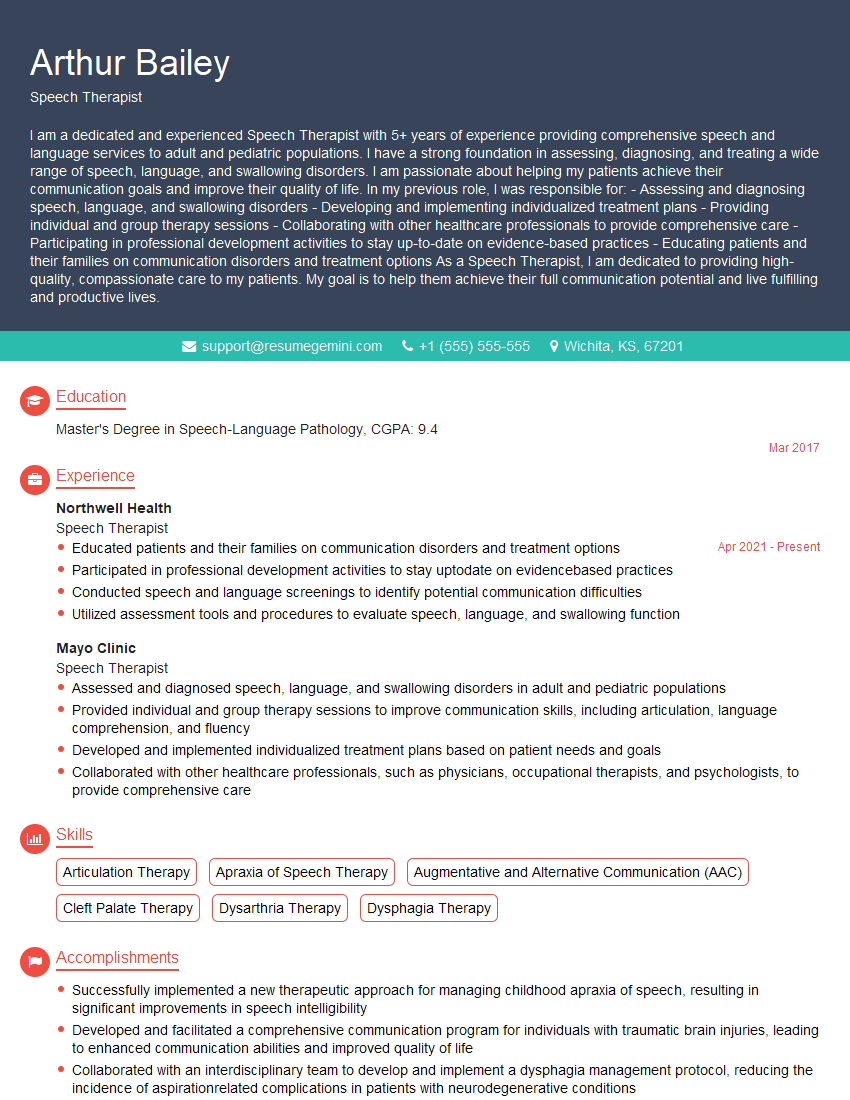
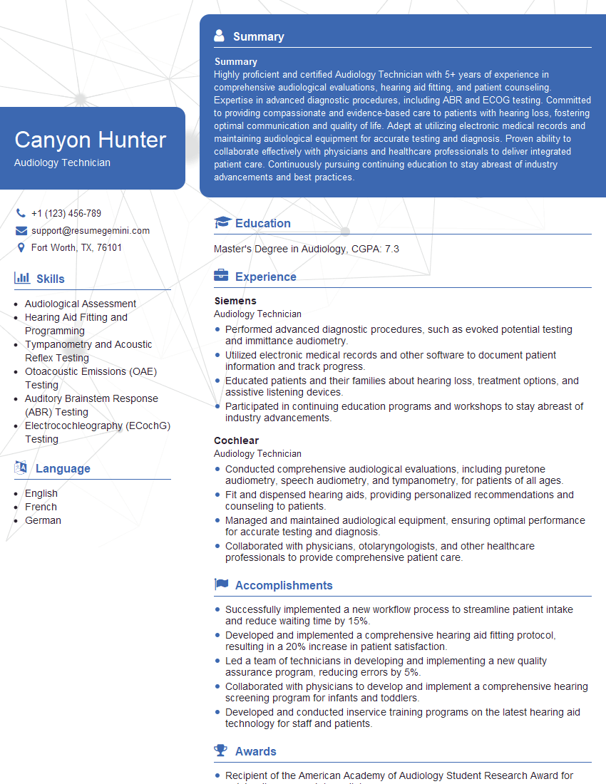
Mastering the Otolaryngology Examination is a significant step towards a successful and rewarding career. Your performance reflects your dedication and expertise, opening doors to prestigious residencies and impactful roles in the field. To further enhance your candidacy, creating an ATS-friendly resume is critical in getting your application noticed. ResumeGemini is a trusted resource that can help you build a compelling resume tailored to showcase your skills and experience effectively. Examples of resumes tailored to Otolaryngology Examination are available to guide you through this process. Invest time in presenting yourself professionally—it’s an investment in your future success.
Explore more articles
Users Rating of Our Blogs
Share Your Experience
We value your feedback! Please rate our content and share your thoughts (optional).
What Readers Say About Our Blog
This was kind of a unique content I found around the specialized skills. Very helpful questions and good detailed answers.
Very Helpful blog, thank you Interviewgemini team.
