Every successful interview starts with knowing what to expect. In this blog, we’ll take you through the top Otologic Surgery interview questions, breaking them down with expert tips to help you deliver impactful answers. Step into your next interview fully prepared and ready to succeed.
Questions Asked in Otologic Surgery Interview
Q 1. Describe your experience with stapedectomy procedures.
Stapedectomy is a microsurgical procedure to correct conductive hearing loss caused by otosclerosis, a condition where the stapes bone in the middle ear becomes fixed, hindering sound transmission. My experience encompasses hundreds of stapedectomies, utilizing both traditional and minimally invasive techniques.
The procedure involves creating a small opening in the eardrum (tympanotomy), removing the diseased stapes superstructure, and replacing it with a prosthesis, usually made of Teflon or titanium. This prosthesis restores the vibration pathway, allowing sound to reach the inner ear effectively. Post-operative care focuses on minimizing infection and monitoring hearing improvement. I’ve found that careful patient selection and meticulous surgical technique are key to maximizing success and minimizing complications like sensorineural hearing loss or facial nerve injury. For example, I recently performed a stapedectomy on a patient with significant hearing loss in one ear, and meticulous technique ensured a successful outcome with minimal discomfort.
Q 2. Explain the surgical techniques used for cholesteatoma removal.
Cholesteatoma removal is a complex procedure aimed at eliminating a cholesteatoma, a destructive skin growth in the middle ear. The surgical approach depends on the extent of the cholesteatoma.
For smaller cholesteatomas, a canal wall-up mastoidectomy might suffice, where the cholesteatoma is removed through the ear canal while preserving the intactness of the bony wall of the ear canal. In cases of larger cholesteatomas or those extending into the mastoid bone, a canal wall-down mastoidectomy is often necessary. This involves removing a portion of the ear canal’s bony wall to allow for complete cholesteatoma removal. The surgical goal is complete removal of all cholesteatoma tissue while preserving hearing function and preventing recurrence. Post-operative care involves careful monitoring for infection, wound healing assessment and sometimes a follow-up procedure for reconstruction.
For example, I recently managed a case of a large cholesteatoma involving the ossicles requiring a canal wall-down mastoidectomy followed by ossiculoplasty for hearing reconstruction. Careful planning and precise execution were crucial for a successful outcome.
Q 3. What are the common complications associated with cochlear implant surgery?
Cochlear implant surgery, while generally safe and effective, carries potential complications. These can be broadly categorized into surgical and post-surgical complications.
- Surgical Complications: These include bleeding, infection, cerebrospinal fluid leak, facial nerve injury, and damage to the labyrinth (inner ear).
- Post-surgical Complications: These can include device malfunction, infection, vertigo (dizziness), tinnitus (ringing in the ears), and varying degrees of hearing improvement. Some patients experience no significant improvement.
Minimizing these risks requires careful patient selection, meticulous surgical technique, and a thorough postoperative management plan including close follow-up appointments. For instance, a patient with pre-existing balance problems might require more intensive post-operative vestibular rehabilitation to aid their recovery.
Q 4. How do you manage Meniere’s disease surgically?
Meniere’s disease, an inner ear disorder causing vertigo, tinnitus, and hearing loss, is surgically managed when medical treatment fails. Surgical options include:
- Endolymphatic sac decompression: This procedure aims to relieve pressure within the inner ear by creating a larger space around the endolymphatic sac, believed to be a key factor in Meniere’s disease.
- Vestibular neurectomy: This involves severing the vestibular nerve, effectively eliminating vertigo but possibly worsening hearing. It is used as a last resort or for patients with predominant vertigo.
- Labyrinthectomy: This more radical procedure involves removal of the inner ear, completely eliminating hearing and balance function on the affected side. It’s rarely used due to its significant consequences.
The choice of surgical intervention depends on the severity of symptoms, the patient’s overall health and preferences, and the potential risks and benefits of each procedure. Careful pre-operative counseling is essential, weighing the possible improvements against the permanent sensory deficits.
Q 5. Discuss your experience with vestibular schwannomas.
Vestibular schwannomas, also known as acoustic neuromas, are benign tumors arising from the vestibular nerve. My experience includes both microsurgical and radiosurgical approaches.
Microsurgery involves removing the tumor through a small incision behind the ear, utilizing specialized microsurgical instruments and techniques to preserve facial nerve function and hearing. This approach is preferred for smaller tumors and when hearing preservation is a priority. Radiosurgery (Gamma Knife or CyberKnife), on the other hand, uses focused radiation to destroy the tumor without open surgery. This technique is better suited for larger tumors, those in difficult locations or for patients with high surgical risk factors. Post-operative care involves monitoring for complications such as hearing loss, facial nerve weakness, and cerebrospinal fluid leak. The choice between microsurgery and radiosurgery is highly individualized and depends on many factors including tumor size, location, and patient health and preferences.
Q 6. Explain the different types of hearing loss and their surgical management.
Hearing loss is classified into conductive, sensorineural, and mixed types.
- Conductive hearing loss: This occurs when sound transmission through the outer or middle ear is impaired. Surgical management includes procedures like tympanoplasty (eardrum repair), ossiculoplasty (middle ear bone reconstruction), and stapedectomy (as discussed earlier).
- Sensorineural hearing loss: This involves damage to the inner ear or auditory nerve. Surgical treatment options are limited, but cochlear implants can provide significant benefit for profound sensorineural hearing loss.
- Mixed hearing loss: This combines elements of both conductive and sensorineural hearing loss. Management strategies involve addressing both components, often with a combination of medical and surgical interventions.
Accurate diagnosis requires a thorough history and physical examination, including pure tone audiometry and other specialized tests. Surgical planning needs careful consideration of the underlying cause and patient’s expectations.
Q 7. Describe your approach to diagnosing and treating otosclerosis.
Otosclerosis is a disease causing progressive conductive hearing loss due to abnormal bone growth in the middle ear. Diagnosis involves a detailed history, physical examination, and audiometric testing. Imaging studies like temporal bone CT scan might be necessary to confirm the diagnosis and assess the extent of the bony involvement.
Treatment depends on the severity of hearing loss and the patient’s preference. For mild hearing loss, medical management with hearing aids might suffice. However, for significant hearing loss, stapedectomy, as described earlier, is the primary surgical intervention. It effectively restores hearing in a large proportion of patients. Post-operative care focuses on preventing infection and monitoring hearing improvement. Successful outcomes usually see significant improvement in hearing and quality of life.
Q 8. How do you assess candidacy for cochlear implantation?
Assessing candidacy for cochlear implantation is a multi-step process requiring a thorough evaluation of the patient’s hearing loss, overall health, and expectations. We begin with a comprehensive audiological assessment to determine the degree and type of hearing loss, confirming profound sensorineural hearing loss unsuitable for amplification. This includes pure-tone audiometry, speech audiometry, and acoustic immittance testing. We then perform imaging studies, typically high-resolution CT scans of the temporal bone, to visualize the cochlea and inner ear structures, assessing for anatomical abnormalities that might hinder implantation. Furthermore, a detailed medical history is obtained to identify any contraindications, such as active middle ear infections, significant neurological conditions, or bleeding disorders. Psychological evaluation might also be necessary to assess the patient’s understanding of the procedure and realistic expectations regarding outcomes. Finally, a multidisciplinary team discussion including the audiologist, surgeon, and speech therapist helps determine candidacy and create a personalized surgical plan. For example, a patient with profound sensorineural hearing loss, suitable anatomy, and realistic expectations would be a good candidate, while a patient with an active infection or significant neurological impairment would not.
Q 9. What are the key differences between ossiculoplasty techniques?
Ossiculoplasty aims to reconstruct the ossicular chain in the middle ear to improve sound transmission. Several techniques exist, each with its strengths and weaknesses.
- Total ossicular replacement prosthesis (TORP): This involves replacing the entire ossicular chain with a prosthetic device. It’s a relatively straightforward procedure suitable for various ossicular defects, but prosthesis malposition or extrusion is a potential complication.
- Partial ossiculoplasty: This technique involves repairing only the damaged portion of the ossicular chain, using either autologous (patient’s own tissue) or alloplastic (synthetic) materials. It is less invasive than TORP but requires precise surgical skill and might not be suitable for all types of ossicular defects. For instance, we might use a small piece of cartilage to reconstruct a partially eroded incus.
- Autologous ossiculoplasty: This uses the patient’s own tissue, such as cartilage or bone, to reconstruct the ossicular chain. It minimizes the risk of rejection or infection, but material availability and the technical challenges of sculpting the graft can make this challenging.
The choice of technique depends on the extent and nature of the ossicular disruption, the surgeon’s experience, and the patient’s specific needs. A thorough preoperative assessment and imaging are crucial for selecting the most appropriate technique.
Q 10. Explain your approach to managing acute mastoiditis.
Acute mastoiditis is a serious infection of the mastoid air cells requiring prompt and aggressive treatment. My approach begins with intravenous antibiotics, tailored to cover common pathogens like Streptococcus pneumoniae and Haemophilus influenzae. Broad-spectrum coverage is initially employed, often adjusted after culture results are available. I perform a thorough otological examination, assessing for signs of middle ear effusion, inflammation, and tenderness over the mastoid. High-resolution CT scans are obtained to visualize the extent of the infection and rule out intracranial complications. Urgent surgical intervention, usually a mastoidectomy, is indicated in cases of significant swelling, severe pain, evidence of intracranial involvement (like sigmoid sinus thrombosis), or lack of response to antibiotics. The surgery aims to drain the infected mastoid air cells, providing adequate ventilation and debridement of necrotic tissue. Postoperative management includes continued intravenous antibiotics, pain control, and close monitoring for signs of complications. For instance, a patient presenting with severe mastoid pain, fever, and evidence of intracranial extension on CT would be an immediate surgical candidate. Conversely, a patient with mild symptoms and good response to antibiotics might be managed conservatively.
Q 11. How do you manage temporal bone fractures?
Management of temporal bone fractures requires a multidisciplinary approach involving neurosurgery and otolaryngology. The initial assessment includes a thorough neurological examination to assess for cranial nerve palsies, cerebrospinal fluid (CSF) leak, and hearing loss. High-resolution CT scans are crucial to delineate the fracture pattern and identify any associated injuries, such as ossicular disruptions or labyrinthine involvement. Management strategies vary depending on the fracture type and associated complications. Non-displaced fractures with minimal associated injuries can be managed conservatively with close observation and serial audiological monitoring. Displaced fractures, CSF leaks, or significant ossicular disruption typically require surgical intervention. Surgical techniques range from simple ossiculoplasty for conductive hearing loss to more complex procedures like craniotomy for CSF leak repair or dural reconstruction. For example, a longitudinal fracture with a conductive hearing loss might be treated with an ossiculoplasty, while a transverse fracture with a CSF leak might necessitate a craniotomy. Postoperative management involves close monitoring for complications such as infection, meningitis, and sensorineural hearing loss.
Q 12. Discuss your experience with translabyrinthine approaches to skull base lesions.
The translabyrinthine approach is a surgical technique used to access lesions of the posterior fossa and petrous apex. I have extensive experience using this approach for various skull base lesions, including glomus tumors, meningiomas, and cholesterol granulomas. This approach involves a posterior-to-anterior opening of the temporal bone, sacrificing the inner ear structures to achieve wide surgical exposure. While it results in permanent hearing loss, it provides excellent access to lesions in the cerebellopontine angle and petrous apex. Preoperative planning involves careful review of high-resolution CT and MRI scans to understand the tumor’s extent and relationship to adjacent structures. Intraoperative neuromonitoring is employed to protect vital cranial nerves during the procedure. The benefits include excellent exposure, relatively low morbidity in experienced hands, and effective tumor removal. A key consideration is the patient’s hearing status prior to surgery, as the translabyrinthine approach is not suitable for patients who place a high value on preserving their hearing. Postoperative care focuses on monitoring for complications like cerebrospinal fluid leakage, infection, and cranial nerve deficits. A thorough understanding of anatomy and meticulous surgical technique are paramount for minimizing risks associated with this approach.
Q 13. How do you counsel patients on the risks and benefits of otologic surgery?
Counseling patients about the risks and benefits of otologic surgery is a crucial aspect of my practice. I strive to provide comprehensive and clear information tailored to each patient’s individual circumstances. Discussions begin by explaining the diagnosis and available treatment options, highlighting the advantages and limitations of each. The risks of otologic surgery are meticulously explained, including potential complications such as bleeding, infection, facial nerve injury, hearing loss (conductive or sensorineural), dizziness, and tinnitus. I emphasize the importance of realistic expectations. For instance, in cochlear implantation, I explain that while hearing improvement is expected, it may not restore normal hearing. I utilize visual aids, diagrams, and patient-friendly language to improve understanding. I encourage patients to ask questions and address their concerns. Furthermore, I often involve family members in these discussions to ensure a shared understanding and support system. The informed consent process is thoroughly documented, confirming the patient’s clear comprehension of the risks, benefits, and alternatives before proceeding with the surgery.
Q 14. Describe your experience with endoscopic ear surgery.
Endoscopic ear surgery is a minimally invasive technique gaining popularity in otology. My experience with this approach includes the management of cholesteatomas, chronic otitis media, and ossicular chain reconstruction. Endoscopic techniques offer several advantages, including improved visualization of the middle ear and mastoid, smaller incisions, reduced trauma, and faster recovery time compared to traditional microsurgical approaches. However, a steep learning curve and specialized equipment are required. Success with endoscopic ear surgery requires meticulous surgical skills, a thorough understanding of three-dimensional anatomy, and the ability to navigate the complex anatomical spaces of the temporal bone. The use of high-definition endoscopes and image-guided navigation systems further enhances the precision and safety of the procedure. While I have extensive experience with microscopic otologic surgery, endoscopic techniques are often preferred for select cases where minimal invasiveness and excellent visualization are paramount. Ongoing research and technological advancements continue to expand the applications of endoscopic ear surgery.
Q 15. What are your preferred methods for monitoring intraoperative hearing?
Intraoperative hearing monitoring is crucial in otologic surgery to minimize the risk of hearing loss. My preferred methods involve a combination of techniques, tailored to the specific surgical procedure. This often begins with preoperative audiometry to establish a baseline. During surgery, I rely heavily on electrocochleography (ECochG), which measures the electrical activity of the cochlea in response to auditory stimuli. This provides real-time feedback on cochlear function. For certain procedures, particularly those involving the stapes, I may also use intraoperative acoustic reflexes testing, monitoring the stapedial reflex to assess the integrity of the ossicular chain. Finally, continuous monitoring of the patient’s response to auditory stimuli, often by having the patient raise their hand or verbally confirm hearing tones during certain stages of surgery is crucial.
Imagine it like this: ECochG is like a sophisticated listening device monitoring the health of the inner ear while acoustic reflex testing gives us an indication of the middle ear’s function. Patient response adds a crucial subjective element. Combining these methods allows for a comprehensive assessment of hearing during the procedure.
Career Expert Tips:
- Ace those interviews! Prepare effectively by reviewing the Top 50 Most Common Interview Questions on ResumeGemini.
- Navigate your job search with confidence! Explore a wide range of Career Tips on ResumeGemini. Learn about common challenges and recommendations to overcome them.
- Craft the perfect resume! Master the Art of Resume Writing with ResumeGemini’s guide. Showcase your unique qualifications and achievements effectively.
- Don’t miss out on holiday savings! Build your dream resume with ResumeGemini’s ATS optimized templates.
Q 16. Explain the role of imaging in the preoperative assessment of otologic pathology.
Preoperative imaging plays a vital role in accurately characterizing otologic pathology. High-resolution computed tomography (CT) scans are essential for visualizing the bony structures of the temporal bone, identifying ossicular chain abnormalities, cholesteatoma size and location, and assessing the extent of erosion in cases like otitis media. Magnetic resonance imaging (MRI) provides superior soft tissue contrast, helping us delineate the extent of inner ear involvement, such as in cases of acoustic neuroma or vestibular schwannomas. Furthermore, advanced techniques like high-resolution CT with 3D reconstruction allow for detailed preoperative planning, especially in complex cases requiring stapedectomy or mastoidectomy.
For example, a patient presenting with hearing loss and dizziness might undergo both CT and MRI scans. The CT will help assess the ossicular chain, while the MRI helps to rule out an inner ear tumor. This integrated approach ensures a precise diagnosis and optimizes surgical planning.
Q 17. How do you interpret audiometric results?
Interpreting audiometric results requires a systematic approach, combining pure-tone audiometry, speech audiometry, and impedance audiometry. Pure-tone audiometry assesses the thresholds of hearing at various frequencies. Speech audiometry measures how well a patient understands speech, helping to differentiate between conductive and sensorineural hearing loss. Impedance audiometry evaluates middle ear function. I look for patterns, such as a conductive hearing loss (problem with the middle ear), sensorineural hearing loss (problem with the inner ear or nerve), or mixed hearing loss. Air-bone gaps are crucial in differentiating conductive from sensorineural components. I also pay attention to speech discrimination scores, which indicate the patient’s ability to understand speech, even if hearing thresholds are within a normal range. A complete analysis requires consideration of the patient’s age, medical history and the results of other diagnostic tests.
For instance, a patient with a significant air-bone gap on pure-tone audiometry suggests a conductive hearing loss, possibly due to otosclerosis or middle ear fluid. This finding would then be confirmed and further characterized with impedance audiometry.
Q 18. Describe your experience with managing post-operative complications.
Managing postoperative complications requires prompt recognition and effective intervention. Common complications include postoperative bleeding, infection, vertigo, and persistent or worsening hearing loss. Postoperative bleeding is addressed with careful hemostasis during surgery and potentially surgical revision if necessary. Infection is treated aggressively with intravenous antibiotics, often guided by culture and sensitivity results. Vertigo is often managed medically with vestibular suppressants and physical therapy. Persistent or worsening hearing loss may necessitate further evaluation and sometimes revision surgery. Close monitoring, patient education, and prompt intervention are critical to managing these complications effectively.
I recall a case where a patient developed a postoperative infection following a mastoidectomy. Immediate initiation of broad-spectrum antibiotics, along with surgical debridement, successfully resolved the infection and prevented further complications. Regular follow-up and open communication with the patient are paramount in addressing any concerns and providing support.
Q 19. How do you utilize advanced imaging techniques (e.g., CT, MRI) in otology?
Advanced imaging techniques are invaluable tools in otology. High-resolution CT provides detailed anatomical information of the temporal bone, crucial for planning complex procedures like cochlear implantation or skull base surgery. 3D CT reconstructions allow for better visualization of intricate structures. MRI is essential for evaluating soft tissue structures like the facial nerve, labyrinth, and tumors, offering superior contrast compared to CT. Diffusion-weighted imaging can help assess tumor viability. These advanced techniques allow for precise preoperative planning, intraoperative navigation, and accurate postoperative assessment.
For example, in a case of an acoustic neuroma, MRI allows precise delineation of tumor size and relationship to cranial nerves, which is crucial in surgical planning. CT would be less helpful for evaluating the tumour itself, but important in assessing the bone.
Q 20. Explain your knowledge of different types of hearing aids and their applications.
There’s a wide range of hearing aids available, each with its own application based on the degree and type of hearing loss. Behind-the-ear (BTE) aids are suitable for various hearing losses, especially severe ones. They are generally robust and have powerful amplification. In-the-ear (ITE) and in-the-canal (ITC) aids are smaller and less visible but might offer less amplification. Completely-in-canal (CIC) aids are the smallest, offering discretion but sometimes less user-friendly. Bone-conduction hearing aids bypass the middle ear and are useful for conductive hearing losses or single-sided deafness. Selection also involves digital versus analog, directional microphones, Bluetooth compatibility, and telecoil technology for phone usage.
Choosing the right hearing aid involves a comprehensive evaluation of the patient’s hearing loss, lifestyle, dexterity, and preferences. I always discuss the pros and cons of each option with my patients, ensuring the selection aligns with their individual needs.
Q 21. How do you manage vertigo and dizziness in your patients?
Managing vertigo and dizziness requires a multi-faceted approach. A thorough history, including the character of the symptoms, triggers, and associated neurological findings, is essential. The diagnosis is often made through clinical evaluation and in some cases, specific tests, such as the head impulse test (HIT), video head impulse test (vHIT), and caloric testing. Medical management may involve vestibular suppressants to reduce symptoms, often during the acute phase. Vestibular rehabilitation therapy (VRT) plays a crucial role in retraining the vestibular system, improving balance and reducing dizziness. In some cases, surgical intervention, such as a vestibular neurectomy, might be considered for refractory cases.
For instance, a patient with benign paroxysmal positional vertigo (BPPV) might benefit from canalith repositioning maneuvers (CRP), a non-surgical treatment aimed at repositioning displaced otoconia within the inner ear. For those with Ménière’s disease, a combination of medical management and VRT are usually the first lines of action. Surgery could be considered as a last resort if medication doesn’t provide adequate relief.
Q 22. What are the current advancements in otologic surgery?
Current advancements in otologic surgery are revolutionizing the field, improving outcomes and minimizing invasiveness. These advancements span several key areas:
- Minimally Invasive Techniques: Endoscopic ear surgery is becoming increasingly prevalent, allowing surgeons to access the middle ear and mastoid through smaller incisions, leading to reduced trauma, faster healing, and less scarring. For example, endoscopic techniques are now widely used in cholesteatoma removal and stapedectomy.
- Image-Guided Surgery: Intraoperative imaging, such as CT scans and fluoroscopy, provides real-time visualization of the delicate structures within the temporal bone, enhancing precision and safety, especially in complex cases. This helps to avoid injury to the facial nerve or inner ear structures.
- Advanced Implants: We’re seeing improved biocompatibility and longevity in middle ear implants, such as ossicular prostheses and implantable hearing aids. These are designed for better integration with the body and more durable performance.
- Robotics and AI: While still emerging, robotic surgery and AI-assisted tools hold immense potential for increasing precision and consistency in otologic procedures. This could lead to more predictable outcomes and potentially reduced complications.
- Regenerative Medicine: Research into tissue engineering and stem cell therapy is exploring possibilities for restoring damaged hearing structures, potentially offering a future where hearing loss can be treated with regenerative techniques rather than solely with implants.
These advancements, taken together, represent a significant step forward in the treatment of ear diseases and hearing loss. They focus on improving surgical precision, minimizing invasiveness, and ultimately, enhancing patient outcomes.
Q 23. Describe your understanding of the anatomy of the temporal bone.
The temporal bone is a complex structure housing critical organs of hearing and balance. Understanding its intricate anatomy is crucial for safe and effective otologic surgery. Key anatomical features include:
- External Ear: Auricle (pinna) and external auditory canal.
- Middle Ear: Tympanic membrane (eardrum), ossicles (malleus, incus, stapes), Eustachian tube, and mastoid air cells.
- Inner Ear: Cochlea (responsible for hearing), semicircular canals and vestibule (responsible for balance), and the internal auditory canal (housing cranial nerves VII and VIII).
- Facial Nerve (CN VII): Traverses the temporal bone in a complex course, vulnerable to injury during surgery. Its location relative to other structures is critical knowledge for any otologic surgeon.
- Internal Auditory Canal (IAC): Contains the facial nerve (CN VII) and the vestibulocochlear nerve (CN VIII).
- Mastoid Air Cells: Air-filled spaces within the mastoid process, connected to the middle ear. Understanding their variations is important for surgical planning and preventing complications.
Surgical approaches must consider the intimate relationships between these structures to minimize the risk of complications such as hearing loss, facial nerve paralysis, or cerebrospinal fluid leakage. A thorough understanding, often reinforced through repeated anatomical dissections and imaging interpretation, is vital for success.
Q 24. Discuss your experience with different types of middle ear implants.
My experience encompasses a wide range of middle ear implants, each chosen based on the specific needs of the patient and the nature of their hearing loss. These include:
- Total Ossicular Replacement Prostheses (TORPs): Used when all three ossicles are damaged or absent. These implants replace the entire ossicular chain, restoring sound conduction from the tympanic membrane to the oval window.
- Partial Ossicular Replacement Prostheses (PORPs): These implants replace only a portion of the ossicular chain, usually the stapes or incus, depending on the extent of the ossicular disruption.
- Stapes Prostheses: Used in cases of otosclerosis or stapes fixation. These implants directly replace the stapes, improving sound transmission to the inner ear.
- Implantable Hearing Aids (BAHA/Bone Anchored Hearing Aids): These are used for individuals with conductive hearing loss who are not suitable candidates for conventional middle ear implants. The implant is surgically attached to the bone, bypassing the middle ear entirely.
The selection of the appropriate implant involves careful consideration of factors such as the type and degree of hearing loss, the anatomical integrity of the middle ear, and the patient’s overall health. Preoperative imaging and meticulous surgical technique are essential for successful implant placement and long-term efficacy.
Q 25. How do you manage facial nerve injuries during otologic surgery?
Facial nerve injury is a serious and potentially devastating complication of otologic surgery. Protecting the facial nerve is paramount. My approach involves several key strategies:
- Precise Surgical Dissection: Careful identification and preservation of the facial nerve throughout the procedure. This relies on a thorough understanding of the facial nerve’s anatomy and its relationship to surrounding structures.
- Intraoperative Monitoring: Using techniques like nerve stimulation to continuously monitor the integrity of the facial nerve during the surgery. This allows for immediate identification and management of any potential injury.
- Microsurgical Techniques: Utilizing magnification and microsurgical instruments to enhance precision and minimize trauma to the nerve.
- Careful Hemostasis: Controlling bleeding to prevent swelling and pressure on the nerve.
- Postoperative Monitoring: Regular assessment of facial nerve function after surgery to detect any signs of injury early.
In the unfortunate event of an intraoperative facial nerve injury, immediate repair is attempted if feasible. Postoperative management may include corticosteroids and/or physical therapy.
Q 26. Explain your knowledge of auditory brainstem responses (ABR).
Auditory Brainstem Responses (ABRs) are electrophysiological tests that measure the electrical activity in the auditory pathway in response to sound stimuli. They are valuable in otologic surgery for several reasons:
- Intraoperative Monitoring: ABR monitoring can be used during surgery to assess the function of the auditory nerve and brainstem, allowing surgeons to identify any potential damage in real-time. This is particularly crucial during procedures near the inner ear.
- Preoperative Assessment: ABRs can help determine the site and severity of hearing loss before surgery, guiding surgical planning and management.
- Postoperative Assessment: ABRs can be used to evaluate the outcome of surgery and assess the integrity of the auditory pathway following intervention.
The test involves placing electrodes on the scalp and presenting clicks or tones to the ear. The resulting waveforms provide information about the timing and amplitude of neural responses. Changes in these waveforms can indicate abnormalities in the auditory pathway.
Q 27. What are the ethical considerations in otologic surgery?
Ethical considerations in otologic surgery are multifaceted and critical for responsible practice. Key areas include:
- Informed Consent: Patients must fully understand the risks, benefits, and alternatives to surgery before giving their consent. This includes discussing potential complications such as hearing loss, facial nerve injury, and infection.
- Beneficence and Non-maleficence: The surgeon’s primary responsibility is to act in the patient’s best interest and avoid causing harm. This requires careful consideration of the risks and benefits of surgery for each individual patient.
- Justice and Equity: Ensuring equitable access to high-quality otologic care, regardless of socioeconomic status or other factors.
- Respect for Persons: Honoring patient autonomy and allowing them to make informed decisions about their treatment.
- Resource Allocation: Making responsible decisions about the allocation of resources, ensuring that expensive procedures are used appropriately and ethically.
These ethical principles must guide every decision made throughout the surgical process, from initial consultation to postoperative follow-up.
Q 28. Describe your experience with patient communication and informed consent in otologic surgery.
Patient communication and informed consent are cornerstones of ethical and effective otologic surgery. My approach involves:
- Thorough Explanation: Clearly explaining the patient’s condition, the proposed surgical procedure, the potential benefits and risks, and reasonable alternatives in a language they understand. Using visual aids (models, diagrams) can be extremely helpful.
- Active Listening: Allowing ample time for the patient to ask questions and address their concerns. Addressing their fears and anxieties openly and honestly.
- Shared Decision-Making: Partnering with the patient to develop a treatment plan that aligns with their values and preferences. It’s not just about providing information; it’s about a collaborative decision-making process.
- Documentation: Meticulous documentation of the consent process, ensuring that the patient’s understanding and agreement are clearly recorded.
- Ongoing Communication: Maintaining open communication with the patient throughout the surgical journey, from preoperative consultation to postoperative follow-up. This strengthens trust and helps manage expectations.
I always strive to ensure that my patients feel informed, empowered, and confident in their decision to undergo otologic surgery. A strong patient-physician relationship is built on trust and open communication; this is fundamental to good surgical outcomes.
Key Topics to Learn for Otologic Surgery Interview
- Basic Otologic Anatomy and Physiology: Mastering the intricate anatomy of the ear, including the external, middle, and inner ear structures, and their physiological functions is paramount. Understanding the complexities of hearing and balance mechanisms is crucial.
- Diagnostic Procedures: Gain a thorough understanding of various diagnostic techniques used in otology, such as audiometry, tympanometry, vestibular testing, and imaging modalities (CT, MRI). Be prepared to discuss their applications and limitations in different clinical scenarios.
- Surgical Techniques: Familiarize yourself with common otologic surgical procedures, including myringoplasty, tympanoplasty, stapedectomy, mastoidectomy, and cochlear implantation. Understand the indications, contraindications, and potential complications of each procedure.
- Otologic Pathology: Develop a comprehensive understanding of common otologic diseases and conditions, such as otitis media, cholesteatoma, otosclerosis, Meniere’s disease, and acoustic neuroma. Know their clinical presentations, diagnostic workups, and management strategies.
- Hearing Loss Management: Be prepared to discuss various approaches to managing hearing loss, including medical management, hearing aids, and surgical interventions. Understand the principles of hearing rehabilitation and patient counseling.
- Balance Disorders: Develop expertise in the diagnosis and management of vestibular disorders, including benign paroxysmal positional vertigo (BPPV), Meniere’s disease, and vestibular neuritis. Be ready to discuss different treatment options, including canalith repositioning maneuvers and medications.
- Pediatric Otology: If relevant to your experience, familiarize yourself with unique considerations in the diagnosis and management of otologic conditions in children.
- Problem-Solving and Case Studies: Practice analyzing clinical scenarios and formulating appropriate diagnostic and treatment plans. Consider various differential diagnoses and weigh the pros and cons of different treatment options.
Next Steps
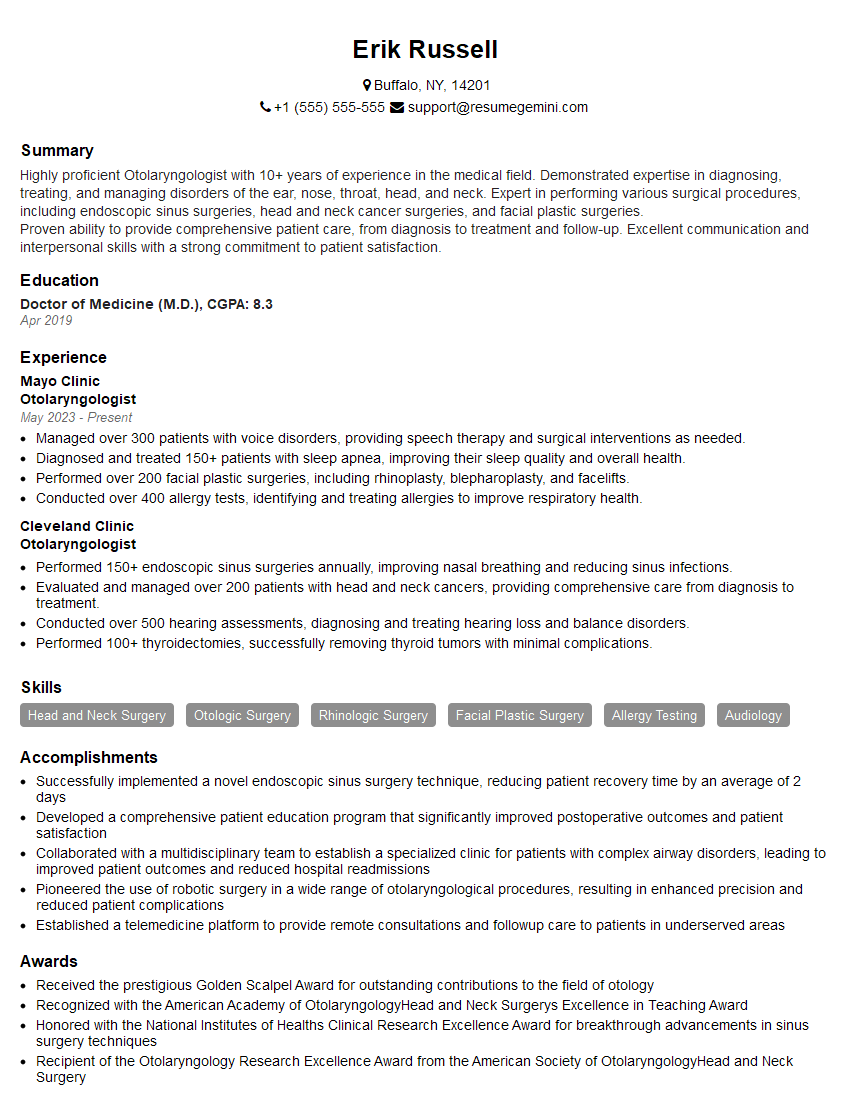
Mastering Otologic Surgery positions you for a rewarding career with significant growth potential, offering opportunities for specialization and leadership roles within the medical field. To enhance your job prospects, crafting a strong, ATS-friendly resume is critical. ResumeGemini is a trusted resource that can help you build a professional and impactful resume tailored to the specific requirements of Otologic Surgery positions. Examples of resumes tailored to Otologic Surgery are available to guide your creation process. Invest in creating a resume that effectively showcases your skills and experience, maximizing your chances of landing your dream job.
Explore more articles
Users Rating of Our Blogs
Share Your Experience
We value your feedback! Please rate our content and share your thoughts (optional).
What Readers Say About Our Blog
This was kind of a unique content I found around the specialized skills. Very helpful questions and good detailed answers.
Very Helpful blog, thank you Interviewgemini team.


