Every successful interview starts with knowing what to expect. In this blog, we’ll take you through the top Pediatric Flatfoot Reconstruction interview questions, breaking them down with expert tips to help you deliver impactful answers. Step into your next interview fully prepared and ready to succeed.
Questions Asked in Pediatric Flatfoot Reconstruction Interview
Q 1. Describe the different types of pediatric flatfoot deformities.
Pediatric flatfoot deformities encompass a spectrum of conditions affecting the alignment of the foot and ankle. They are broadly categorized based on the underlying cause and the flexibility of the deformity.
- Flexible Flatfoot: This is the most common type. The arch collapses when weight-bearing but returns when the child is non-weight-bearing. It’s often asymptomatic and related to laxity of the ligaments supporting the foot.
- Rigid Flatfoot: The arch remains collapsed even when non-weight-bearing, indicating a more structural problem. This may be due to tarsal coalition (fusion of two or more tarsal bones), vertical talus (a birth defect), or other bony abnormalities.
- Congenital Vertical Talus: This is a severe, rigid flatfoot deformity present at birth where the talus bone is positioned vertically, causing a rocker-bottom appearance.
- Metatarsus adductus: This is a forefoot deformity where the front part of the foot turns inward. While not strictly a flatfoot, it often co-exists and contributes to the overall deformity.
- Calcaneovalgus: A congenital condition where the heel is turned outward and the foot is in a flat position.
Understanding the specific type is crucial for appropriate diagnosis and treatment planning.
Q 2. Explain the clinical presentation of flexible versus rigid flatfoot in children.
The clinical presentation differs significantly between flexible and rigid flatfoot:
- Flexible Flatfoot: Often presents with a painless, flexible flatfoot arch that collapses with weight-bearing. Children may show pronation (inward rolling of the foot) and may have associated symptoms like ankle pain or fatigue. The foot is usually supple and easily manipulated.
- Rigid Flatfoot: Typically presents with a consistently flattened arch, even when non-weight-bearing. It is often associated with limited ankle and subtalar joint motion. Pain is more frequently encountered and may be significant, affecting daily activities. The foot feels stiff and less adaptable.
A thorough physical exam, including assessment of flexibility, range of motion, and gait analysis, is crucial in differentiating these types. Imaging studies like X-rays may be needed to confirm the diagnosis and rule out underlying structural issues.
Q 3. What are the non-surgical management options for pediatric flatfoot?
Non-surgical management is the first-line approach for most children with flexible flatfoot. The goal is to support the foot and improve function, not necessarily to correct the appearance. Options include:
- Observation and Monitoring: Many children with flexible flatfoot improve spontaneously as they grow and their ligaments strengthen. Regular follow-up appointments are key.
- Orthotics: Custom-made or prefabricated arch supports help to improve foot alignment and reduce strain on the muscles and ligaments. These are particularly helpful for children with pain or significant pronation.
- Physical Therapy: Exercises focused on strengthening the muscles of the foot and lower leg can improve foot function and stability. These might include stretching and strengthening exercises, and gait retraining.
- Bracing: In some cases, bracing might be used to correct the deformity and provide support, particularly if severe pronation or pain is present. This would often be a short-term solution while awaiting other treatment.
Non-surgical treatments prioritize conservative measures, aiming to manage symptoms and encourage natural improvement.
Q 4. Discuss the indications for surgical intervention in pediatric flatfoot.
Surgical intervention for pediatric flatfoot is usually reserved for cases that fail to respond to conservative management or present with significant functional limitations or pain. Indications include:
- Rigid flatfoot deformity: When the flatfoot is rigid and doesn’t improve with non-surgical approaches.
- Significant pain: Persistent pain despite conservative treatment which affects the child’s daily activities.
- Progressive deformity: If the flatfoot is worsening despite conservative measures.
- Severe functional limitations: If the deformity significantly limits the child’s ability to walk, run, or participate in activities.
- Significant hindfoot valgus: This is when the hindfoot is tilted excessively inwards.
- Symptomatic tarsal coalition: A fusion of two or more tarsal bones causing pain and limitation of motion.
The decision to proceed with surgery is made on a case-by-case basis, after careful consideration of the child’s age, the severity of the deformity, and the potential risks and benefits of the procedure.
Q 5. What are the commonly used surgical techniques for correcting pediatric flatfoot?
Several surgical techniques can correct pediatric flatfoot, each tailored to the specific deformity. Common procedures include:
- Medial Displacement Calcaneal Osteotomy (MDCO): This involves cutting and repositioning the heel bone to improve alignment.
- Lateral Column Lengthening: This lengthens the outer bones of the foot to restore the arch.
- Triple Arthrodesis: This fuses three joints in the hindfoot (subtalar, talonavicular, and calcaneocuboid joints) to provide stability. This is usually reserved for older children or teens with severe, rigid deformities and is less commonly performed in younger children.
- Soft tissue procedures: These involve releasing tight ligaments or tendons around the foot and ankle to improve flexibility. Often these are performed in conjunction with bony procedures.
The choice of surgical technique depends on several factors, including the child’s age, the severity of the deformity, and the surgeon’s experience and preference.
Q 6. Explain the principles of medial displacement calcaneal osteotomy.
Medial Displacement Calcaneal Osteotomy (MDCO) is a common surgical technique used to correct flatfoot deformity by realigning the heel bone. The principle is to move the heel bone medially (inward) and plantarflex it (downward), thereby restoring the medial longitudinal arch.
This is achieved by making a carefully planned osteotomy (surgical cut) in the calcaneus. The bone is then repositioned and secured with screws or pins. The medial displacement of the calcaneus helps to reduce hindfoot valgus (outward tilting of the heel) and improves the position of the talonavicular joint, the key joint in the midfoot. This procedure aims to correct the collapse of the arch by restoring the natural alignment of the bones.
Post-operatively, the foot is immobilized in a cast for several weeks to allow the bone to heal. Physical therapy is usually needed to restore full range of motion and function.
Q 7. Describe the procedure for a lateral column lengthening.
Lateral column lengthening procedures aim to correct flatfoot deformities by lengthening the outer bones of the foot, specifically the cuboid and/or the lateral cuneiform. This helps to restore the arch’s height and width, combating the effects of the collapse.
The procedure typically involves making an osteotomy in one or both of these bones. Special techniques are then used to create a gap and lengthen the bone. This gap allows for controlled lengthening, which is usually achieved by inserting a bone graft or by gradual distraction using external fixators. The lengthened bones are fixed in place with screws or pins.
This procedure is often combined with soft tissue procedures such as tendon releases to ensure a balanced correction. Post-operatively, the foot is immobilized in a cast or brace to allow for healing, and physical therapy is vital for regaining full mobility.
Q 8. What are the potential complications of surgical correction of pediatric flatfoot?
Surgical correction of pediatric flatfoot, while often beneficial, carries potential complications. These can be broadly categorized into problems related to the surgical procedure itself, the anesthesia, and the patient’s individual response to the surgery and recovery process.
- Infection: A serious risk, requiring prompt antibiotic treatment and potentially further surgery.
- Nerve damage: Injury to the nerves around the ankle and foot can cause numbness, tingling, or weakness. Careful surgical technique minimizes this risk, but it’s a possibility.
- Non-union or malunion of bones: The bones may not heal properly or may heal in an incorrect position, requiring revision surgery.
- Hardware complications: If screws or plates are used, they can loosen, break, or irritate the surrounding tissues, necessitating removal.
- Recurrence of flatfoot deformity: Although rare with proper surgical planning and execution, the flatfoot can sometimes recur.
- Anesthesia-related complications: These are rare but potentially serious and include allergic reactions or respiratory issues.
- Delayed wound healing: This can prolong recovery and increase the risk of infection.
- Stiffness and limited range of motion: This is a common post-operative finding but usually improves with physical therapy.
- Compartment syndrome: This is a serious condition involving increased pressure within the muscle compartments of the leg, necessitating immediate surgical intervention. This is rare but must be carefully monitored.
The exact risks vary depending on the specific surgical procedure, the surgeon’s experience, and the child’s overall health.
Q 9. How do you assess the outcome of pediatric flatfoot surgery?
Assessing the outcome of pediatric flatfoot surgery is a multi-faceted process that goes beyond simply measuring the angles on X-rays. We aim for both functional and cosmetic improvement.
- Clinical examination: This includes assessing the child’s range of motion, gait pattern, and overall foot alignment.
- Radiographic assessment: X-rays are used to measure the angles of the foot bones and assess the alignment of the joints. We use standardized measurements to track improvement.
- Functional outcome scores: Standardized questionnaires and scales, like the Foot Function Index or the AOFAS Ankle-Hindfoot Scale (adapted for children), help to quantify the child’s ability to participate in daily activities. We often include parent and patient reports.
- Patient-reported outcome measures (PROMs): These assess the child’s pain levels, functional limitations, and overall satisfaction with the surgery. This is crucial, as the child’s experience is paramount.
- Longitudinal follow-up: Regular check-ups are essential to monitor progress and address any potential problems that may arise over time. This can extend over several years to ensure long-term success.
We don’t solely focus on the perfect anatomical alignment; instead, we prioritize restoring function and reducing pain so the child can participate normally in their activities. For example, a slight residual deformity is acceptable if the child is pain-free and able to run and play without limitation.
Q 10. What are the common post-operative complications and their management?
Common post-operative complications following pediatric flatfoot surgery include infection, wound healing problems, nerve injury, stiffness, and recurrence of the deformity as already discussed. Management varies depending on the specific complication.
- Infection: This is addressed with antibiotics, wound debridement (surgical removal of infected tissue), and possibly drainage of any abscesses. Severe cases may require further surgery.
- Wound healing problems: These can include delayed healing or wound breakdown. Management involves wound care, dressings, and potentially surgical revision.
- Nerve injury: Treatment focuses on supportive measures such as physical therapy and pain management. In rare cases, further surgery may be necessary.
- Stiffness and limited range of motion: This is often managed with physical therapy, including range of motion exercises and strengthening programs.
- Recurrence of deformity: If the flatfoot deformity recurs, revision surgery may be considered. This is far less common with modern techniques, but it remains a consideration.
Careful monitoring during the post-operative period is critical for early detection and prompt management of these complications. Regular follow-up appointments are crucial, allowing for early intervention and avoidance of more serious issues.
Q 11. How do you counsel parents regarding the risks and benefits of surgery?
Counseling parents about pediatric flatfoot surgery involves a delicate balance of providing accurate information, addressing concerns, and ensuring shared decision-making.
The conversation begins with a thorough explanation of the child’s condition, including the severity of the deformity, its potential impact on their future function, and the non-surgical options explored. We then discuss the proposed surgical procedure, outlining the potential benefits – improved foot alignment, reduced pain, and improved function.
Crucially, we spend significant time discussing the potential risks and complications, presented in an honest and transparent manner. We use simple language, avoiding medical jargon, and we often provide visual aids such as diagrams or images.
We emphasize that surgery is not always the best option and that the decision should be made jointly, based on the child’s specific needs and the family’s preferences. We answer all questions patiently and encourage open communication. For example, I frequently share stories of past patients and their outcomes, showcasing the positives and also the challenges. We ensure we tailor this discussion to the individual child and their unique concerns.
Q 12. Describe your approach to managing pain in pediatric flatfoot patients.
Managing pain in pediatric flatfoot patients requires a multi-modal approach tailored to the child’s age, developmental stage, and the specific pain experienced.
- Pharmacological management: Pain medication, such as acetaminophen or ibuprofen, is often prescribed in the initial post-operative period. Opioids are rarely necessary in pediatric flatfoot surgery.
- Physical therapy: Early mobilization and physical therapy are essential for pain management and restoring function. This includes range-of-motion exercises, strengthening exercises, and gait training.
- Non-pharmacological methods: These include ice packs, elevation of the foot, rest, and distraction techniques such as reading or playing games.
- Psychological support: Addressing the child’s emotional concerns and anxiety is important, as these factors can influence their pain experience. This may involve providing age-appropriate explanations, creating a supportive environment and working with child life specialists.
We emphasize a proactive approach, preventing pain rather than simply treating it. Pain management should be individualized, and we regularly assess its effectiveness, adapting the plan as needed to ensure the child’s comfort and recovery.
Q 13. How do you assess for and manage potential infections post-operatively?
Post-operative infection is a serious concern in any surgical procedure, and pediatric flatfoot surgery is no exception. Careful assessment and proactive management are crucial.
- Regular wound checks: We carefully examine the surgical site at each post-operative visit, looking for signs of infection such as redness, swelling, warmth, tenderness, pus, or increased pain.
- Laboratory tests: Blood tests may be performed if an infection is suspected to identify the causative organism and guide antibiotic therapy.
- Wound cultures: If infection is suspected, a sample from the wound is sent to the laboratory for culture and sensitivity testing to determine the most effective antibiotic.
- Antibiotic therapy: Appropriate antibiotics are administered based on the results of the culture and sensitivity testing.
- Surgical debridement: If the infection is severe and unresponsive to antibiotics, surgical removal of infected tissue may be necessary.
Prophylactic antibiotics are sometimes administered before surgery to minimize the risk of infection. Maintaining meticulous surgical technique, strict aseptic precautions in the operating room, and proper post-operative wound care are also essential in preventing infection.
Q 14. What are the long-term outcomes of surgical correction of pediatric flatfoot?
Long-term outcomes of pediatric flatfoot surgery are generally positive for most patients. The majority experience significant improvements in foot alignment, pain relief, and functional ability.
However, the long-term success depends on various factors, including the child’s age at the time of surgery, the severity of the initial deformity, the surgical technique used, and adherence to post-operative care recommendations. Longitudinal follow-up, spanning years, is critical to fully understand the long-term effects.
While the majority of children achieve satisfactory results, some may experience residual deformity, stiffness, or recurrent pain. The possibility of revision surgery remains, albeit rare. These follow-up studies help us refine our surgical approaches and improve our understanding of long-term outcomes. It also helps individual patients understand the range of possible outcomes, including those that might deviate from the most favorable results. This transparency helps in setting realistic expectations.
Q 15. How do you differentiate between congenital and acquired flatfoot?
Differentiating between congenital and acquired flatfoot relies on understanding the onset of the condition. Congenital flatfoot is present at birth, often due to a developmental anomaly affecting the bones and soft tissues of the foot. This might involve abnormalities in the tarsal coalition (fusion of bones), or variations in the shape and arrangement of bones. In contrast, acquired flatfoot develops after birth, usually as a result of factors like excessive pronation (inward rolling of the foot), muscle weakness (e.g., from cerebral palsy), ligament laxity, or trauma. A detailed history, taking into account the child’s developmental milestones and any potential injury or family history of foot problems, is crucial for accurate diagnosis.
Imagine a child born with a noticeably flat foot – this strongly suggests a congenital cause. On the other hand, a child who develops a flat foot progressively over time after years of seemingly normal development may have an acquired flatfoot, possibly related to rapid growth or increased activity.
Career Expert Tips:
- Ace those interviews! Prepare effectively by reviewing the Top 50 Most Common Interview Questions on ResumeGemini.
- Navigate your job search with confidence! Explore a wide range of Career Tips on ResumeGemini. Learn about common challenges and recommendations to overcome them.
- Craft the perfect resume! Master the Art of Resume Writing with ResumeGemini’s guide. Showcase your unique qualifications and achievements effectively.
- Don’t miss out on holiday savings! Build your dream resume with ResumeGemini’s ATS optimized templates.
Q 16. Discuss the role of bracing in the management of pediatric flatfoot.
Bracing plays a significant role in the conservative management of pediatric flatfoot, especially in flexible flatfoot (where the arch returns when the child is on their toes). The goal isn’t necessarily to completely correct the arch but rather to support the foot, reduce pain, and guide proper bone and muscle development. Braces such as custom-molded orthotics or commercially available supportive shoes provide medial arch support and may help improve foot posture and function. Early intervention with bracing is often recommended, especially in younger children where the bones are still developing and more responsive to external forces. The effectiveness of bracing is highly individual and depends on the severity of the flatfoot, the age of the child, and the compliance in wearing the brace consistently.
Think of a brace as scaffolding for a building under construction – it supports the structure until it’s strong enough to stand on its own. In this analogy, the child’s foot is the building and the brace is the scaffolding providing support and guiding normal development.
Q 17. What are the radiographic features used to evaluate pediatric flatfoot?
Radiographic evaluation is essential in assessing the structural aspects of pediatric flatfoot. Key features to examine on X-rays include:
- Calcaneal inclination angle (Kite’s angle): This measures the angle between the long axis of the calcaneus and the ground. A decreased angle indicates a valgus (outward turning) heel.
- Talocalcaneal angle: This assesses the relationship between the talus and calcaneus. An increased angle suggests subtalar joint instability.
- Cyma line: This smooth curve along the medial aspect of the foot should be intact; disruption suggests deformity.
- Navicular drop: This measures the difference in the navicular height between weight-bearing and non-weight-bearing positions. An increased drop suggests arch collapse.
- Presence of coalitions (fusion): X-rays can identify bony fusions between tarsal bones.
These measurements help in understanding the severity and type of flatfoot, guiding treatment decisions. For example, a significant increase in the talocalcaneal angle might suggest a more complex case requiring surgical intervention compared to a milder case showing only a slight decrease in Kite’s angle.
Q 18. Explain the importance of gait analysis in the evaluation of pediatric flatfoot.
Gait analysis plays a crucial role in understanding how the flatfoot affects the child’s movement and overall biomechanics. It’s more than just looking at the foot itself; it examines how the entire body compensates for the flatfoot during walking and running. Gait analysis typically uses video recording and pressure sensors to assess various parameters such as:
- Stride length and cadence: Does the flatfoot affect the child’s walking efficiency?
- Foot contact pattern: Is there excessive pronation or supination (inward or outward rolling of the foot)?
- Joint angles and range of motion: How do the ankles, knees, and hips move during gait?
- Muscle activation patterns: Are specific muscles overworking or underworking to compensate for the flatfoot?
This information allows clinicians to determine the functional impact of the flatfoot and to tailor treatment strategies. For example, gait analysis may reveal compensatory movements in the knees and hips indicating a need for more comprehensive interventions than simple orthotics. It helps us assess treatment outcomes as well, by comparing pre and post-operative gait parameters.
Q 19. What are the different types of implants used in pediatric flatfoot surgery?
The choice of implant in pediatric flatfoot surgery depends on the specific deformity and the surgeon’s preference. However, several commonly used implants include:
- Screws: Used for fixation in procedures like medial displacement calcaneal osteotomy (MDCO).
- Plates: Offer more robust fixation for complex osteotomies or fusions.
- Bioabsorbable implants: These gradually dissolve over time, eliminating the need for a second procedure to remove them. They are particularly attractive in children because they adapt to the growing bone.
- Growth plates: Sometimes used in cases requiring manipulation of the growth plates to correct specific aspects of the deformity. These are specialized and require advanced surgical planning and execution.
The use of minimally invasive techniques is often preferred in children to minimize tissue trauma and facilitate faster recovery.
Q 20. Describe your experience with minimally invasive techniques for pediatric flatfoot correction.
My experience with minimally invasive techniques for pediatric flatfoot correction has been very positive. Techniques like percutaneous procedures, smaller incisions, and the use of image guidance allow for precise correction with reduced surgical trauma. These methods typically result in less post-operative pain, shorter hospital stays, and quicker rehabilitation. The development and refinement of these techniques have been significant in recent years. For instance, we often utilize fluoroscopy or CT guidance to precisely place screws or guide osteotomies with greater accuracy and minimize invasiveness. We find that children recover significantly faster and with less discomfort compared to open procedures.
A specific example would be using a minimally invasive approach for a calcaneal osteotomy. Instead of a large incision, we use small incisions with specialized instruments. This leads to faster healing, less scarring, and reduced risk of complications.
Q 21. How do you address the challenges of managing flatfoot in children with neuromuscular disorders?
Managing flatfoot in children with neuromuscular disorders presents unique challenges. These children often have associated muscle weakness, spasticity, or joint contractures that complicate the presentation and management of flatfoot. Treatment strategies often involve a multidisciplinary approach involving orthotists, physical therapists, and possibly other specialists such as neurologists. Surgical interventions might be considered but need very careful consideration of the overall condition. The goal is often to improve function and reduce pain rather than achieving perfect anatomical correction. The use of serial casting, bracing, or tendon lengthening surgeries might be utilized selectively and conservatively in these cases to achieve optimal results without compromising other aspects of the child’s overall health.
For instance, a child with cerebral palsy and severe muscle weakness might benefit from a custom-made ankle-foot orthosis (AFO) rather than a surgical intervention. The AFO helps improve gait and reduces the strain on the foot and ankle. In other cases, selective tendon lengthening or surgical procedures may be deemed necessary depending on the severity and associated complications. The management plan must be tailored to the individual needs of each child and carefully coordinated amongst the multidisciplinary team involved.
Q 22. What are the ethical considerations in deciding on surgical intervention in pediatric flatfoot?
Ethical considerations in pediatric flatfoot surgery are paramount. We must always prioritize the child’s well-being above all else. The decision to operate should never be taken lightly. Before recommending surgery, we must carefully weigh the potential benefits against the risks. This involves a thorough discussion with the parents and the child (age-appropriately), outlining the potential benefits (improved function, reduced pain, better cosmetic appearance), risks (infection, nerve injury, non-union, growth plate damage, recurrence), and alternatives (conservative management, bracing, physical therapy). Informed consent is crucial; parents must fully understand the procedure, its implications, and the possibility of complications. We also consider the child’s age and developmental stage, acknowledging that surgery might carry different ethical implications for a young child versus an adolescent. For example, a minor might not fully grasp the long-term effects of surgery, making careful parental involvement especially critical. In situations of borderline cases, conservative management is always favored initially. Open communication with the family is key to ensuring ethical decision-making.
Q 23. Explain your understanding of the growth plate and its implications in surgical planning.
The growth plate, or physis, is a cartilaginous area at the end of long bones responsible for longitudinal growth. In pediatric flatfoot reconstruction, precise surgical planning is critical because damage to the growth plate can significantly impact the child’s future foot development. The location and type of surgical intervention must carefully consider the proximity to growth plates, aiming to minimize their disruption. Techniques like minimally invasive procedures, precise osteotomy placement, and the use of plates and screws designed to avoid the physis are employed to preserve growth. For example, during a calcaneal osteotomy, we strategically plan the incision and screw placement to avoid the posterior calcaneal physis to prevent growth retardation. Failure to consider the growth plate’s location can lead to premature fusion, resulting in leg length discrepancy, angular deformities, and altered foot mechanics. Pre-operative imaging (X-rays, CT scans) is crucial to accurately identify the location of growth plates and guide surgical planning. Post-operative imaging is then employed to monitor for any potential issues.
Q 24. How do you manage a case of recurrent flatfoot deformity after initial surgery?
Recurrent flatfoot deformity after initial surgery is challenging. Management depends on the cause of recurrence, the type of initial surgery, and the child’s age and skeletal maturity. A thorough clinical examination, including gait analysis, and advanced imaging studies (X-rays, MRI) are essential to assess the extent of recurrence and identify any underlying issues, like inadequate correction of the initial deformity or secondary changes like ligament laxity. Conservative treatment, including bracing, physical therapy, and orthotics, is often attempted first, depending on the severity of the recurrence. If conservative management fails, a revision surgery may be necessary. This might involve different surgical techniques than the initial procedure, such as additional ligament reconstruction, or adjustments to the previous osteotomy. The choice of revision surgery is highly individualized and depends on the specific circumstances. It’s a complex process requiring a high level of surgical expertise and meticulous pre-operative planning.
Q 25. Describe your approach to pre-operative planning and patient selection for surgery.
Pre-operative planning for pediatric flatfoot surgery is a meticulous process. Patient selection begins with a detailed history, focusing on the onset, progression, and symptoms of the deformity. A comprehensive physical examination includes assessing the range of motion, muscle strength, and flexibility of the foot and ankle. We then employ advanced imaging (X-rays, MRI, CT scans) to evaluate the bone structure, cartilage, and soft tissues, identifying the severity and type of deformity and assessing the condition of the growth plates. This information helps determine the appropriate surgical technique. We also consider the child’s overall health, activity level, and expectations for treatment. Patients with severe pain, significant functional limitations, or progressive deformity are usually good candidates for surgery. Patients with mild symptoms or significant underlying medical conditions might be better suited to conservative management. The decision is always made collaboratively with the family, ensuring they fully understand the risks and benefits of surgery.
Q 26. What is your experience with different types of footwear recommendations for pediatric flatfoot?
Footwear recommendations are an important part of both pre- and post-operative management of pediatric flatfoot. Pre-operatively, supportive footwear with good arch support and firm soles can help manage symptoms and delay surgery in some cases. Post-operatively, footwear recommendations are tailored to the individual’s needs and the type of surgery performed. In general, we recommend comfortable, well-fitting shoes with sufficient arch support and cushioning. Shoes that are too tight or too loose can hinder healing and increase discomfort. Specialized orthotics, custom-made or commercially available, are often prescribed to provide additional support and correct foot alignment. The choice of footwear and orthotics should consider the child’s age, activity level, and the specific demands placed on their feet. In cases of severe deformity or after complex surgeries, custom-molded orthotics are often necessary to provide the optimal level of support and alignment. The goal is to improve foot function, reduce pain, and prevent further deformity.
Q 27. How do you monitor a patient’s progress during the rehabilitation phase?
Monitoring patient progress during the rehabilitation phase is critical. It involves regular follow-up appointments that include clinical examinations, focusing on pain levels, range of motion, gait, and functional assessments. Regular X-rays are employed to monitor bone healing and growth plate integrity. Physical therapy plays a significant role; we track their progress in exercises aimed at improving strength, flexibility, and balance. The child’s ability to participate in normal activities is also an indicator of recovery. We use functional scales (like the Foot Function Index) to objectively measure improvements in daily activities. Parents also play a vital role, providing feedback on the child’s progress and any concerns. If the progress is not as expected, we adjust the treatment plan accordingly, which might include additional physical therapy, modifications to orthotics, or further surgical intervention if necessary. The rehabilitation phase is crucial for a successful outcome, and close monitoring ensures optimal results.
Q 28. Discuss the importance of multidisciplinary care in the management of complex pediatric flatfoot cases.
Multidisciplinary care is essential, especially in complex pediatric flatfoot cases. A team approach ensures comprehensive management. The team typically includes an orthopedic surgeon specializing in pediatric foot and ankle surgery, a physical therapist experienced in pediatric rehabilitation, a podiatrist, and sometimes a pediatric rheumatologist or geneticist, depending on the underlying conditions. The orthopedic surgeon guides the surgical aspects, while the physical therapist develops and monitors the rehabilitation program. Podiatrists offer expertise in orthotics and conservative management. Other specialists are brought in based on specific needs of the case. This collaborative approach allows for a holistic assessment, customized treatment plan, and effective monitoring of the patient’s progress. Regular communication between team members ensures everyone is informed and working towards the same goal – providing the child with the best possible outcome. This multidisciplinary approach significantly improves patient outcomes and ensures the best quality of care.
Key Topics to Learn for Pediatric Flatfoot Reconstruction Interview
- Etiology and Pathophysiology of Pediatric Flatfoot: Understanding the various causes of flatfoot in children, including developmental, neuromuscular, and connective tissue disorders. This includes mastering the biomechanics involved.
- Clinical Examination and Diagnosis: Proficiently performing a comprehensive physical examination, interpreting radiographic imaging (X-rays, CT scans), and utilizing other diagnostic tools to accurately assess the severity and type of flatfoot deformity.
- Non-Operative Management Strategies: Knowing the indications, limitations, and effectiveness of conservative treatments such as orthotics, bracing, and physical therapy. Consider the patient’s age and growth potential.
- Surgical Techniques and Indications: Developing a thorough understanding of different surgical procedures used in pediatric flatfoot reconstruction, including their respective indications and contraindications. This includes understanding the principles of tendon transfers, osteotomies, and arthrodesis.
- Post-Operative Care and Rehabilitation: Familiarity with appropriate post-operative management, including casting, bracing, weight-bearing restrictions, and physical therapy protocols to optimize patient outcomes and minimize complications.
- Complications and Management: Recognizing potential complications associated with pediatric flatfoot reconstruction (e.g., infection, non-union, malunion, nerve injury) and implementing effective management strategies.
- Growth Plate Considerations: Understanding the impact of surgical interventions on the child’s growth plates and the importance of preserving growth potential.
- Long-Term Outcomes and Follow-up: Appreciation for the long-term functional and aesthetic outcomes of surgical intervention and the importance of comprehensive follow-up care.
- Ethical Considerations and Shared Decision-Making: Understanding the ethical implications of surgical intervention in children, involving parents in the decision-making process, and managing patient and family expectations.
Next Steps
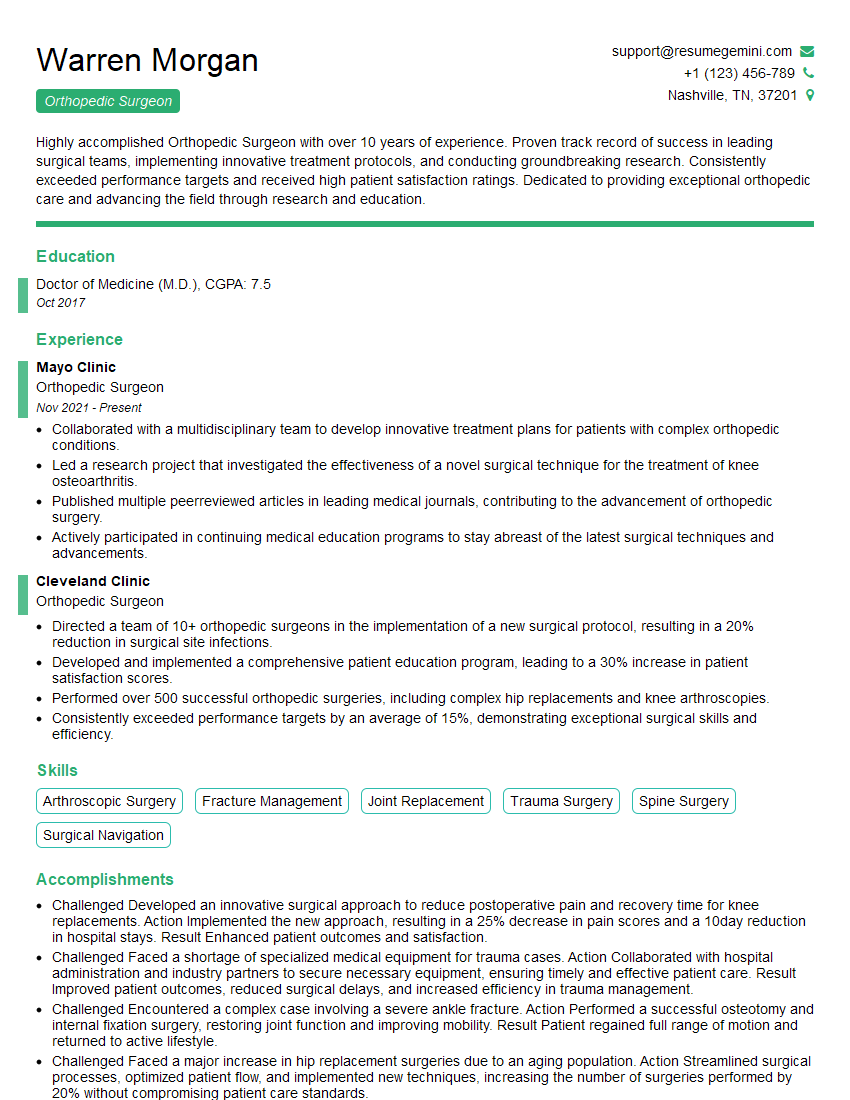
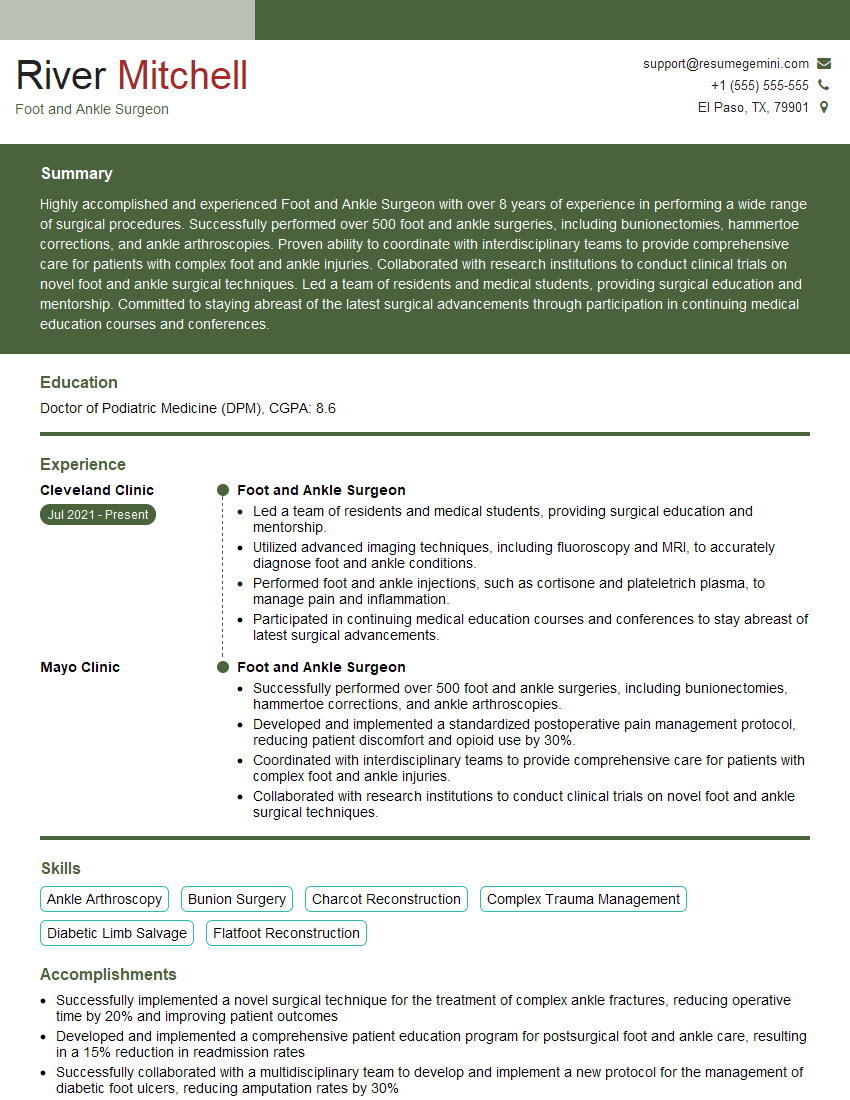
Mastering Pediatric Flatfoot Reconstruction significantly enhances your career prospects in pediatric orthopedics, demonstrating a specialized skill set highly valued by employers. A strong, ATS-friendly resume is crucial for showcasing your expertise and securing interviews. ResumeGemini is a trusted resource to help you build a professional and impactful resume that highlights your unique qualifications. Examples of resumes tailored to Pediatric Flatfoot Reconstruction are available to guide you. Invest in your future; craft a resume that reflects your dedication and expertise.
Explore more articles
Users Rating of Our Blogs
Share Your Experience
We value your feedback! Please rate our content and share your thoughts (optional).
What Readers Say About Our Blog
This was kind of a unique content I found around the specialized skills. Very helpful questions and good detailed answers.
Very Helpful blog, thank you Interviewgemini team.

