Interviews are opportunities to demonstrate your expertise, and this guide is here to help you shine. Explore the essential Skin Lesion Removal interview questions that employers frequently ask, paired with strategies for crafting responses that set you apart from the competition.
Questions Asked in Skin Lesion Removal Interview
Q 1. Describe your experience with various skin lesion removal techniques (e.g., excision, shave biopsy, electrocautery).
My experience encompasses a wide range of skin lesion removal techniques, each chosen based on the lesion’s characteristics and the patient’s individual needs.
- Excision: This is a common method for removing most skin lesions. It involves surgically cutting out the lesion and a small margin of surrounding healthy tissue. The sample is then sent for pathology to confirm the diagnosis. I frequently use this for suspicious lesions that need complete removal for histological analysis. For example, a suspicious mole that shows concerning features on visual inspection would typically be excised.
- Shave Biopsy: This technique uses a razor-like instrument to shave off a superficial layer of the lesion. It’s ideal for lesions that are raised and relatively small, and is often used for rapid assessment of something like a wart or seborrheic keratosis. However, it’s not suitable for deep lesions or those needing complete removal. The depth of the shave must be carefully considered and balanced against the need for diagnostic accuracy.
- Electrocautery: This involves using a heated instrument to destroy the lesion using heat. It’s often used for smaller, benign lesions like warts or small skin tags. While it’s quick and generally effective for these types of lesions, it is not suitable for more complex lesions or where tissue sampling is required for histological analysis. For instance, it might be used for a superficial, benign lesion but not for something that needs full excisional biopsy.
The choice of technique is always tailored to the individual case, factoring in factors like lesion size, depth, location, and the patient’s overall health.
Q 2. Explain the process of obtaining informed consent for skin lesion removal procedures.
Obtaining informed consent is crucial for ethical and legal reasons. It ensures the patient understands the procedure, its benefits, risks, and alternatives. The process typically involves:
- Detailed Explanation: I clearly explain the nature of the lesion, the proposed procedure, the likely outcome, and the potential benefits and risks (including bleeding, infection, scarring, and the rare possibility of nerve damage).
- Alternative Treatments: I discuss alternative treatment options, if any, such as watchful waiting, topical treatments, or different surgical techniques.
- Answering Questions: I thoroughly answer any questions the patient may have, ensuring they understand the information before proceeding.
- Documentation: I meticulously document the consent process, including the patient’s understanding and agreement to the procedure. This includes written documentation signed by both the patient and myself.
A simple analogy is like buying a car – you wouldn’t buy it without understanding its features, cost, and potential issues. Informed consent works the same way for medical procedures, empowering the patient to make an informed decision.
Q 3. How do you differentiate between benign and malignant skin lesions during a physical examination?
Differentiating between benign and malignant skin lesions during a physical examination relies on careful observation using the ABCDEs of melanoma (although this is not solely for melanoma detection) and other clinical features:
- Asymmetry: Benign lesions tend to be symmetrical; malignant ones are often asymmetrical.
- Border: Benign lesions usually have well-defined borders, while malignant ones often have irregular, notched, or blurred borders.
- Color: Benign lesions are usually uniform in color; malignant lesions may have variations in color (e.g., shades of brown, black, red, white, or blue).
- Diameter: Malignant lesions are often larger than 6 millimeters (the size of a pencil eraser), though early melanomas can be smaller.
- Evolving: Changes in size, shape, color, or other characteristics over time are a major warning sign of malignancy.
Beyond the ABCDEs, other factors like the lesion’s texture (smooth, rough, crusted), elevation (flat, raised, or nodular), and the presence of bleeding or ulceration help in assessment. However, a physical exam alone isn’t definitive. A biopsy and histological examination are crucial for a conclusive diagnosis.
Q 4. What are the key steps in performing a Mohs micrographic surgery procedure?
Mohs micrographic surgery is a precise technique used to remove skin cancers, especially those with a high risk of recurrence, like basal cell carcinoma and squamous cell carcinoma. The key steps are:
- Anesthesia: Local anesthesia is administered to numb the area.
- Excision: The visible part of the tumor is carefully excised, and the tissue is labeled and sent to the lab.
- Mapping: The excised tissue is mapped and carefully examined under a microscope.
- Stage-by-stage Removal: If cancer cells are found at the edge of the specimen, the surgeon removes additional tissue from the corresponding area, repeating the process until the margins are clear.
- Reconstruction: Once clear margins are achieved, the wound is repaired using sutures, skin grafts, or flaps, depending on the size and location of the lesion.
Mohs surgery offers a high cure rate and minimizes the removal of healthy tissue, making it particularly beneficial for lesions in cosmetically sensitive areas.
Q 5. Explain the importance of proper wound closure techniques after skin lesion removal.
Proper wound closure is essential for minimizing scarring, preventing infection, and promoting optimal healing. The technique depends on the size, depth, and location of the wound. Methods include:
- Simple Sutures: Used for smaller, easily approximated wounds.
- Advanced Sutures: More complex closure techniques may be necessary for larger or more irregular wounds.
- Skin Grafts: For larger defects, a skin graft may be necessary to cover the wound and promote healing.
- Skin Flaps: These are used for larger or deeper defects, where local tissue is advanced to cover the wound.
Careful attention to wound closure minimizes scarring and improves cosmetic outcomes. The type of suture material is considered along with wound tension and tension-relieving techniques to minimize scar hypertrophy.
Q 6. How do you manage potential complications like bleeding, infection, or scarring after a procedure?
Managing potential complications is a critical part of the process.
- Bleeding: Direct pressure is usually sufficient to control minor bleeding. In more significant cases, sutures or cauterization may be needed.
- Infection: Prophylactic antibiotics are sometimes used, and patients are instructed on proper wound care, including keeping the wound clean and dry and monitoring for signs of infection (increased pain, swelling, redness, or pus).
- Scarring: While some scarring is inevitable, techniques like meticulous wound closure, early mobilization, and the use of silicone sheeting can minimize scarring and improve cosmetic outcomes. In cases of hypertrophic scarring, steroid injections or laser therapy may be needed.
Post-operative follow-up is crucial for early detection and management of any complications. I regularly see patients in the follow up period to check wound healing and answer any concerns.
Q 7. Describe your experience with different types of skin grafts and flaps.
My experience includes using various skin grafts and flaps for wound reconstruction after skin lesion removal, particularly in cases of larger defects or lesions in cosmetically sensitive areas.
- Split-thickness skin grafts: These grafts include a portion of the dermis, and the donor site heals spontaneously. They are versatile but have a higher risk of contracture and may not match the surrounding skin perfectly.
- Full-thickness skin grafts: These grafts include all layers of the dermis and epidermis and provide a better cosmetic result. However, the donor site requires more extensive closure and may have a larger scar. They are typically harvested from an inconspicuous site such as the groin.
- Local flaps: These involve moving adjacent skin to cover the defect. They offer a good cosmetic outcome as the tissue is from the same area, but they may create a secondary defect that needs closure.
- Free flaps: These are complex procedures that involve transferring a flap of tissue from a distant site, often requiring microsurgical techniques for revascularization. Free flaps are suitable for the reconstruction of large defects with significant tissue loss. A microvascular anastomosis is required.
The choice of graft or flap depends on the size and location of the defect, the quality of the surrounding tissue, and the patient’s overall health and expectations. It is important to discuss the pros and cons of each with the patient prior to selecting the most appropriate method.
Q 8. How do you determine the appropriate depth of excision for a suspicious lesion?
Determining the appropriate depth of excision for a suspicious lesion is crucial for complete removal and minimizing recurrence. It’s not a one-size-fits-all approach but depends heavily on several factors, including the lesion’s type, size, depth of invasion (as assessed clinically and, if available, with imaging such as dermoscopy or ultrasound), and location.
For example, a superficial basal cell carcinoma might only require a few millimeters of depth, while a deeply invasive melanoma may necessitate a much deeper excision, potentially including underlying subcutaneous fat. The goal is to achieve clear surgical margins, meaning that the excised tissue completely surrounds the lesion. We use visual inspection during the procedure to assess margins, and pathologic examination of the excised tissue confirms the adequacy of the margins.
We often use Mohs micrographic surgery for high-risk lesions, such as recurrent or large lesions, to ensure complete removal with minimal scarring. This technique involves the precise removal of thin layers of tissue, with immediate frozen section analysis, to guide subsequent excision. This process allows us to map the extent of the tumor, ensuring complete resection. In simpler cases, we’d follow established guidelines for excision depth based on lesion type and size.
Q 9. What are the common histopathological findings for various skin lesions?
Histopathological findings vary greatly depending on the type of skin lesion. A proper diagnosis is absolutely critical for determining the appropriate treatment course and prognosis. Here are some common examples:
- Benign lesions: These often show features like orderly cellular arrangement, minimal atypia (abnormal cell growth), and no evidence of invasion into surrounding tissues. Examples include seborrheic keratoses (showing keratin-filled structures), nevi (moles, showing melanocytes in a nest-like pattern), and fibromas (showing collagen fibers).
- Malignant lesions: These display characteristics such as disordered cellular growth, significant atypia, and invasion into surrounding tissues. Examples include:
- Basal cell carcinoma (BCC): Often shows nests or cords of basaloid cells, sometimes with palisading (a pattern of tightly packed cells resembling a picket fence).
- Squamous cell carcinoma (SCC): Typically demonstrates keratin pearls (areas of keratinized cells) and cellular atypia.
- Melanoma: Characterized by varying degrees of cellular atypia, asymmetry, and often demonstrates invasion into the dermis and sometimes beyond. The depth of invasion (Breslow thickness) is a critical prognostic factor.
It is important to note that the microscopic analysis is complex and needs to be interpreted by a qualified dermatopathologist. The description above provides a general overview. Specific features can often indicate subtypes and influence the treatment plan.
Q 10. Describe your experience with managing patients with complex skin cancer cases.
My experience with complex skin cancer cases involves a multi-disciplinary approach. This often includes collaboration with medical and radiation oncologists, plastic surgeons, and dermatopathologists. I’ve managed cases involving extensive melanomas requiring wide local excision and sentinel lymph node biopsy, as well as recurrent basal cell carcinomas needing Mohs micrographic surgery for complete resection.
One challenging case I recall involved a patient with a large, recurrent squamous cell carcinoma on the face, which required multiple Mohs procedures and subsequent reconstruction with a local flap. Careful planning, meticulous surgical technique, and close communication with the reconstructive surgeon were vital for achieving a positive outcome, restoring both the patient’s physical appearance and mental wellbeing. The key in these cases is to prioritize patient safety, achieve complete tumor removal, and minimize morbidity, while keeping the patient informed and involved in the decision-making process at every stage.
Q 11. How do you counsel patients about the risks and benefits of different treatment options?
Patient counseling is a cornerstone of my practice. I always begin by explaining the diagnosis in clear, non-technical terms, ensuring the patient understands the nature of their lesion and the implications for their health. I then outline the available treatment options, including the risks and benefits of each, tailored to the specific patient and lesion. This includes discussing potential complications such as scarring, infection, and nerve damage, as well as the likelihood of recurrence.
For example, I might explain that while excision is generally safe and effective, it can leave a scar, especially in cosmetically sensitive areas. Alternatively, I might explain that cryotherapy, though less invasive, might not be as effective for larger or deeply invasive lesions. Ultimately, the choice of treatment is made collaboratively, with the patient’s preferences, values, and understanding fully considered. I always encourage patients to ask questions and ensure they feel comfortable with the chosen course of action.
Q 12. Explain your approach to diagnosing and managing skin infections associated with skin lesion removal.
Skin infections following skin lesion removal are a serious concern. I employ a proactive approach to prevent them, using sterile techniques during the procedure and providing clear post-operative instructions regarding wound care. Early diagnosis is crucial, and I emphasize the importance of patient vigilance regarding signs of infection such as increasing pain, redness, swelling, purulent discharge, or fever.
Should an infection occur, I’ll typically assess the severity and choose appropriate treatment. This might range from topical antibiotics for minor infections to oral antibiotics or even intravenous antibiotics for more severe cases. In cases of cellulitis (a spreading infection of the skin and underlying tissues), prompt treatment is essential to prevent systemic spread. Wound cultures may be obtained to identify the specific pathogen and guide antimicrobial therapy. For severe or resistant infections, consultation with an infectious disease specialist might be necessary.
Q 13. Describe your experience with cryosurgery and its applications in skin lesion removal.
Cryosurgery is a valuable technique for removing certain types of skin lesions. It involves freezing the tissue with liquid nitrogen, causing cell destruction. Its advantages include minimal bleeding, a relatively quick procedure, and minimal scarring for smaller lesions. However, it’s not suitable for all lesions, and the depth of freezing needs to be carefully controlled to ensure complete removal without causing excessive tissue damage.
I typically use cryosurgery for benign lesions such as actinic keratoses, seborrheic keratoses, and some types of warts. The technique involves applying liquid nitrogen to the lesion using a spray or probe. The freezing cycle is carefully monitored, and multiple freeze-thaw cycles may be needed to achieve complete destruction of the targeted tissue. Post-treatment monitoring is crucial to ensure proper healing and assess the effectiveness of the treatment. It’s important to note that the cosmetic outcome can vary and is less predictable compared to surgical excision.
Q 14. How do you ensure the proper handling and processing of tissue specimens for pathology?
Proper handling and processing of tissue specimens are critical for accurate pathological diagnosis. This involves careful specimen labeling, fixation, and transport. Each specimen must be meticulously labeled with the patient’s name, date, time of excision, lesion location, and any other relevant information. It’s essential to use a permanent marker to avoid label smudging or fading.
Formalin is generally used as a fixative, and the tissue is immersed in an appropriate volume of formalin solution to ensure proper preservation. The specimen is then carefully packaged to prevent leakage during transport to the pathology laboratory. Timely delivery is critical. Any delay can affect the quality of the microscopic analysis, hindering an accurate diagnosis. Compliance with laboratory guidelines is crucial for maintaining the integrity of the specimen and ensuring accurate and reliable results.
Q 15. What are the latest advancements in skin lesion removal techniques?
Advancements in skin lesion removal are constantly evolving, driven by a need for minimally invasive techniques with improved cosmetic outcomes and reduced scarring. Here are some key developments:
- Improved Laser Technology: Newer lasers offer more precise targeting of lesions with less collateral damage to surrounding tissue. For example, fractional CO2 lasers allow for controlled ablation with reduced downtime and improved healing.
- Radiofrequency Ablation: This technique uses radio waves to heat and destroy lesions, minimizing scarring and bleeding. It’s particularly useful for smaller lesions and those in cosmetically sensitive areas.
- Mohs Micrographic Surgery Enhancements: Improvements in microscopic techniques and digital imaging are enhancing the precision and efficiency of Mohs surgery, a specialized procedure for removing skin cancers with minimal tissue excision.
- Cryotherapy Advancements: While a long-standing technique, improved cryotherapy devices provide more controlled freezing and thawing cycles, leading to better outcomes and less scarring.
- Shave Excision and Curettage Refinements: These simpler techniques are being enhanced by better instruments and improved training, leading to better precision and reduced complications.
- Non-invasive Options: Research is ongoing into non-invasive techniques, including topical therapies and photodynamic therapy, which may be suitable for specific types of lesions.
The choice of technique is always tailored to the individual patient and lesion characteristics.
Career Expert Tips:
- Ace those interviews! Prepare effectively by reviewing the Top 50 Most Common Interview Questions on ResumeGemini.
- Navigate your job search with confidence! Explore a wide range of Career Tips on ResumeGemini. Learn about common challenges and recommendations to overcome them.
- Craft the perfect resume! Master the Art of Resume Writing with ResumeGemini’s guide. Showcase your unique qualifications and achievements effectively.
- Don’t miss out on holiday savings! Build your dream resume with ResumeGemini’s ATS optimized templates.
Q 16. Describe your experience with laser therapy for skin lesion removal.
My experience with laser therapy for skin lesion removal is extensive. I have utilized various laser types, including CO2, Erbium:YAG, and pulsed dye lasers, depending on the lesion type, size, location, and patient skin type. For example, CO2 lasers are excellent for precise removal of raised lesions like warts or actinic keratoses. Erbium:YAG lasers are gentler and often preferred for more superficial lesions or on delicate skin. Pulsed dye lasers are primarily used for vascular lesions. I always assess the patient’s skin type and carefully select the parameters (wavelength, pulse duration, energy fluence) to minimize side effects like scarring and hyperpigmentation. Post-treatment care, including wound management and sun protection, is crucial for optimal healing.
I have found laser therapy to be highly effective, particularly when combined with other techniques such as topical treatments or photodynamic therapy. Patient satisfaction with the cosmetic results of laser therapy is generally high. I always discuss potential risks and benefits with each patient beforehand.
Q 17. How do you assess a patient’s suitability for specific skin lesion removal techniques?
Assessing a patient’s suitability for a specific skin lesion removal technique requires a comprehensive evaluation that considers several key factors:
- Lesion characteristics: Size, depth, type (benign or malignant), location, and clinical appearance are all crucial in determining the appropriate technique.
- Patient factors: Age, skin type, medical history (including allergies, bleeding disorders, and medication use), and overall health are vital considerations.
- Cosmetically sensitive areas: Lesions on the face or other highly visible areas might necessitate techniques that minimize scarring.
- Patient preferences: Open communication with patients regarding their concerns, expectations, and preferences is essential.
For instance, a small, superficial benign lesion on the arm might be easily removed with cryotherapy, while a large, deep, suspicious lesion on the face might require Mohs micrographic surgery to ensure complete removal and minimize scarring. Each case is unique, and a customized approach is essential for optimal patient outcomes.
Q 18. What are the key considerations for managing lesions on cosmetically sensitive areas?
Managing lesions on cosmetically sensitive areas requires a meticulous approach that prioritizes minimizing scarring and achieving optimal cosmetic results. Key considerations include:
- Technique selection: Techniques like laser therapy, radiofrequency ablation, or Mohs surgery with precise closure are often favored over techniques that might result in more noticeable scarring.
- Precise excision and wound closure: Surgical skills and attention to detail are essential for minimizing the visible scar.
- Post-operative care: Meticulous wound care, including appropriate dressings, sun protection, and possibly silicone gel sheeting, can significantly impact the final scar appearance.
- Patient education: Educating patients about realistic expectations regarding scarring and the importance of post-operative care is vital.
For example, removing a lesion on the face might involve using a minimally invasive technique like laser therapy and then employing advanced suturing techniques to minimize visible scarring.
Q 19. How do you address patient anxiety and concerns regarding skin lesion removal procedures?
Addressing patient anxiety and concerns is paramount. I begin by establishing a trusting relationship through open communication and empathetic listening. I explain the procedure in simple terms, addressing their specific concerns. Visual aids, such as photographs or diagrams, can be incredibly helpful. I openly discuss potential risks, benefits, and alternatives to the procedure. I also offer the opportunity to ask questions and provide realistic expectations. In some cases, anxiety-reducing strategies such as pre-operative counseling or medication might be considered.
For instance, I might show a patient before-and-after photos of similar procedures to alleviate fears about scarring. I always reassure patients that their comfort and well-being are my top priorities. This approach helps build trust and ensures a positive patient experience.
Q 20. What is your approach to documentation and record-keeping for skin lesion removal procedures?
Thorough documentation is critical for legal, medical, and quality assurance purposes. My approach involves meticulous record-keeping, including:
- Detailed patient history: This includes medical history, allergies, medications, and relevant past procedures.
- Lesion description: Precise documentation of the lesion’s location, size, shape, color, and clinical characteristics.
- Procedure details: Type of procedure, technique used, anesthesia employed, and any complications encountered.
- Photographs: Before, during, and after photographs are crucial for visualizing the lesion and the outcome.
- Histopathology report (if applicable): Results from any biopsies performed are meticulously documented.
- Post-operative instructions: Clear and concise written instructions are provided to the patient regarding wound care, sun protection, and follow-up appointments.
All this information is recorded in the patient’s electronic health record, ensuring easy access and efficient management of patient data. This detailed record-keeping is essential for continuity of care and legal protection.
Q 21. Describe your experience with performing skin biopsies.
Performing skin biopsies is a routine part of my practice. The technique varies depending on the lesion’s characteristics and location. I commonly perform shave biopsies, punch biopsies, and excisional biopsies. Shave biopsies are ideal for superficial lesions, punch biopsies for smaller, deeper lesions, and excisional biopsies for larger lesions or those that require complete removal. Proper technique is crucial to ensure an adequate sample for histopathological examination while minimizing patient discomfort and scarring. I use appropriate anesthesia, and sterile technique is paramount to prevent infection. Following the procedure, I provide patients with detailed instructions for wound care and arrange for follow-up to review the biopsy results.
Accurate biopsy technique is essential for ensuring the pathologist has a representative sample, leading to an accurate diagnosis. I always obtain informed consent from the patient before performing a biopsy and discuss the possible implications of the results.
Q 22. How do you differentiate between various types of skin cancers?
Differentiating between skin cancers relies heavily on a combination of visual examination, biopsy, and histopathological analysis. The most common types are basal cell carcinoma (BCC), squamous cell carcinoma (SCC), and melanoma. BCCs typically present as pearly or waxy nodules, often with visible blood vessels. SCCs might appear as firm, crusted lesions that bleed easily. Melanomas are characterized by the ABCDEs: Asymmetry, Border irregularity, Color variation, Diameter greater than 6mm, and Evolving size, shape, or color. However, it’s crucial to remember that atypical presentations exist, highlighting the importance of biopsy for definitive diagnosis. For example, a melanoma might appear as a small, flat lesion mimicking a freckle, making early detection challenging.
A biopsy involves removing a small sample of the lesion for microscopic examination. The pathologist analyzes the tissue to identify the type of cancer cells, their depth of invasion (important for staging), and other key features like ulceration or lymphovascular invasion, which helps guide treatment decisions.
Q 23. What are the key differences between surgical excision and other lesion removal methods?
Surgical excision is the gold standard for many skin lesions, especially cancerous ones. It involves completely removing the lesion and a margin of healthy surrounding tissue to ensure complete eradication of cancerous cells. Other methods include shave excision (removal of the lesion at skin surface level), curettage (scooping out the lesion), electrosurgery (using electrical current to destroy tissue), and cryotherapy (freezing the lesion with liquid nitrogen).
The key difference lies in the extent of tissue removal and the suitability for various lesion types and locations. Surgical excision provides the best opportunity for histopathological examination of the excised tissue, crucial for confirming diagnosis and assessing margins. Other methods might be faster or less invasive for smaller, benign lesions, but they might not be as effective for deeper or larger lesions, particularly cancerous ones. For instance, a large BCC might necessitate surgical excision to ensure complete removal, while a small, superficial seborrheic keratosis might be adequately treated with cryotherapy.
Q 24. Describe your experience with the use of local anesthesia during skin lesion removal.
My experience with local anesthesia in skin lesion removal is extensive and involves a variety of techniques tailored to the patient’s needs and the lesion’s size and location. I commonly use lidocaine with or without epinephrine. Epinephrine helps constrict blood vessels, reducing bleeding during the procedure and improving visibility. The injection technique itself is crucial; using a small gauge needle and injecting slowly and carefully minimizes patient discomfort. I always communicate with the patient throughout the process, explaining each step and addressing any concerns. I might use topical anesthetic cream prior to injection to further numb the area, particularly for sensitive individuals or larger lesions. For example, a lesion on the face might require a more delicate approach, perhaps utilizing multiple small injections to minimize distortion.
Patient comfort is paramount. I assess the patient’s pain tolerance and adjust the anesthetic technique accordingly. In some cases, I might use a nerve block technique to anesthetize a larger area, which is especially beneficial for multiple lesions in close proximity. Post-injection, I carefully monitor the patient for any signs of allergic reaction or adverse effects from the anesthetic.
Q 25. How do you monitor a patient’s post-operative course after a skin lesion removal procedure?
Post-operative monitoring involves a multi-faceted approach. Immediately following the procedure, I assess the patient for bleeding, pain, and any signs of infection. I provide detailed wound care instructions and schedule a follow-up appointment within a week. During the follow-up, I assess wound healing, look for signs of infection (redness, swelling, increased pain, pus), and evaluate the cosmetic outcome. Any concerning findings—excessive bleeding, significant pain, or signs of infection—prompt further investigation and appropriate intervention, such as antibiotics or additional surgical debridement.
I also emphasize the importance of patient communication. Patients are encouraged to contact my office immediately if they experience any unexpected symptoms. Regular follow-up appointments are crucial for monitoring wound healing, especially in cases of larger or more complex lesions. For instance, a patient with a large excision might require more frequent follow-ups to monitor for excessive scarring or wound dehiscence (separation of the wound edges).
Q 26. What are the key elements of proper wound care after skin lesion removal?
Proper wound care is crucial for optimal healing and minimizing scarring. It typically involves keeping the wound clean and dry, applying a sterile dressing as needed (depending on the depth and location of the wound), and avoiding excessive manipulation of the wound. Patients are instructed to gently cleanse the wound with mild soap and water, pat it dry, and apply a thin layer of antibiotic ointment. Sterile dressings help protect the wound from infection and promote a moist healing environment. I provide detailed instructions tailored to the specific procedure and the patient’s individual circumstances. For example, a wound on a highly mobile area might require a more robust dressing to prevent reopening.
Patients are advised to avoid activities that might traumatize the wound. Swimming, strenuous exercise, and exposure to harsh chemicals should be avoided during the healing period. They are also informed about potential signs of infection, and instructed to seek medical attention if these signs arise.
Q 27. How do you handle unexpected complications during a skin lesion removal procedure?
Unexpected complications during a skin lesion removal procedure are rare but require prompt and decisive action. These could include excessive bleeding, nerve damage, infection, or unexpected depth of lesion. Excessive bleeding is addressed by applying pressure, using electrocautery to seal blood vessels, or if necessary, surgical repair. Nerve damage is identified through neurological assessment and managed with appropriate pain management and physical therapy. Infection is treated promptly with antibiotics. If the lesion’s depth is greater than anticipated, I would adjust the surgical technique and ensure complete removal while minimizing further tissue damage.
A key element in handling unexpected complications is meticulous planning and preparation. Having the appropriate instruments and supplies readily available, coupled with a thorough understanding of the procedure and potential complications, ensures I can effectively manage any situation. Documentation of the entire procedure, including the complication and its management, is crucial for both legal and medical purposes.
Q 28. Describe your experience with the management of keloids and hypertrophic scars.
Keloids and hypertrophic scars are a significant concern following skin lesion removal, especially in patients with a predisposition. Keloids extend beyond the original wound boundaries, while hypertrophic scars remain within the wound but are raised and red. Management involves a multi-pronged approach. Minimizing tension on the wound during healing is important; this might involve using specialized sutures or dressings. Following surgical excision, I often recommend silicone gel sheeting or pressure garments to flatten the scar and reduce inflammation. For more aggressive cases, corticosteroid injections directly into the scar tissue can help reduce inflammation and improve cosmetic appearance. Other options include laser therapy, cryotherapy, or surgical excision, but these are generally considered for more recalcitrant cases.
Patient education is critical. I explain the potential for keloid or hypertrophic scar formation and discuss preventive measures. It’s important to manage expectations; complete elimination of the scar might not always be achievable, but significant improvement is often possible with the appropriate management strategy.
Key Topics to Learn for Skin Lesion Removal Interview
- Types of Skin Lesions: Understanding benign vs. malignant lesions, common types (e.g., nevi, keratoses, basal cell carcinomas), and their clinical presentations.
- Diagnostic Techniques: Mastering dermoscopy, biopsy techniques, and interpreting histopathological reports for accurate diagnosis.
- Treatment Modalities: Familiarizing yourself with surgical excision, shave excision, curettage, cryotherapy, laser therapy, and other relevant removal techniques, including their indications and contraindications.
- Anesthesia and Pain Management: Understanding local anesthesia techniques and strategies for managing patient comfort during procedures.
- Wound Care and Closure: Knowledge of proper wound closure techniques (sutures, staples, adhesives), dressing selection, and post-operative care instructions.
- Complications and Management: Identifying potential complications (e.g., bleeding, infection, scarring), and strategies for their prevention and management.
- Patient Communication and Counseling: Developing strong communication skills to effectively explain procedures, address patient concerns, and provide appropriate pre- and post-operative instructions.
- Legal and Ethical Considerations: Understanding informed consent, documentation practices, and adherence to relevant medical regulations.
- Advanced Techniques and Technologies: Researching the latest advancements in skin lesion removal, such as Mohs micrographic surgery or advanced laser technologies.
Next Steps
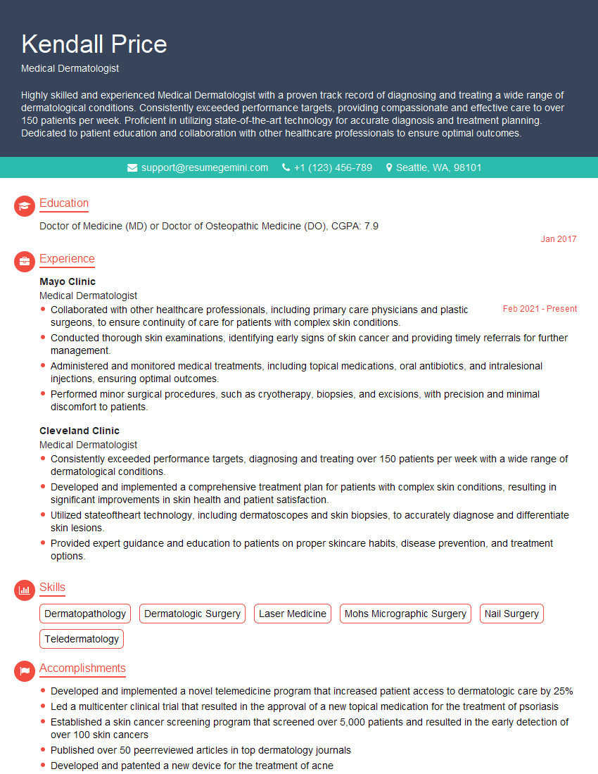
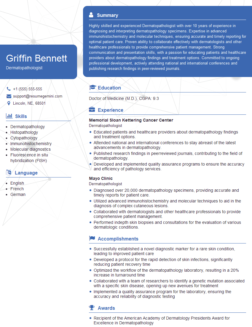
Mastering Skin Lesion Removal techniques significantly enhances your career prospects in dermatology and related fields, opening doors to specialized roles and increased earning potential. A well-crafted resume is crucial for showcasing your skills and experience to potential employers. Building an ATS-friendly resume increases the likelihood of your application being noticed and considered. ResumeGemini is a trusted resource that can help you create a compelling and effective resume, ensuring your qualifications shine. Examples of resumes tailored to Skin Lesion Removal professionals are available within ResumeGemini to help guide you.
Explore more articles
Users Rating of Our Blogs
Share Your Experience
We value your feedback! Please rate our content and share your thoughts (optional).
What Readers Say About Our Blog
This was kind of a unique content I found around the specialized skills. Very helpful questions and good detailed answers.
Very Helpful blog, thank you Interviewgemini team.
