Every successful interview starts with knowing what to expect. In this blog, we’ll take you through the top Spinal Stenosis interview questions, breaking them down with expert tips to help you deliver impactful answers. Step into your next interview fully prepared and ready to succeed.
Questions Asked in Spinal Stenosis Interview
Q 1. Describe the pathophysiology of lumbar spinal stenosis.
Lumbar spinal stenosis is a narrowing of the spinal canal in the lower back, causing pressure on the spinal cord and nerves. This pressure results in pain, numbness, and weakness in the legs and feet. The pathophysiology is complex and multifaceted, but it primarily involves a decrease in the available space within the spinal canal. This narrowing can be caused by several factors, often acting in combination:
Degenerative changes: Age-related wear and tear on the intervertebral discs (the cushions between vertebrae) leads to disc desiccation (drying out) and bulging or herniation. Osteophytes (bone spurs) also form along the edges of the vertebrae, further reducing the canal’s diameter. Facet joint hypertrophy (enlargement) also contributes, as these joints become arthritic and bony growths restrict space.
Ligamentum flavum thickening: The ligamentum flavum, a ligament supporting the spine, can thicken and buckle into the spinal canal, further constricting the space for the nerves.
Spondylolisthesis: This is the forward slippage of one vertebra over another, which can significantly reduce the spinal canal space.
The combination of these changes leads to compression of the nerve roots exiting the spinal cord, causing the characteristic symptoms of lumbar spinal stenosis. Imagine a crowded hallway – the hallway represents the spinal canal, and the people represent the nerves. If the hallway becomes too narrow, the people will feel squeezed and uncomfortable, just as nerves do in spinal stenosis.
Q 2. Differentiate between degenerative and congenital spinal stenosis.
The key difference between degenerative and congenital spinal stenosis lies in their etiology (cause):
Degenerative spinal stenosis is the most common type and is primarily caused by age-related changes in the spine as described above. It’s a gradual process that develops over time due to wear and tear.
Congenital spinal stenosis is present at birth. It’s due to an anomaly in the development of the spine, leading to a smaller-than-normal spinal canal. Individuals with congenital stenosis may not experience symptoms until later in life when other degenerative changes further narrow the already restricted space. Think of it like a pre-existing narrow hallway – even minor changes can cause significant crowding later on.
In essence, degenerative stenosis is acquired, while congenital stenosis is inherent. Both can manifest with similar clinical presentations, but their underlying causes are vastly different.
Q 3. What are the common clinical presentations of cervical spinal stenosis?
Cervical spinal stenosis, a narrowing of the spinal canal in the neck, presents with symptoms that typically affect the arms and hands, unlike lumbar stenosis which affects the legs and feet. Common clinical presentations include:
Neck pain: Often a dull ache, worsened by certain movements.
Radiculopathy (nerve root compression): Pain, numbness, tingling, or weakness radiating down one or both arms. Specific dermatomes (areas of skin supplied by a single nerve root) are often affected, providing clues to the level of nerve compression.
Myelopathy (spinal cord compression): This is more serious and can involve gait disturbances (difficulty walking), clumsiness of hands, balance problems, and even bowel or bladder dysfunction. It’s a sign that the spinal cord itself is being significantly compressed.
Weakness: Muscle weakness in the arms or hands.
Spasticity: Increased muscle tone, leading to stiffness.
The severity of symptoms depends on the degree of stenosis and the extent of nerve or cord compression. Some individuals may experience only mild discomfort, while others may have debilitating symptoms that interfere significantly with daily life.
Q 4. Explain the diagnostic imaging modalities used in evaluating spinal stenosis.
Several diagnostic imaging modalities are used to evaluate spinal stenosis, each offering unique advantages:
X-rays: Provide a basic view of the bone structure, showing bone spurs (osteophytes), spondylolisthesis, and other bony abnormalities that contribute to stenosis. However, x-rays don’t visualize soft tissues like discs and ligaments well.
CT scans (Computed Tomography): Offer detailed cross-sectional images of the bones and soft tissues, giving a more precise assessment of the spinal canal size and the degree of narrowing. CT myelograms involve injecting contrast dye into the spinal canal to better visualize the spinal cord and nerve roots.
MRI scans (Magnetic Resonance Imaging): Provide superior visualization of soft tissues, including the spinal cord, nerve roots, intervertebral discs, and ligaments. This allows for the precise identification of the structures contributing to stenosis and assessment of the extent of nerve compression.
The choice of modality depends on the clinical presentation and the specific information needed. Often, a combination of imaging techniques provides the most complete picture.
Q 5. Discuss the role of MRI in identifying spinal stenosis.
MRI is the gold standard imaging technique for identifying spinal stenosis because of its exceptional ability to visualize soft tissues. Unlike X-rays or CT scans, which primarily show bone, MRI clearly demonstrates:
The size of the spinal canal: MRI precisely measures the dimensions of the spinal canal, allowing quantification of the degree of narrowing.
The condition of the intervertebral discs: Disc bulging, herniation, and desiccation are readily visible, providing crucial information about their contribution to stenosis.
The ligamentum flavum: Thickening or buckling of this ligament can be clearly identified.
The spinal cord and nerve roots: MRI shows the anatomy of the spinal cord and nerve roots and reveals compression or displacement caused by the stenosis.
The presence of edema (swelling): This indicates inflammation and nerve irritation.
This detailed information helps in determining the severity of stenosis, identifying the contributing factors, and guiding treatment planning.
Q 6. How do you interpret findings on an MRI showing spinal stenosis?
Interpreting MRI findings in spinal stenosis requires careful attention to several aspects:
Spinal canal diameter: Measurement of the sagittal diameter (anterior-posterior) of the spinal canal at various levels helps determine the degree of narrowing. This is often compared to established normative values.
Presence and severity of disc herniation or bulging: The location and size of herniated or bulging discs are noted. Their position relative to the nerve roots is crucial, indicating the potential for compression.
Ligamentum flavum thickening: The thickness and extent of thickening of this ligament are assessed. Significant thickening can significantly compromise spinal canal space.
Facet joint hypertrophy: The size and extent of the facet joint osteophytes are noted, indicating their contribution to stenosis.
Spinal cord and nerve root compression: The degree of compression on the spinal cord and nerve roots is assessed. The presence of edema or signal abnormalities in the spinal cord or nerve roots indicates inflammation and injury.
Spondylolisthesis: The degree of slippage of one vertebra over another is measured if present.
The radiologist’s report combines these findings to provide a comprehensive description of the stenosis, helping clinicians make informed decisions regarding treatment.
Q 7. What are the conservative treatment options for spinal stenosis?
Conservative treatment options for spinal stenosis aim to reduce pain, improve function, and avoid surgery. These often involve a multi-modal approach:
Pain medications: Over-the-counter analgesics (like ibuprofen or acetaminophen) and prescription medications (such as NSAIDs or opioids) may be used to manage pain.
Physical therapy: Exercises to strengthen core muscles, improve flexibility, and improve posture can help alleviate pain and improve function. This also often incorporates modalities like ultrasound and heat therapy.
Epidural steroid injections: These injections deliver corticosteroids directly to the inflamed area in the spinal canal, reducing inflammation and pain. They’re often used for short-term pain relief.
Weight management: Weight loss can reduce pressure on the spine and alleviate symptoms.
Lifestyle modifications: Avoiding activities that aggravate symptoms, using assistive devices (like canes or walkers), and pacing activities can help manage pain and improve quality of life.
Alternative therapies: Some patients find relief through alternative therapies such as acupuncture, chiropractic care, or massage therapy. The evidence supporting their effectiveness varies.
The goal is to find the combination of conservative treatments that best suits the individual’s needs and provides the most effective pain relief and functional improvement.
Q 8. Describe the indications for surgical intervention in spinal stenosis.
Surgical intervention for spinal stenosis is indicated when conservative management, such as physical therapy, medication, and injections, fails to provide adequate pain relief and improve neurological function. The decision is highly individualized and depends on the severity of symptoms, the patient’s overall health, and the extent of spinal stenosis. Specific indications include:
- Intractable pain: Severe, persistent pain that significantly impacts daily life and doesn’t respond to non-surgical treatments.
- Progressive neurological deficits: Worsening weakness, numbness, or bowel/bladder dysfunction indicating nerve compression.
- Cauda equina syndrome: A serious condition requiring immediate surgery, characterized by bowel/bladder dysfunction, saddle anesthesia (numbness in the saddle area), and severe leg weakness. This is a surgical emergency.
- Severe spinal instability: Cases where the spine is unstable, risking further damage or neurological compromise.
- Failed conservative management: When non-surgical approaches have been tried for a sufficient duration without satisfactory results.
It’s crucial to emphasize that surgery isn’t always the best option. A thorough assessment of the patient’s condition, including imaging studies (MRI, CT), neurological examination, and a comprehensive discussion of risks and benefits, is essential before considering surgical intervention.
Q 9. Explain the different surgical techniques used to treat spinal stenosis.
Several surgical techniques can address spinal stenosis, each tailored to the specific location and severity of the stenosis. The choice depends on factors like the patient’s age, overall health, and the anatomical specifics of the stenosis. Common techniques include:
- Laminectomy: This involves removing a portion of the lamina (the bony arch of the vertebra) to decompress the spinal canal and relieve pressure on the nerves. It’s one of the most common procedures for spinal stenosis.
- Laminoplasty: Instead of removing the lamina, this technique involves spreading or hinged opening of the lamina to create more space. This procedure preserves the structural integrity of the vertebra and can potentially lead to faster recovery.
- Foraminotomy: This procedure widens the intervertebral foramen (the opening where spinal nerves exit the spine) to relieve pressure on the nerve roots. It’s often performed in conjunction with a laminectomy.
- Discectomy: This involves the removal of a portion or all of a herniated disc to alleviate nerve compression. This is often necessary when disc herniation contributes to the stenosis.
- Spinal fusion: In cases of significant spinal instability, fusion involves connecting two or more vertebrae together with bone grafts or metal implants to stabilize the spine. This is typically a more extensive procedure with a longer recovery period.
Minimally invasive techniques are also frequently used. These procedures involve smaller incisions, leading to less tissue damage, reduced pain, and faster recovery. These often utilize specialized instruments and imaging guidance.
Q 10. What are the potential complications of spinal stenosis surgery?
Spinal stenosis surgery, while generally effective, carries potential complications like any major surgery. These complications can be broadly classified into:
- Infection: Wound infection, epidural abscess, or meningitis are serious possibilities, although rare with proper aseptic techniques.
- Bleeding: Hematoma formation can cause pressure on the spinal cord, requiring emergency intervention.
- Nerve damage: Although the goal is decompression, there’s a risk of inadvertently injuring nerves during the surgery, leading to new or worsened neurological deficits.
- CSF leak: Leakage of cerebrospinal fluid can lead to headaches and other complications.
- Dural tear: A tear in the dura mater (the protective membrane surrounding the spinal cord) may require repair.
- Hardware failure: In fusion surgeries, the implanted hardware may fail over time, requiring revision surgery.
- Pseudarthrosis: Failure of the fusion to heal properly can lead to persistent pain and instability.
- Adjacent segment disease: Increased stress on segments of the spine adjacent to the fusion site may lead to degeneration and stenosis in those areas over time.
The incidence of these complications varies depending on the specific surgical technique, the surgeon’s experience, and the patient’s overall health. Pre-operative assessment helps minimize risks and thorough post-operative monitoring aids in early detection and management of complications.
Q 11. Discuss post-operative management of spinal stenosis patients.
Post-operative management of spinal stenosis patients is crucial for optimal recovery and minimizing complications. It typically involves:
- Pain management: Medication, including analgesics and potentially opioids, is used to control post-operative pain. This is transitioned to less potent medication as the patient recovers.
- Physical therapy: A tailored rehabilitation program helps improve strength, flexibility, and range of motion. It focuses on core strengthening, posture correction, and gait retraining.
- Wound care: Regular monitoring and dressing changes are crucial to prevent infection.
- Neurological monitoring: Regular neurological examinations assess for any new or worsening neurological deficits.
- Activity modification: Gradual return to normal activities is essential, guided by the surgeon’s recommendations and the patient’s progress.
- Follow-up appointments: Regular visits allow the surgeon to monitor healing, assess the effectiveness of the surgery, and address any complications.
The recovery period varies depending on the extent of the surgery and the patient’s overall health, but it generally involves several weeks of restricted activity followed by a gradual increase in physical activity.
Q 12. How do you assess the effectiveness of treatment for spinal stenosis?
Assessing the effectiveness of treatment for spinal stenosis involves a multi-faceted approach, focusing on both subjective and objective measures. We consider:
- Pain reduction: This is often the primary outcome measure. We assess changes in pain intensity using validated pain scales (e.g., Visual Analog Scale, Numeric Rating Scale).
- Improved neurological function: We assess any improvements in strength, sensation, bowel/bladder function, and gait using neurological examinations.
- Functional improvement: We assess the patient’s ability to perform daily activities using functional outcome measures, such as the Oswestry Disability Index.
- Quality of life: Patient-reported outcome measures (PROMs) such as SF-36 assess the overall impact of the condition and treatment on their daily lives.
- Imaging studies: Post-operative imaging (MRI, CT) may be used to assess the surgical decompression and the stability of the spine, although these findings don’t always correlate perfectly with clinical outcomes.
The combination of these measures provides a comprehensive assessment of treatment success, ensuring that both pain relief and improved function are considered.
Q 13. What are the common complications associated with spinal stenosis?
Common complications associated with spinal stenosis can arise gradually or acutely. These include:
- Neurogenic claudication: Pain, cramping, and numbness in the legs and buttocks, especially when walking (better with rest or bending forward).
- Back pain: Aching, stiffness, or pain in the lower back.
- Leg weakness: Difficulty walking, climbing stairs, or standing for prolonged periods.
- Numbness and tingling: Loss of sensation or tingling in the legs, feet, or buttocks.
- Bowel and bladder dysfunction: In severe cases, this can occur, indicating compression of the cauda equina (a bundle of nerve roots at the end of the spinal cord).
- Spinal instability: Movement of the vertebrae beyond their normal range, potentially leading to further pain and neurological problems.
The severity of these complications varies widely depending on the degree of spinal stenosis and the individual patient’s response to the condition. Early diagnosis and management are key to preventing long-term complications.
Q 14. How do you differentiate spinal stenosis from other spinal conditions?
Differentiating spinal stenosis from other spinal conditions requires a thorough history, physical examination, and imaging studies (MRI is typically the gold standard). Key differences include:
- Spinal stenosis presents with symptoms related to nerve root compression, often manifesting as neurogenic claudication (leg pain relieved by bending forward), back pain, and progressive neurological deficits (weakness, numbness).
- Spondylolisthesis involves the forward slippage of one vertebra over another, potentially causing nerve compression, but often presenting with more localized pain and a different pattern of neurological symptoms.
- Disc herniation involves the protrusion of the intervertebral disc material, often causing localized pain and radiculopathy (nerve pain radiating down the leg) in a specific dermatome, rather than the more generalized symptoms of stenosis.
- Degenerative disc disease is a broader term encompassing age-related changes in the intervertebral discs, which may contribute to stenosis but doesn’t necessarily cause the same clinical picture.
- Spinal tumors typically present with more rapid progression of symptoms, potentially involving severe pain, neurological deficits, and systemic symptoms.
Detailed neurological examination, including assessment of reflexes, muscle strength, and sensation, helps pinpoint the specific source of compression. Imaging studies are crucial for visualizing the spinal anatomy and identifying the underlying cause of the symptoms.
Q 15. Describe the role of epidural steroid injections in spinal stenosis.
Epidural steroid injections (ESIs) are a common treatment for spinal stenosis, a condition causing narrowing of the spinal canal. They aim to reduce inflammation and pain around the spinal nerves. The procedure involves injecting a corticosteroid medication directly into the epidural space, the area surrounding the spinal cord and nerve roots. This medication acts as an anti-inflammatory, decreasing swelling that’s compressing the nerves and causing pain. Think of it like this: the inflamed area is like a swollen hose, restricting water flow. The steroid injection helps reduce the swelling, allowing for better ‘flow’ – in this case, nerve function, reducing pain and improving mobility.
How it works: The corticosteroid reduces inflammation, providing temporary pain relief. It doesn’t address the underlying anatomical narrowing, but it buys time for other treatments to work, like physical therapy, or allows patients to better participate in those treatments. The relief is often temporary, lasting weeks to months, and multiple injections may be necessary. The benefits need to be carefully weighed against potential side effects like infection, bleeding, or nerve damage, although these are relatively uncommon when performed by experienced professionals.
Career Expert Tips:
- Ace those interviews! Prepare effectively by reviewing the Top 50 Most Common Interview Questions on ResumeGemini.
- Navigate your job search with confidence! Explore a wide range of Career Tips on ResumeGemini. Learn about common challenges and recommendations to overcome them.
- Craft the perfect resume! Master the Art of Resume Writing with ResumeGemini’s guide. Showcase your unique qualifications and achievements effectively.
- Don’t miss out on holiday savings! Build your dream resume with ResumeGemini’s ATS optimized templates.
Q 16. What are the different types of spinal stenosis?
Spinal stenosis can be categorized in a few ways, primarily by location and cause. We often classify it as:
- Cervical Spinal Stenosis: Narrowing in the neck region, affecting the nerves supplying the arms and hands. This often presents with neck pain, radiating arm pain (radiculopathy), and hand weakness or numbness.
- Thoracic Spinal Stenosis: Narrowing in the mid-back (thoracic spine). This is less common than cervical or lumbar stenosis and often presents with back pain and potentially radiating pain to the chest or abdomen.
- Lumbar Spinal Stenosis: Narrowing in the lower back (lumbar spine), the most common type. This usually results in pain in the lower back, buttocks, and legs (neurogenic claudication), often worsened by standing or walking and relieved by sitting or bending forward.
Further classification can be based on the cause of the narrowing:
- Degenerative Stenosis: The most common type, caused by age-related changes like disc degeneration, osteophyte formation (bone spurs), and ligamentum flavum hypertrophy (thickening of the ligament).
- Congenital Stenosis: Present from birth due to a malformation of the spinal canal.
- Acquired Stenosis: Develops after a trauma, tumor, or infection.
Understanding the specific type and cause helps determine the best treatment plan.
Q 17. Explain the role of physical therapy in managing spinal stenosis.
Physical therapy plays a crucial role in managing spinal stenosis, focusing on improving strength, flexibility, and overall function. It’s often the cornerstone of conservative management. The goals include:
- Pain Management: Exercises can strengthen the muscles supporting the spine, reducing stress on the affected nerves and relieving pain.
- Improved Mobility: Stretching and range-of-motion exercises improve flexibility and reduce stiffness. Specific exercises may target improving posture and gait.
- Increased Strength and Endurance: Strengthening exercises, tailored to the individual’s condition, enhance stability and reduce reliance on pain medications.
- Posture Correction: Poor posture can exacerbate symptoms; therapists guide patients toward proper posture and body mechanics.
- Education and Lifestyle Modifications: Therapists educate patients on proper lifting techniques, ergonomic workstation setups, and activities to avoid.
Example: A patient with lumbar stenosis might benefit from core strengthening exercises to improve spinal stability, stretches to increase flexibility in the hip and hamstring muscles, and gait training to improve walking efficiency.
Q 18. What are the limitations of conservative treatment for spinal stenosis?
While conservative treatments like physical therapy, medication, and ESIs are often effective, they have limitations for spinal stenosis. These limitations include:
- Limited Effectiveness in Severe Cases: In patients with significant neural compression, conservative treatments might not provide adequate pain relief or functional improvement.
- Temporary Relief: Many conservative treatments, such as ESIs, provide temporary relief, requiring repeated interventions.
- Slow Progression: It can take time to see significant improvements with physical therapy, and some patients may not be able to commit to the necessary regimen.
- Side Effects of Medications: Long-term use of pain medications can lead to side effects, including gastrointestinal problems, dependency, or other adverse reactions.
- Lack of Structural Correction: Conservative treatments do not address the underlying anatomical narrowing of the spinal canal.
If conservative treatment fails to provide sufficient relief after a reasonable trial period, surgical intervention may be considered.
Q 19. Discuss the use of bracing in the management of spinal stenosis.
Bracing is less frequently used for spinal stenosis compared to other spinal conditions like fractures or post-surgery support. It’s not a primary treatment modality. However, in certain situations, a brace might be considered:
- Post-Surgery: A brace may be used temporarily after surgery to provide support and stability while the spine heals.
- Pain Management: In some cases, a brace can provide temporary relief from pain by limiting spinal motion, but this is typically only a short-term measure and not a long-term solution.
- As an Adjunct to Other Therapies: A brace may be used in conjunction with other treatments such as physical therapy to enhance stability and reduce pain during certain activities.
It’s crucial to understand that bracing alone does not address the underlying cause of spinal stenosis. The benefits should be carefully evaluated against potential downsides such as muscle weakness from disuse and potential skin issues.
Q 20. How do you counsel patients about the risks and benefits of surgery for spinal stenosis?
Counseling patients about spinal stenosis surgery requires a thorough discussion of risks and benefits, tailored to the individual patient’s condition and preferences. I always start by emphasizing that surgery is not always necessary and that conservative treatment should be given a fair trial. The conversation covers:
- Benefits: Potential improvements in pain, mobility, and overall quality of life. I use realistic expectations, explaining that surgery doesn’t guarantee complete pain relief but often significantly improves symptoms.
- Risks: I discuss potential complications such as infection, bleeding, nerve damage, and the possibility that surgery may not provide sufficient relief. The specific risks vary depending on the surgical approach and the patient’s overall health.
- Alternatives: We review the effectiveness of non-surgical options and the reasons why surgery is being considered.
- Recovery: I detail the expected recovery time, including hospitalization, rehabilitation, and return to normal activities. This helps set realistic expectations and manage the patient’s anxieties.
- Long-Term Outcomes: I discuss the likelihood of long-term success based on the patient’s specific case and the surgeon’s experience.
I use plain language, avoiding jargon, and encourage patients to ask questions. Shared decision-making is paramount: the patient needs to understand the risks and benefits to make an informed choice.
Q 21. What are the long-term outcomes of surgical treatment for spinal stenosis?
The long-term outcomes of spinal stenosis surgery are generally positive for many patients, but individual results vary. Success is defined by improvement in pain, neurological function, and overall quality of life. Factors influencing long-term outcomes include:
- Severity of Stenosis: Patients with more severe stenosis may have a slower or less complete recovery.
- Surgical Technique: The type of surgery performed impacts the outcome; some procedures are more successful in specific situations.
- Patient Factors: Age, overall health, compliance with post-operative rehabilitation, and pre-existing conditions influence recovery.
- Post-operative Rehabilitation: Active participation in physical therapy is critical for optimal recovery and long-term results.
Studies show that a significant percentage of patients experience substantial pain relief and improved function after surgery. However, some individuals may still experience residual pain or require additional treatment over time. It’s important for patients to understand that surgery aims to improve their quality of life, not eliminate all symptoms forever.
Q 22. Describe the role of minimally invasive surgery in treating spinal stenosis.
Minimally invasive surgery (MIS) for spinal stenosis offers a less traumatic alternative to traditional open surgery. Instead of making large incisions, MIS utilizes smaller incisions, smaller instruments, and advanced imaging techniques like fluoroscopy to access and repair the affected area of the spine. This approach leads to several advantages.
- Reduced tissue trauma: Smaller incisions mean less muscle damage, resulting in less post-operative pain and faster recovery times.
- Shorter hospital stays: Patients often go home the same day or within a day or two of the procedure.
- Less blood loss: The smaller incisions minimize bleeding during surgery.
- Faster return to normal activities: Patients can typically resume their daily activities much sooner compared to open surgery.
For example, a patient with lumbar spinal stenosis causing significant leg pain might benefit from MIS techniques like foraminotomy (enlarging the openings where nerves exit the spine) or discectomy (removing a portion of a herniated disc). The choice of specific MIS procedure depends on the individual’s anatomy, the location and severity of the stenosis, and other health factors.
Q 23. What are the emerging trends in the treatment of spinal stenosis?
Emerging trends in spinal stenosis treatment are focused on improving outcomes and minimizing invasiveness. Key trends include:
- Advanced imaging techniques: High-resolution MRI and CT scans are allowing for more precise diagnosis and surgical planning, leading to more effective treatments.
- Robotic-assisted surgery: Robots are improving surgical precision, minimizing invasiveness, and potentially reducing complication rates.
- Personalized medicine: Tailoring treatment plans to individual patient characteristics, including genetics and lifestyle factors, is gaining traction.
- Focus on non-surgical treatments: There’s a growing emphasis on optimizing non-surgical options, such as physical therapy, medication management, and epidural injections, before resorting to surgery. This involves better patient education and targeted interventions.
- Regenerative medicine (discussed further in a later question): This holds great promise for stimulating the body’s natural healing processes to repair damaged spinal structures.
These advancements aim to improve patient outcomes, reduce recovery time, and enhance quality of life for individuals with spinal stenosis.
Q 24. Discuss the role of regenerative medicine in treating spinal stenosis.
Regenerative medicine offers exciting possibilities for spinal stenosis treatment by focusing on repairing damaged tissues rather than just removing them. Several approaches are being explored:
- Stem cell therapy: Injecting stem cells into the affected area could stimulate the regeneration of cartilage and other spinal tissues. This is still in the research phase but shows promising results in animal studies.
- Growth factor therapy: Using growth factors to stimulate tissue repair and regeneration is another area of active research.
- Platelet-rich plasma (PRP) therapy: Concentrated platelets from the patient’s own blood are injected into the affected area to promote healing. While not a cure, it can potentially help reduce pain and inflammation.
The clinical evidence for the effectiveness of these regenerative therapies in treating spinal stenosis is still limited, and large-scale clinical trials are needed to confirm their efficacy and safety. However, they represent a significant advancement in potentially offering long-term solutions, potentially reducing the need for more invasive procedures in the future.
Q 25. Explain the importance of patient education in managing spinal stenosis.
Patient education is crucial in managing spinal stenosis. It empowers patients to actively participate in their care and improve their outcomes. Effective patient education involves several key elements:
- Understanding the condition: Patients need a clear explanation of spinal stenosis, its causes, and how it affects their bodies.
- Treatment options: Discussions should cover non-surgical and surgical options, including the risks, benefits, and limitations of each approach.
- Lifestyle modifications: Patients should learn about strategies to manage their symptoms, such as exercises, weight management, posture correction, and ergonomic adjustments.
- Pain management: Education on pain management techniques, including medication, physical therapy, and alternative therapies, is essential.
- Realistic expectations: Openly discussing the limitations of treatment and setting realistic expectations regarding recovery is crucial for patient satisfaction.
For example, a patient newly diagnosed with spinal stenosis might benefit from a detailed handout explaining the condition, a demonstration of appropriate exercises, and a list of resources for pain management support groups.
Q 26. How do you assess patient satisfaction after spinal stenosis treatment?
Assessing patient satisfaction after spinal stenosis treatment is a multi-faceted process involving both objective and subjective measures. We employ various methods:
- Standardized questionnaires: Validated questionnaires such as the Oswestry Disability Index (ODI) and the Short Form-36 (SF-36) assess physical function, pain levels, and quality of life. Changes in scores from pre-treatment to post-treatment indicate improvement.
- Clinical assessments: Regular clinical evaluations assess neurological function, range of motion, and pain levels.
- Patient interviews: Direct conversations with patients allow us to gain a deeper understanding of their experience, address concerns, and identify areas for improvement in their care.
- Follow-up appointments: Scheduled follow-up appointments provide opportunities to monitor progress, address complications, and gauge patient satisfaction.
By combining these approaches, we can obtain a comprehensive picture of patient satisfaction and identify areas where we can refine our treatment strategies.
Q 27. Describe your experience with managing complex cases of spinal stenosis.
Managing complex cases of spinal stenosis often requires a multidisciplinary approach. I have extensive experience in dealing with cases involving:
- Severe stenosis with neurological deficits: These cases require careful surgical planning and execution to minimize the risk of further neurological damage.
- Spinal deformities: Cases involving scoliosis or other spinal deformities necessitate a more complex surgical approach often involving advanced techniques to address both the stenosis and the deformity.
- Revision surgeries: Patients who have undergone previous spinal surgeries may require more complex revision procedures to address complications or recurrence of stenosis.
- Patients with multiple comorbidities: Managing patients with other medical conditions such as diabetes, heart disease, or obesity requires careful coordination with other specialists to optimize treatment safety and efficacy.
For example, a patient with severe lumbar stenosis causing cauda equina syndrome (a serious neurological condition) requires immediate surgical intervention. In such cases, close collaboration with neurosurgery and anesthesiology colleagues is paramount to ensure successful outcomes.
Q 28. What are your strategies for communicating effectively with patients and their families about spinal stenosis?
Effective communication with patients and their families regarding spinal stenosis is critical. My strategy involves:
- Clear and concise explanations: I use plain language to explain the condition, treatment options, and potential risks and benefits.
- Active listening: I take the time to listen to patients’ concerns and answer their questions thoroughly.
- Visual aids: Using diagrams, models, and imaging studies helps patients understand their condition and the proposed treatment plan.
- Involving family members: I encourage the involvement of family members in the decision-making process, especially when patients are struggling to comprehend complex medical information.
- Empathy and compassion: Showing empathy and understanding helps to build trust and rapport with patients and their families.
- Written materials: Providing written materials summarizing the discussion reinforces key information and ensures patients have access to the details after the consultation.
I believe that open, honest, and compassionate communication fosters a strong doctor-patient relationship which is crucial for successful management of this often complex condition.
Key Topics to Learn for Spinal Stenosis Interview
- Anatomy and Physiology: Understand the anatomical structures involved in spinal stenosis, including vertebrae, intervertebral discs, spinal cord, and nerve roots. Master the physiological mechanisms leading to stenosis and its impact on nerve function.
- Pathophysiology: Develop a strong grasp of the different types of spinal stenosis (degenerative, congenital, etc.) and their underlying causes. Be prepared to discuss the inflammatory processes and degenerative changes that contribute to the condition.
- Clinical Presentation: Familiarize yourself with the typical symptoms of spinal stenosis, including neurogenic claudication, back pain, radiculopathy, and weakness. Understand how these symptoms vary based on the location and severity of the stenosis.
- Diagnostic Imaging: Learn to interpret common imaging modalities used to diagnose spinal stenosis, such as X-rays, CT scans, and MRI. Understand the strengths and limitations of each technique and how they inform treatment decisions.
- Treatment Options: Be prepared to discuss conservative management strategies (e.g., physical therapy, medication) and surgical interventions (e.g., laminectomy, foraminotomy). Understand the indications, contraindications, and potential complications of each approach.
- Differential Diagnosis: Develop the ability to differentiate spinal stenosis from other conditions that present with similar symptoms, such as disc herniation, spinal tumors, and spondylolisthesis.
- Post-operative Care and Rehabilitation: Understand the principles of post-operative management and rehabilitation for patients undergoing spinal stenosis surgery. This includes pain management, physical therapy, and functional recovery.
- Research and Current Trends: Stay updated on recent advancements in the diagnosis and treatment of spinal stenosis. This demonstrates your commitment to ongoing learning and professional development.
Next Steps
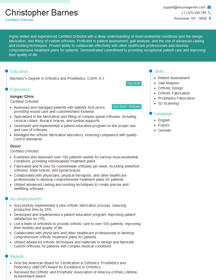
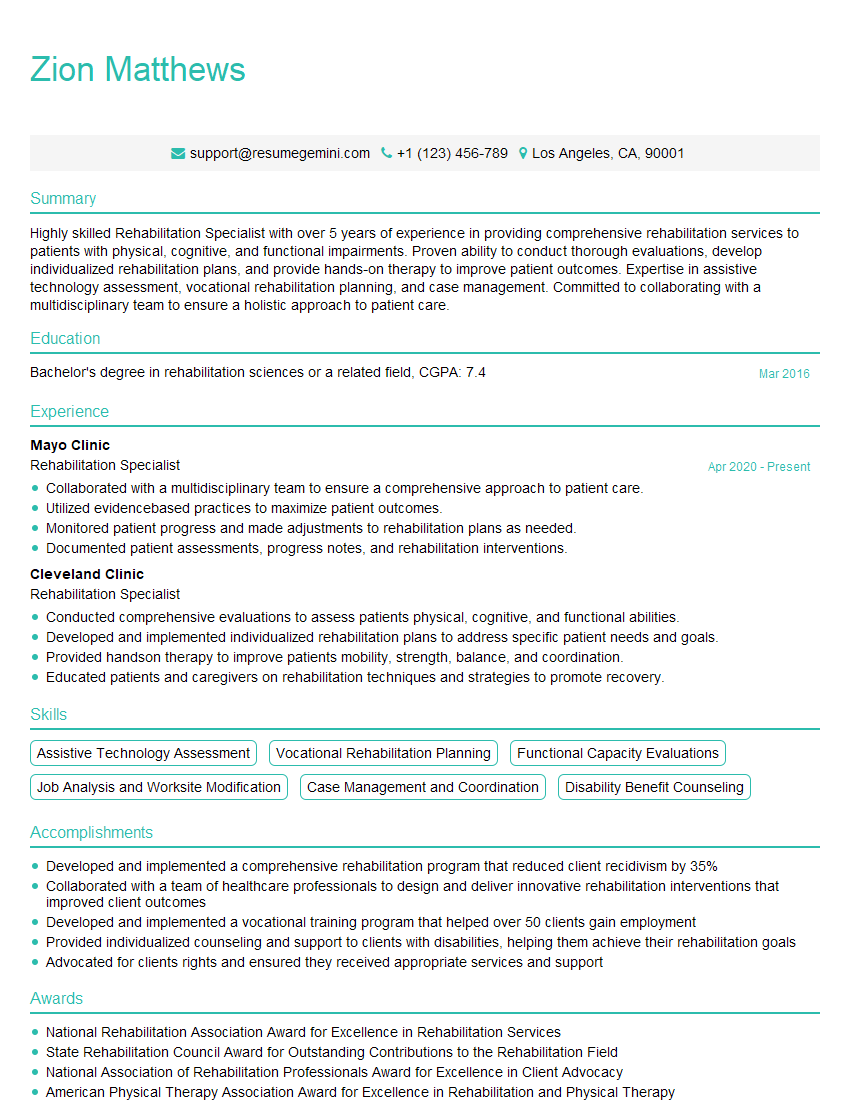
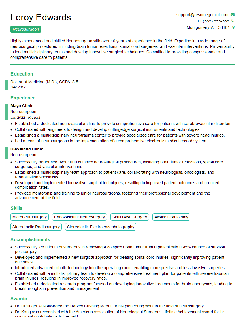
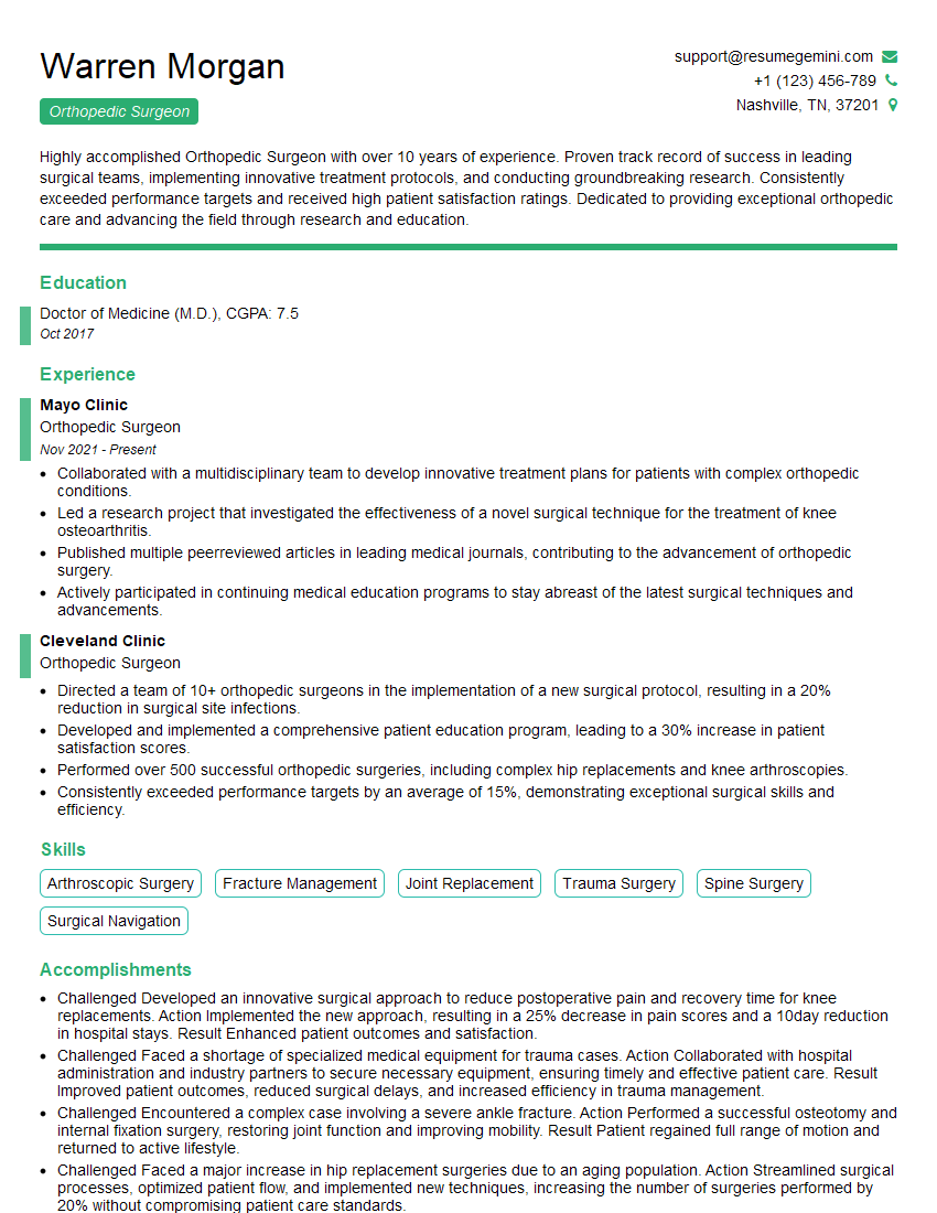
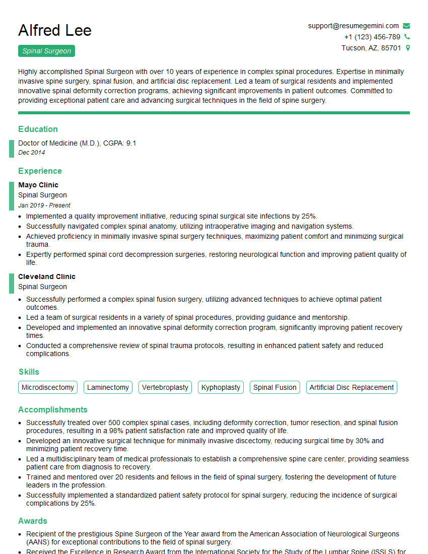
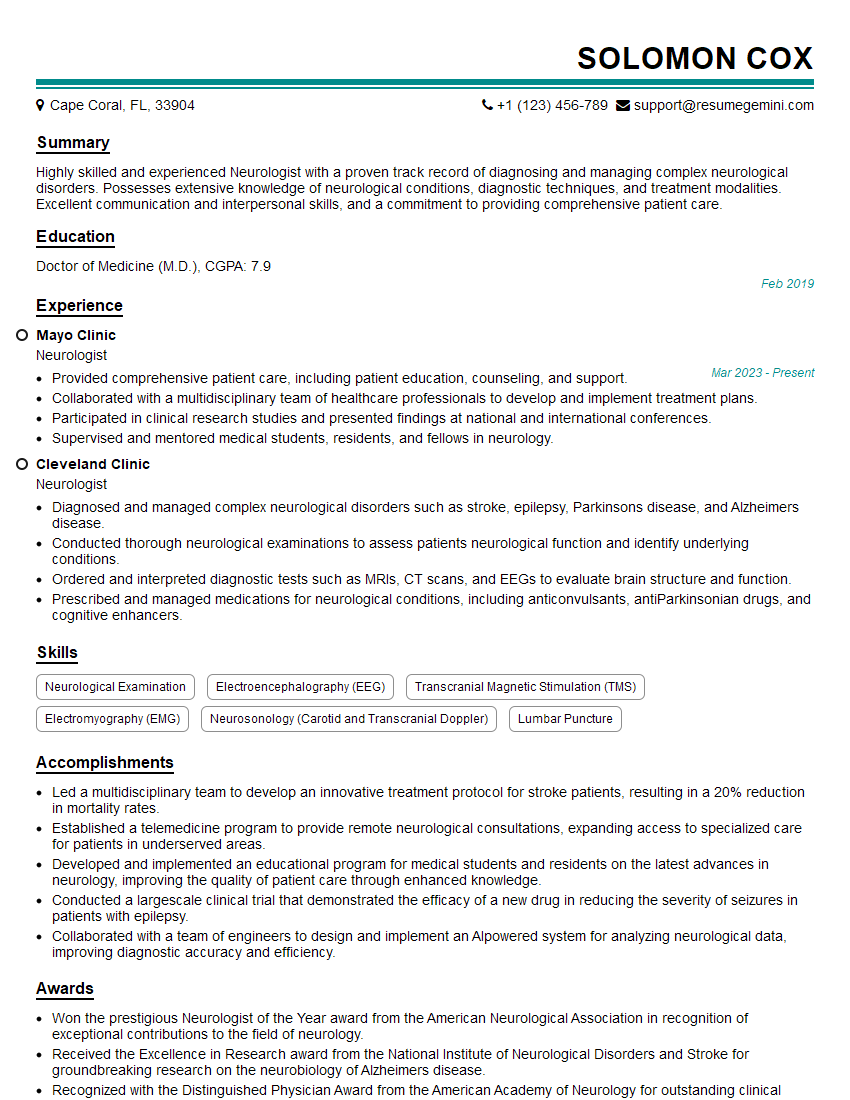
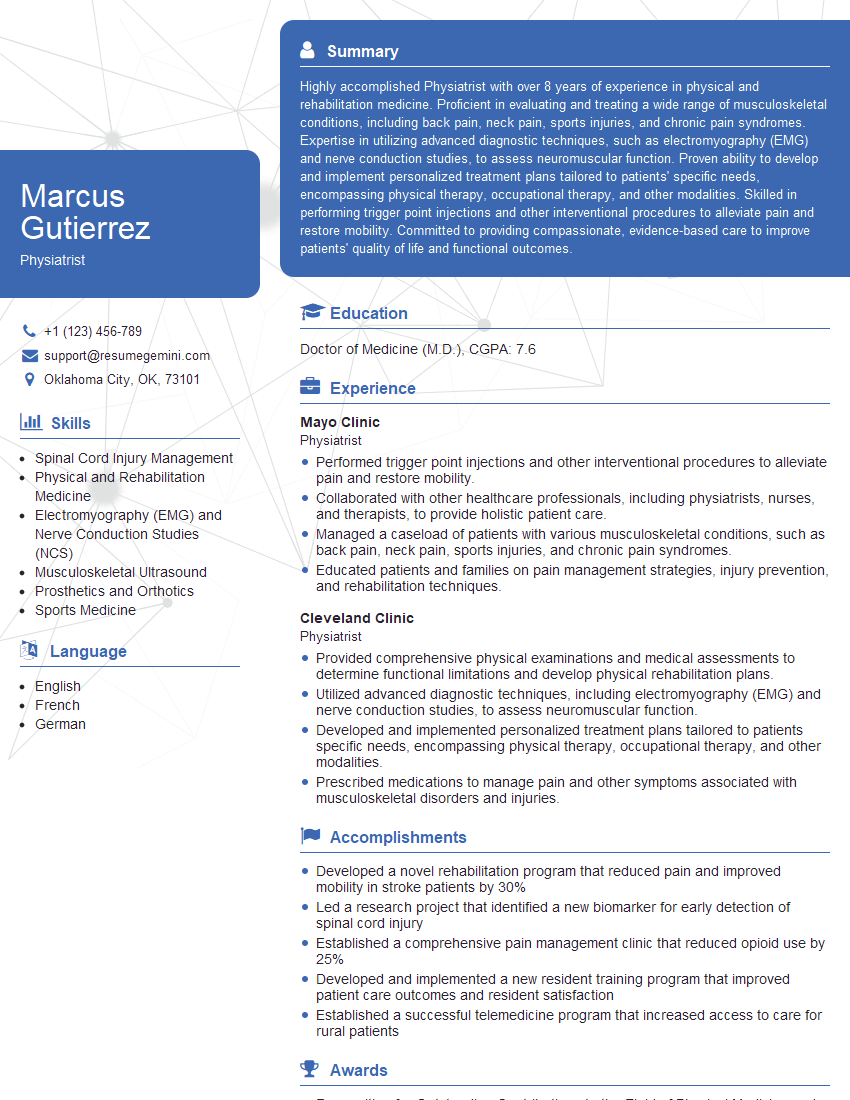
Mastering spinal stenosis is crucial for career advancement in the medical and healthcare fields. A deep understanding of this common condition demonstrates a high level of clinical expertise and problem-solving skills, highly valued by employers. To maximize your job prospects, create an ATS-friendly resume that highlights your relevant skills and experience. ResumeGemini is a trusted resource that can help you build a professional and impactful resume, tailored to the specific requirements of the jobs you are targeting. Examples of resumes tailored to Spinal Stenosis are available to help you get started.
Explore more articles
Users Rating of Our Blogs
Share Your Experience
We value your feedback! Please rate our content and share your thoughts (optional).
What Readers Say About Our Blog
This was kind of a unique content I found around the specialized skills. Very helpful questions and good detailed answers.
Very Helpful blog, thank you Interviewgemini team.
