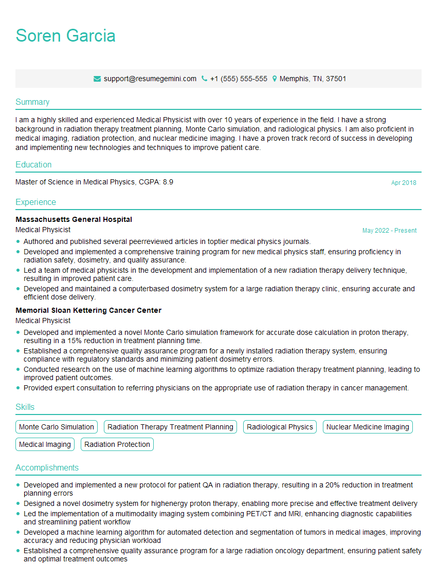
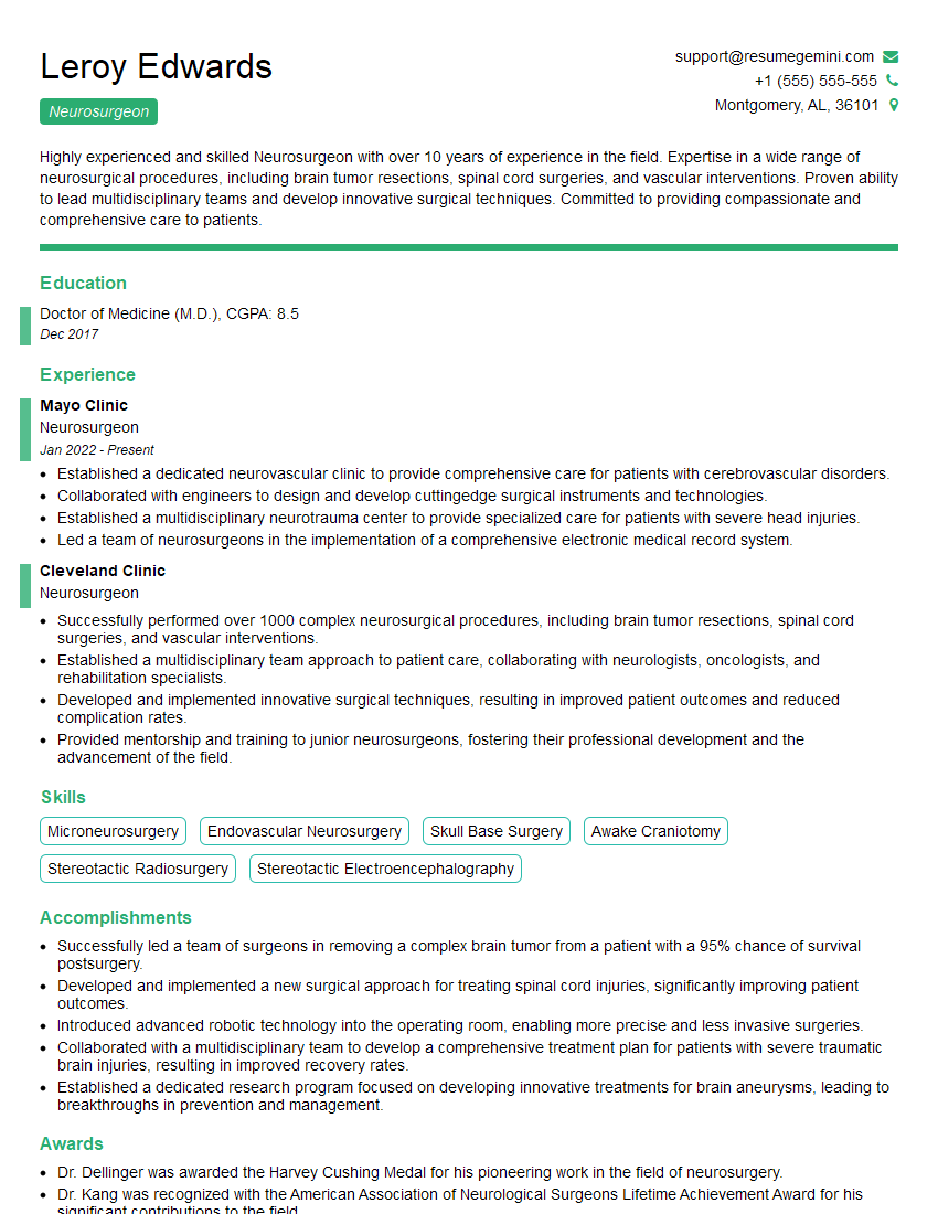
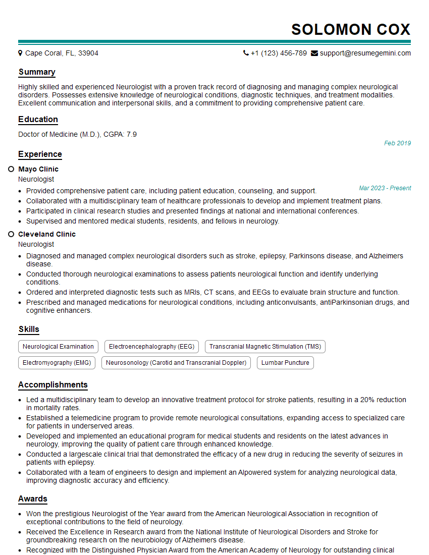
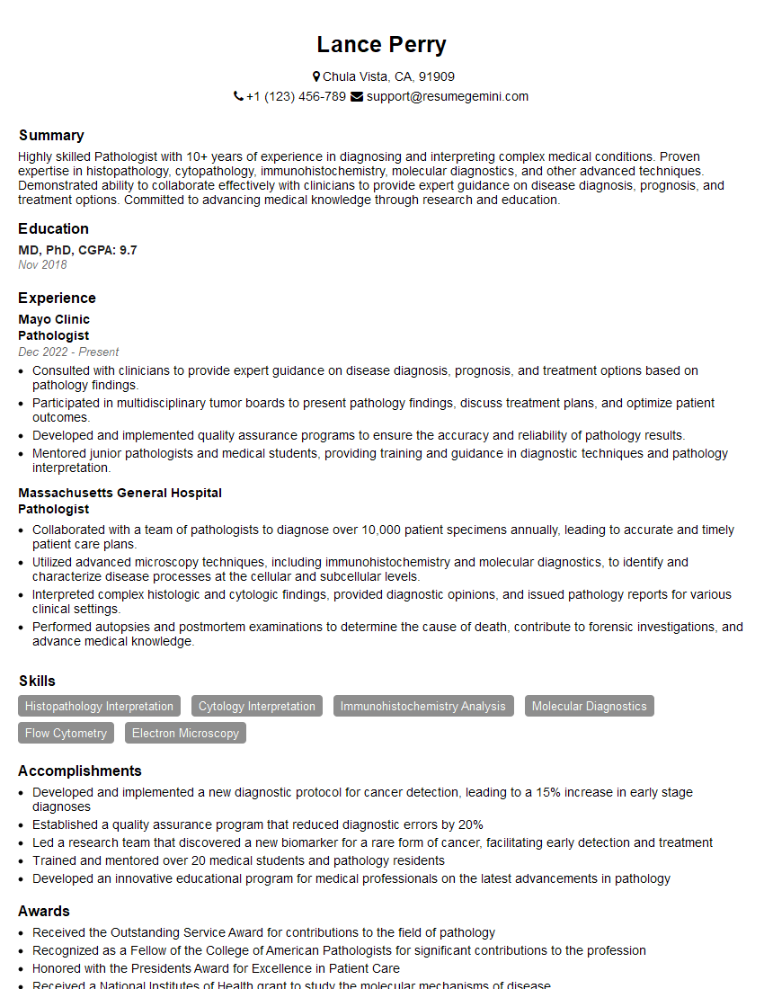
Unlock your full potential by mastering the most common Spinal Tumor interview questions. This blog offers a deep dive into the critical topics, ensuring you’re not only prepared to answer but to excel. With these insights, you’ll approach your interview with clarity and confidence.
Questions Asked in Spinal Tumor Interview
Q 1. Describe the different types of spinal tumors (e.g., intramedullary, extramedullary, benign, malignant).
Spinal tumors are classified based on their location relative to the spinal cord and their biological behavior (benign or malignant). Think of the spine like a layered cake:
- Intramedullary tumors grow within the spinal cord itself. These are often less common but can be incredibly serious due to their direct impact on nerve tissue. Examples include ependymomas and astrocytomas.
- Extramedullary tumors develop outside the spinal cord, but within the spinal canal. These can be further categorized as:
- Intradural extramedullary tumors: Located within the dura mater (the tough outer membrane surrounding the spinal cord). Meningiomas and schwannomas are common examples.
- Extradural tumors: Grow outside the dura mater, often originating from the vertebrae, bone marrow, or nearby tissues. Metastases (cancer spread from another part of the body) are the most common extradural spinal tumors.
- Benign tumors are generally slow-growing and non-cancerous. While they can still cause problems due to their size and location, they are less likely to spread to other parts of the body.
- Malignant tumors are cancerous and can spread (metastasize) to other areas, often requiring aggressive treatment.
Understanding this classification system is crucial for determining the appropriate diagnostic and treatment strategies.
Q 2. Explain the diagnostic process for a suspected spinal tumor, including imaging techniques and biopsy procedures.
Diagnosing a spinal tumor involves a multi-step process, beginning with a thorough neurological examination to assess symptoms. Imaging techniques play a critical role:
- Magnetic Resonance Imaging (MRI): This is the gold standard for visualizing spinal tumors. MRI provides detailed images of both soft tissues (spinal cord, nerves) and bone, allowing precise localization and characterization of the tumor.
- Computed Tomography (CT) scan: While less detailed for soft tissue than MRI, CT scans are excellent for evaluating bone involvement, particularly in cases of suspected vertebral metastases.
- Myelography: A less frequently used technique that involves injecting contrast dye into the spinal fluid to enhance the visibility of the spinal cord and its surrounding structures.
A biopsy is often necessary to confirm the diagnosis and determine the specific type of tumor. This involves surgically removing a small sample of the tumor tissue for microscopic examination by a pathologist. There are various biopsy techniques, chosen depending on the tumor’s location and accessibility, minimizing risk to the spinal cord.
Q 3. Discuss the common symptoms associated with spinal tumors and how they vary depending on location and size.
Symptoms of spinal tumors vary greatly depending on their location, size, and growth rate. Imagine a wire being squeezed – the closer to the source of the pressure, the more severe the effect.
- Pain: Often the first symptom, which can be localized to the back or radiate down the limbs (radiculopathy). The pain might worsen at night or with movement.
- Weakness or paralysis: Depending on the location and size, tumors can compress the spinal cord, causing weakness, numbness, tingling (paresthesia), or even paralysis in the affected limbs.
- Bowel or bladder dysfunction: Tumors affecting the lower spinal cord can lead to problems with bowel and bladder control (incontinence or retention).
- Gait disturbances: Difficulty with walking, balance, and coordination.
- Sensory changes: Numbness, tingling, or changes in temperature sensation.
For example, a small intramedullary tumor might initially present with subtle sensory changes, while a large extradural tumor could quickly cause severe pain and paralysis. Early diagnosis is vital due to potential for rapid neurological decline.
Q 4. What are the surgical approaches for removing spinal tumors, and what are the risks associated with each?
Surgical removal is the primary treatment for many spinal tumors, aimed at relieving pressure on the spinal cord and nerves. The approach depends on the tumor’s location and size:
- Anterior approach: Surgeons access the tumor from the front of the spine, often requiring the removal of a portion of the vertebra. This allows for excellent visualization and is particularly suitable for tumors located in the front of the spinal canal.
- Posterior approach: Access is gained from the back, involving removing a portion of the lamina (the bony arch of the vertebra). This is useful for tumors located in the posterior aspect of the spinal canal.
- Combined anterior and posterior approach: Sometimes necessary for tumors requiring extensive removal.
Risks associated with spinal tumor surgery include:
- Bleeding
- Infection
- Nerve damage
- CSF leak (leakage of cerebrospinal fluid)
- Instability of the spine (requiring instrumentation)
- Recurrence of the tumor
The choice of surgical technique is crucial and must be meticulously planned, considering the specific anatomy and characteristics of the tumor, as well as the patient’s overall health.
Q 5. Explain the role of radiation therapy in the management of spinal tumors.
Radiation therapy plays a vital role in managing spinal tumors, either as the primary treatment or in conjunction with surgery. It’s like using a targeted beam to shrink or destroy cancerous cells.
- External beam radiation therapy: High-energy radiation beams are directed at the tumor from outside the body. This is often used for larger tumors that cannot be completely removed surgically, or for tumors that have a high risk of recurrence.
- Stereotactic radiosurgery (SRS): A highly precise form of radiation therapy delivering a high dose of radiation to a small area, minimizing damage to surrounding tissues. Useful for small, well-defined tumors.
Radiation therapy can help reduce tumor size, alleviate pain, improve neurological function, and potentially prolong survival. However, side effects such as fatigue, skin irritation, and radiation myelopathy (damage to the spinal cord) are possible.
Q 6. Describe the role of chemotherapy in the management of spinal tumors.
Chemotherapy is used primarily for malignant spinal tumors, especially when they have spread to other parts of the body (metastasized). It works systemically, targeting cancer cells throughout the body.
Chemotherapy drugs are administered intravenously or orally, reaching the spinal tumor through the bloodstream. The choice of chemotherapy regimen depends on the tumor type, its aggressiveness, and the patient’s overall health. It may be used alone, or more commonly, in combination with other treatments like radiation therapy or surgery. It is important to note that the effectiveness of chemotherapy can vary, and side effects such as nausea, vomiting, hair loss, and bone marrow suppression are common.
Q 7. Discuss the potential complications of spinal tumor surgery.
Spinal tumor surgery carries potential complications, some immediate and some appearing later. It’s crucial to understand these risks before proceeding:
- Infection: A risk with any surgery, especially one involving the spine, and can lead to serious consequences.
- Bleeding: Major blood vessels and nerves are near the spine, making bleeding a significant concern.
- Nerve damage: Surgery can inadvertently injure nerves, leading to weakness, numbness, or paralysis.
- CSF leak: Leakage of cerebrospinal fluid can cause headaches and other neurological symptoms.
- Instability of the spine: Removal of bone or other structures may lead to spinal instability, requiring additional surgery (e.g., spinal fusion) for stabilization.
- Pain: Post-surgical pain is common, but persistent or severe pain can be a significant complication.
- Neurological deficits: These range from minor sensory changes to major paralysis, and their severity depends on the location of the tumor and the extent of surgical intervention.
- Recurrence of the tumor: Even with successful surgical removal, the tumor may recur in the future.
Post-operative care is crucial for minimizing these complications and ensuring optimal patient recovery. Physical therapy and rehabilitation play a vital role in restoring function and improving the patient’s quality of life.
Q 8. How do you manage spinal instability after tumor resection?
Managing spinal instability after tumor resection is crucial to prevent neurological deterioration and improve patient outcomes. The extent of instability depends on the location and size of the tumor, the amount of bone removed during surgery, and the patient’s pre-existing condition. Our approach is tailored to each patient’s unique needs.
We initially assess the degree of instability using imaging techniques like X-rays and CT scans. If significant instability is present, surgical stabilization is often necessary. This involves using various spinal instrumentation techniques (discussed in the next question) to fuse the affected vertebrae together, effectively creating a rigid support structure. In less severe cases, a brace may suffice to provide external support and allow the spine to heal.
For example, a patient with a large tumor involving multiple vertebral bodies might require a complex posterior spinal fusion with instrumentation, potentially including rods, screws, and hooks. Conversely, a patient with a smaller tumor and minimal bone resection might only need a short fusion with less extensive instrumentation, perhaps even supplemented by bracing.
Q 9. What are the different types of spinal instrumentation used for stabilization?
Several types of spinal instrumentation are used for stabilization after spinal tumor resection, each chosen based on the specific anatomical location, extent of resection, and patient factors.
- Rods and Screws: These are commonly used for posterior spinal fusions. Pedicle screws are inserted into the bony pedicles of the vertebrae, providing strong fixation. Connecting rods then provide structural support across the affected segments.
- Hooks: These are used in conjunction with rods to provide additional fixation points, especially in cases where pedicle screw placement is challenging.
- Plates and Screws: Anterior approaches may involve the use of plates and screws to stabilize the vertebrae from the front.
- Interbody cages: These are implanted between vertebral bodies to restore disc height and provide structural support. They can be filled with bone graft to promote fusion.
- Vertebral body replacements: In cases of extensive bone loss, artificial vertebral body replacements can be used to restore vertebral height and stability.
The choice of instrumentation is a complex decision, involving careful consideration of the patient’s anatomy, the surgical approach, and the desired level of stability. For instance, a minimally invasive approach might employ smaller screws and less extensive instrumentation.
Q 10. Explain the principles of surgical navigation and its role in spinal tumor surgery.
Surgical navigation systems use image guidance to help surgeons precisely locate and remove spinal tumors. This technology significantly improves accuracy, minimizes damage to surrounding neural structures, and allows for more complete tumor resection. Think of it as a GPS for surgery.
The principles involve pre-operative imaging (CT or MRI) that’s registered with the patient intra-operatively. Sensors track the surgical instruments in real time, and this information is overlaid onto the pre-operative images on a monitor. The surgeon can see the precise location of the instruments relative to the tumor and surrounding anatomy, allowing for safer and more targeted resection.
For example, in a case involving a tumor adjacent to the spinal cord, navigation helps the surgeon avoid inadvertently damaging the delicate nerve structures. This leads to improved neurological outcomes and potentially reduces post-operative complications.
Q 11. How do you assess the neurological status of a patient with a spinal tumor pre- and post-operatively?
Neurological assessment is crucial before and after spinal tumor surgery. Pre-operatively, it helps determine the baseline neurological function and identifies the extent of any existing deficits. Post-operatively, it helps assess the effectiveness of the surgery and detect any potential complications.
We use a standardized neurological examination that includes:
- Motor strength: Assessing muscle strength in different muscle groups.
- Sensory function: Testing touch, pain, temperature, and proprioception (sense of body position).
- Reflexes: Evaluating deep tendon reflexes.
- Sphincter function: Assessing bowel and bladder control.
- Pain assessment: Evaluating pain levels.
The findings are meticulously documented. Any changes in neurological status between pre- and post-operative assessments help us evaluate the success of the procedure and guide post-operative management. For instance, a decline in motor strength post-operatively could indicate a surgical complication requiring intervention.
Q 12. Describe the use of intraoperative neuromonitoring during spinal tumor surgery.
Intraoperative neuromonitoring (IONM) involves continuously monitoring the function of the spinal cord and nerves during surgery. This real-time feedback helps surgeons avoid injuring these structures and provides immediate warning of any potential complications. Think of it as a ‘live’ neurological exam during the procedure.
Various methods are used, including:
- Somatosensory evoked potentials (SSEPs): Assess the integrity of sensory pathways.
- Motor evoked potentials (MEPs): Assess the integrity of motor pathways.
- Electromyography (EMG): Measures the electrical activity of muscles.
IONM is particularly valuable in complex cases where the tumor is close to critical neural structures. Any significant changes in the monitored signals during surgery alert the surgical team to potential nerve damage, allowing for immediate adjustments to the surgical technique to minimize further injury. It significantly improves patient safety and minimizes neurological complications.
Q 13. What are the common histological types of spinal tumors and their associated prognoses?
Spinal tumors are classified histologically, meaning by their cell type and microscopic appearance. This classification is vital for determining prognosis and treatment strategy.
- Meningiomas: These are benign tumors arising from the meninges (protective layers surrounding the brain and spinal cord). They usually have a good prognosis with surgical resection.
- Schwannomas: These benign tumors originate from Schwann cells, which produce the myelin sheath around nerves. They also typically have a good prognosis with surgical removal.
- Neurofibromas: Benign tumors arising from nerve tissue. Prognosis depends on the specific type and location.
- Ependymomas: These are tumors originating in the ependyma, the lining of the spinal cord’s central canal. Prognosis varies depending on the grade and location.
- Metastatic tumors: These are cancers that have spread from other parts of the body to the spine. Prognosis is highly dependent on the primary cancer’s type and stage.
It’s important to note that prognosis is complex and depends not only on the histological type but also on factors such as the tumor’s size, location, extent of spinal cord compression, and the patient’s overall health.
Q 14. Discuss the importance of multidisciplinary team management in spinal tumor cases.
Multidisciplinary team (MDT) management is essential in spinal tumor cases, ensuring a comprehensive approach that considers all aspects of the patient’s care. It’s not just about surgery; it’s about a holistic approach that optimizes outcomes.
A typical MDT would include:
- Neurosurgeon: Plans and performs the surgery.
- Oncologist: Provides expertise on cancer treatment, including chemotherapy and radiation therapy.
- Radiologist: Provides imaging expertise for diagnosis and treatment planning.
- Pathologist: Analyzes tissue samples to determine the tumor type and grade.
- Orthopedic surgeon: May be involved in cases requiring complex spinal reconstruction.
- Pain management specialist: Helps manage pain before, during, and after surgery.
- Rehabilitation specialist: Develops a rehabilitation plan to help patients regain function.
The MDT approach ensures that the patient receives the best possible care, combining the expertise of specialists from different disciplines. It allows for a coordinated and individualized treatment plan tailored to the patient’s specific needs, leading to improved outcomes and quality of life.
Q 15. Explain the role of genetic testing in the management of spinal tumors.
Genetic testing plays a crucial role in the management of spinal tumors by helping us understand the tumor’s characteristics and guide treatment decisions. For example, identifying specific genetic mutations can help determine if a tumor is benign or malignant, predict its aggressiveness, and even personalize therapy.
In some cases, genetic testing can reveal mutations that make the tumor susceptible to targeted therapies, which may be more effective and less toxic than traditional chemotherapy. For instance, identifying a BRAF mutation in a meningioma might suggest the use of BRAF inhibitors. Conversely, understanding the genetic profile can sometimes predict resistance to certain treatments, allowing us to avoid unnecessary side effects.
The process typically involves taking a tissue sample (biopsy) from the tumor, extracting DNA, and then analyzing it for specific mutations. The results are then used to create a personalized treatment plan in conjunction with imaging and other clinical findings.
Career Expert Tips:
- Ace those interviews! Prepare effectively by reviewing the Top 50 Most Common Interview Questions on ResumeGemini.
- Navigate your job search with confidence! Explore a wide range of Career Tips on ResumeGemini. Learn about common challenges and recommendations to overcome them.
- Craft the perfect resume! Master the Art of Resume Writing with ResumeGemini’s guide. Showcase your unique qualifications and achievements effectively.
- Don’t miss out on holiday savings! Build your dream resume with ResumeGemini’s ATS optimized templates.
Q 16. How do you manage patients with spinal cord compression due to a tumor?
Managing spinal cord compression from a tumor is a critical time-sensitive situation requiring a multidisciplinary approach. The primary goal is to rapidly decompress the spinal cord to prevent permanent neurological damage. Think of it like relieving pressure on a squeezed water pipe – the quicker you relieve the pressure, the less damage you’ll likely see.
This often involves a combination of strategies. High-dose corticosteroids are frequently used first to reduce inflammation and swelling around the spinal cord, providing temporary relief and buying time for definitive treatment. The second step usually involves surgery, either to remove the tumor completely or partially, depending on its location, size, and the patient’s overall health. In cases where surgery is too risky or impossible, radiation therapy may be used to shrink the tumor and relieve compression.
Post-surgery, ongoing monitoring is vital to assess neurological function and detect any complications. Rehabilitation therapy, including physiotherapy and occupational therapy, often plays a crucial role in helping patients regain strength and mobility.
Q 17. What are the long-term complications of spinal tumor surgery and treatment?
Long-term complications following spinal tumor surgery and treatment can vary greatly depending on the type and location of the tumor, the extent of surgery, and the individual’s overall health. These complications can be neurological, orthopedic, or related to the treatment itself.
- Neurological complications: These can include persistent pain, weakness, numbness, bowel or bladder dysfunction, and even paralysis, depending on the severity of the compression prior to treatment and the extent of surgical damage.
- Orthopedic complications: Spinal instability, scoliosis (curvature of the spine), and persistent pain at the surgical site are possible. The surgical procedure itself can sometimes weaken the spine.
- Treatment-related complications: Radiation therapy can lead to long-term damage to surrounding tissues, causing pain, inflammation, and even fractures. Chemotherapy can have systemic side effects, affecting organs beyond the spine.
It’s important to note that not every patient experiences these complications, and careful surgical planning and post-operative care significantly reduce the risks.
Q 18. Describe the different types of spinal implants used in spinal tumor surgery.
The choice of spinal implant in spinal tumor surgery depends heavily on the specific needs of each patient, considering factors like the location and extent of the tumor, the stability of the spine after tumor removal, and the patient’s overall health.
We use several types:
- Vertebral body replacement (VBR): These implants are used to replace damaged vertebrae that have been removed during surgery. They are available in various materials like titanium or carbon fiber composites, designed to restore spinal stability and height.
- Interbody fusion cages: These are cages placed between vertebrae to promote bone fusion, stabilizing the spine after tumor resection. They may be filled with bone graft material to enhance the fusion process.
- Plates and screws: These are used for stabilization, usually in conjunction with other implants, to reinforce the spine after significant tumor resection, especially in areas of instability.
- Rods and hooks: These are employed to provide more extensive spinal stabilization, often used for larger tumors or in cases where significant bone has been removed.
The selection of the most suitable implant is a meticulous process involving careful preoperative planning and evaluation.
Q 19. Explain the principles of minimally invasive techniques in spinal tumor surgery.
Minimally invasive techniques in spinal tumor surgery aim to achieve the same therapeutic goals as traditional open surgery while minimizing tissue trauma, reducing blood loss, decreasing pain, shortening hospital stays, and speeding up recovery. Think of it as keyhole surgery – smaller incisions with less disruption to surrounding healthy tissue.
These techniques often utilize specialized instruments and imaging guidance (like fluoroscopy or navigation systems) to allow surgeons to reach the tumor through smaller incisions. Examples include image-guided biopsies, laser ablation (for certain tumors), and minimally invasive surgical approaches to decompress the spinal cord.
The advantages of minimally invasive surgery are numerous, but it’s crucial to understand that not every spinal tumor is suitable for this approach. The tumor’s location, size, and characteristics, as well as the patient’s overall health, must be carefully considered to determine the best surgical strategy.
Q 20. How do you counsel patients and their families about spinal tumor diagnosis and treatment?
Counseling patients and their families about a spinal tumor diagnosis and treatment requires sensitivity, empathy, and clear communication. The conversation should begin by acknowledging the emotional impact of the diagnosis, providing a supportive and safe space for questions and concerns.
I explain the diagnosis in clear, understandable terms, avoiding excessive medical jargon. I carefully outline the treatment options, their benefits, risks, and potential side effects, emphasizing shared decision-making. I involve the patient in creating a personalized treatment plan that aligns with their values and goals. Realistic expectations about recovery and potential long-term effects are also discussed.
I strongly encourage patients to bring family members or support persons to these sessions, recognizing that having a trusted companion present can make a significant difference in understanding and processing complex medical information. I regularly follow up with patients, addressing any emerging concerns and providing ongoing support throughout the treatment journey.
Q 21. Discuss the use of targeted therapy in spinal tumor management.
Targeted therapy in spinal tumor management represents a significant advancement in cancer treatment. Unlike traditional chemotherapy, which affects all rapidly dividing cells, targeted therapies focus on specific molecules or pathways involved in tumor growth and survival. This approach minimizes damage to healthy cells and reduces side effects.
For instance, certain types of spinal tumors, such as meningiomas, may harbor specific genetic mutations that make them susceptible to inhibitors targeting those specific mutations. Another example involves using tyrosine kinase inhibitors for specific types of sarcomas.
The selection of a targeted therapy is highly dependent on the specific type and genetic profile of the spinal tumor. Biopsies are crucial for determining which targeted therapies might be most effective and for monitoring response to treatment. While targeted therapies are often highly effective, they are not always curative, and their use is always considered in the context of the patient’s overall health and other treatment options.
Q 22. Explain the role of immunotherapy in the treatment of spinal tumors.
Immunotherapy plays an increasingly important role in treating spinal tumors, particularly those that are malignant. Unlike traditional therapies like surgery, radiation, and chemotherapy that directly target and kill cancer cells, immunotherapy harnesses the body’s own immune system to fight the tumor. This is achieved through various approaches.
- Checkpoint inhibitors: These drugs block proteins that prevent the immune system from attacking cancer cells. For example, they might target PD-1 or CTLA-4 pathways. While not yet extensively used in *all* spinal tumor types, their efficacy in other cancers suggests promising future applications.
- Adoptive cell transfer: This involves removing immune cells (like T-cells) from the patient, modifying them in a lab to better target the tumor, and then reintroducing them into the body. This is a highly specialized approach and is currently more common in research settings for spinal tumors.
- Oncolytic viruses: These are engineered viruses that selectively infect and destroy cancer cells while leaving healthy cells unharmed. This is an area of active research, with potential applications for spinal tumors.
The effectiveness of immunotherapy varies greatly depending on the type and stage of the spinal tumor, the patient’s overall health, and the specific immunotherapy used. It’s often used in combination with other treatments.
Q 23. How do you differentiate between benign and malignant spinal tumors on imaging?
Differentiating between benign and malignant spinal tumors on imaging requires careful analysis of several features. While imaging alone isn’t definitive, it provides crucial clues.
- Size and shape: Benign tumors tend to be well-defined with smooth borders, while malignant tumors are often irregular and poorly defined, potentially invading surrounding tissues.
- Bone involvement: Malignant tumors frequently cause significant bone destruction or erosion, whereas benign tumors may show minimal or no bone involvement. Look for features like lytic lesions (bone destruction) or sclerotic lesions (increased bone density).
- Signal intensity: On MRI, different tissues have unique signal intensities (brightness). Malignant tumors often have different signal intensities compared to normal spinal cord and bone, potentially showing enhancement after contrast administration. Benign tumors may have a more homogenous signal.
- Extra-osseous extension: Malignant tumors often extend beyond the bone into soft tissues (paraspinal muscles, etc.), a feature less common in benign tumors.
Imaging modalities like MRI and CT scans are essential, often used together. A detailed radiological report along with clinical information is vital for proper diagnosis. Ultimately, a biopsy is typically necessary to confirm the diagnosis and determine the specific type of tumor.
Q 24. Discuss the management of recurrent spinal tumors.
Managing recurrent spinal tumors is complex and depends on several factors, including the type of tumor, its location, the patient’s overall health, and the previous treatments received.
- Re-evaluation: The first step involves a comprehensive re-evaluation of the patient, including imaging (MRI, CT), neurological examination, and possibly biopsy to determine the extent of recurrence.
- Treatment options: Depending on the factors above, the management plan might involve:
- Surgery: Surgical resection is a possibility if the recurrence is localized and accessible, aiming to remove as much tumor as safely possible.
- Radiation therapy: Stereotactic radiosurgery (SRS) or fractionated radiotherapy may be employed to target the recurrence.
- Chemotherapy: Systemic chemotherapy might be considered, depending on the tumor type and its sensitivity to chemotherapy agents.
- Immunotherapy: Emerging therapies like immunotherapy may offer additional treatment options.
- Palliative care: In cases where curative treatment isn’t feasible, palliative care is paramount to manage pain, neurological symptoms, and improve the patient’s quality of life.
The decision-making process typically involves a multidisciplinary team, including neurosurgeons, oncologists, radiation oncologists, and palliative care specialists.
Q 25. Describe your experience with specific surgical techniques (e.g., laminectomy, vertebrectomy).
My experience encompasses a wide range of spinal surgical techniques, including laminectomy and vertebrectomy. These are often used in spinal tumor surgery, but the choice depends on the tumor’s location, size, and the extent of spinal involvement.
- Laminectomy: This involves removing a portion of the lamina (the posterior bony arch of a vertebra) to decompress the spinal cord or nerve roots. It’s frequently used for tumors affecting the posterior elements of the spine. I’ve performed numerous laminectomies for intramedullary tumors (within the spinal cord) and extradural tumors (outside the dura mater).
- Vertebrectomy: This is a more extensive procedure, involving the partial or complete removal of one or more vertebrae. This is necessary when the tumor involves the vertebral body itself and requires more aggressive resection for decompression and tumor removal. I have extensive experience with anterior and posterior approaches to vertebrectomy, often incorporating instrumentation and fusion techniques to stabilize the spine post-operatively. For example, in cases of extensive vertebral body involvement from chordomas or metastatic disease, a combined anterior-posterior approach may be necessary.
Patient selection, meticulous surgical planning, and meticulous intraoperative monitoring are paramount in these complex surgeries. The goal is maximal tumor resection while minimizing neurological damage.
Q 26. Explain how you would manage a patient with a rapidly growing spinal tumor presenting with severe neurological deficits.
Managing a patient with a rapidly growing spinal tumor and severe neurological deficits necessitates an immediate and aggressive approach. This is a critical situation demanding rapid intervention.
- Emergency assessment: The patient needs immediate neurological assessment to determine the severity and location of deficits. Imaging (MRI) is crucial to delineate the tumor’s extent and its effect on the spinal cord or nerve roots.
- Stabilization: This is paramount. If the spinal column is unstable, surgical stabilization is often needed prior to tumor resection to prevent further neurological compromise. This might involve placement of rods, screws, and other instrumentation.
- Surgical decompression: If the neurological deficits are directly attributable to spinal cord compression, urgent surgical decompression is typically indicated. The goal here is to alleviate pressure on the spinal cord or nerve roots, even if complete tumor removal isn’t immediately feasible. The extent of surgery—laminectomy, vertebrectomy, or other approaches—depends on the specific anatomical location and the nature of the tumor.
- Adjunctive therapies: Post-operative management might involve high-dose steroids to reduce edema (swelling) around the spinal cord, pain management, and potentially further therapies (radiation, chemotherapy, immunotherapy) after the initial stabilization and decompression.
This scenario highlights the importance of a timely, well-coordinated multidisciplinary approach involving neurosurgery, oncology, and intensive care.
Q 27. Discuss the use of palliative care in patients with spinal tumors.
Palliative care plays a vital role in the management of patients with spinal tumors, especially those with advanced disease where curative treatment isn’t a realistic goal. Its focus is on improving the patient’s quality of life and addressing their physical, emotional, and spiritual needs.
- Pain management: Spinal tumors can cause significant pain, requiring a multi-modal approach using medication (opioids, NSAIDs, other analgesics), nerve blocks, radiation therapy, and other interventions.
- Symptom control: Palliative care addresses other symptoms such as bowel or bladder dysfunction, weakness, fatigue, and psychological distress.
- Emotional and spiritual support: This is crucial. Palliative care teams provide counseling, support groups, and spiritual guidance to help patients and their families cope with the emotional burden of the illness.
- Advance care planning: Palliative care facilitates discussions about end-of-life wishes, ensuring the patient’s preferences are honored.
It’s important to understand that palliative care isn’t about giving up; it’s about optimizing the patient’s comfort and overall well-being throughout their journey, integrating seamlessly with curative treatments when possible.
Q 28. What are the current research trends in the treatment of spinal tumors?
Research in spinal tumor treatment is constantly evolving, driven by the need for improved outcomes and reduced toxicity. Several key areas are under active investigation:
- Targeted therapies: Identifying specific molecular targets within spinal tumor cells allows for the development of drugs that specifically target these molecules, potentially minimizing side effects on healthy cells.
- Immunotherapy advancements: Ongoing research aims to improve the effectiveness of immunotherapy approaches, including novel checkpoint inhibitors and adoptive cell transfer techniques specifically tailored for spinal tumors.
- Minimally invasive surgical techniques: Developing and refining less invasive surgical approaches reduces the trauma associated with spinal surgery, leading to faster recovery times and improved patient outcomes. This includes advances in robotic surgery and image-guided techniques.
- Improved radiation delivery: Research focuses on more precise and targeted radiation delivery methods, like proton beam therapy, reducing damage to surrounding healthy tissues.
- Combination therapies: Investigating optimal combinations of surgery, radiation, chemotherapy, and immunotherapy to enhance treatment efficacy and improve patient survival.
- Biomarkers: Identifying biomarkers that can predict tumor behavior and response to therapy enables personalized treatment approaches tailored to individual patients.
These research advancements offer hope for improved treatments and better outcomes for patients with spinal tumors in the future.
Key Topics to Learn for Spinal Tumor Interview
- Tumor Classification & Grading: Understanding the different types of spinal tumors (primary vs. metastatic, benign vs. malignant), their grading systems, and implications for treatment planning.
- Imaging Techniques: Proficiency in interpreting various imaging modalities such as MRI, CT scans, and myelograms to identify tumor location, size, and extent of spinal cord compression.
- Surgical Approaches: Familiarity with different surgical techniques used in spinal tumor resection, including minimally invasive approaches and complex spinal reconstructive procedures.
- Radiation Therapy & Chemotherapy: Knowledge of the role of radiation therapy and chemotherapy in the management of spinal tumors, both as primary treatment and adjuvant therapy.
- Pre-operative and Post-operative Care: Understanding the crucial aspects of patient management before and after surgery, including neurologic monitoring, pain management, and rehabilitation.
- Case Studies & Problem Solving: Ability to analyze clinical scenarios, formulate differential diagnoses, and propose appropriate management strategies for various types of spinal tumors.
- Neurosurgical Oncology Principles: A grasp of the broader context of neurosurgical oncology, encompassing tumor biology, genetics, and multidisciplinary treatment approaches.
- Current Research & Advances: Staying abreast of the latest advancements in the diagnosis and treatment of spinal tumors, including novel therapies and surgical techniques.
Next Steps
Mastering the intricacies of spinal tumor management is crucial for career advancement in neurosurgery and related fields. A strong understanding of this complex area opens doors to specialized roles and enhances your contribution to patient care. To maximize your job prospects, creating an ATS-friendly resume is essential. ResumeGemini is a trusted resource that can help you build a professional and impactful resume that highlights your skills and experience effectively. Examples of resumes tailored to Spinal Tumor expertise are available within ResumeGemini to guide you. Take the next step in your career journey – invest in your resume today.
Explore more articles
Users Rating of Our Blogs
Share Your Experience
We value your feedback! Please rate our content and share your thoughts (optional).
What Readers Say About Our Blog
This was kind of a unique content I found around the specialized skills. Very helpful questions and good detailed answers.
Very Helpful blog, thank you Interviewgemini team.
