The right preparation can turn an interview into an opportunity to showcase your expertise. This guide to Spondylolisthesis interview questions is your ultimate resource, providing key insights and tips to help you ace your responses and stand out as a top candidate.
Questions Asked in Spondylolisthesis Interview
Q 1. Describe the different types of spondylolisthesis.
Spondylolisthesis is classified into several types, primarily based on the underlying cause of the vertebral slippage. Think of it like different ways a vertebra can slip forward on the one below it.
- Dysplastic Spondylolisthesis: This is a congenital condition, meaning it’s present at birth. A defect in the pars interarticularis (a part of the vertebra) causes instability and slippage. Imagine a weak link in a chain causing the chain to break and slip.
- Isthmic Spondylolisthesis: This is the most common type. It results from a fracture (stress fracture) or a defect in the pars interarticularis. Repeated stress, like in sports, can lead to this fracture, making the vertebra unstable.
- Degenerative Spondylolisthesis: This is related to wear and tear of the spine, mostly affecting older adults. The facet joints (the joints between the vertebrae) degenerate, losing stability, causing the vertebra to slip. Think of it like old car parts wearing down.
- Traumatic Spondylolisthesis: This occurs due to a significant injury, like a fracture, that causes vertebral displacement.
- Pathologic Spondylolisthesis: This is caused by underlying diseases that weaken the bone, such as tumors or infections. This weakens the structural integrity of the vertebra leading to slippage.
Understanding these different types is crucial for determining the best treatment approach.
Q 2. Explain the pathophysiology of spondylolytic spondylolisthesis.
Isthmic spondylolisthesis, the most common type, has a specific pathophysiology. It begins with a stress fracture or defect in the pars interarticularis – a small segment of bone connecting parts of the vertebra. This fracture, often occurring gradually due to repetitive stress, leads to instability in the vertebra. As the pars interarticularis weakens, the vertebra above it can gradually slip forward on the vertebra below. Imagine a tiny crack in a bone that eventually creates a break, allowing the bone segments to separate and slip. This slippage can gradually increase over time, sometimes significantly affecting spinal alignment and causing pain.
The repetitive stress leading to this fracture is often seen in athletes involved in activities like gymnastics, weightlifting, or football – sports that involve hyperextension or repetitive loading of the lumbar spine.
Q 3. What are the common clinical presentations of spondylolisthesis?
The clinical presentation of spondylolisthesis varies greatly depending on the severity of the slippage and individual factors. Many individuals are asymptomatic and discovered incidentally during imaging studies. However, common symptoms include:
- Low back pain: This is often the most prominent symptom, ranging from mild aches to severe, debilitating pain.
- Buttock pain: Pain can radiate to the buttocks.
- Hamstring tightness or pain: This is caused by nerve root irritation.
- Neurological symptoms: In severe cases, nerve root compression can lead to numbness, tingling, weakness, or even bowel or bladder dysfunction (cauda equina syndrome).
- Spinal deformity: Significant slippage can lead to a noticeable change in the spinal curvature, sometimes resulting in an increased lumbar lordosis (inward curve).
- Leg pain (sciatica): Nerve compression can also manifest as leg pain radiating down the leg.
The severity of symptoms doesn’t always correlate with the degree of slippage. A small amount of slippage can cause significant pain, while a larger slippage might be asymptomatic.
Q 4. Discuss the role of imaging (X-ray, MRI, CT) in diagnosing spondylolisthesis.
Imaging plays a crucial role in diagnosing spondylolisthesis and assessing its severity.
- X-rays: These are the primary imaging modality. Lateral (side) views of the spine are essential for measuring the degree of slippage, using the Meyerding grading system (grades I-V, with V being the most severe). X-rays also reveal the presence of a pars defect in isthmic spondylolisthesis. The presence of degenerative changes in degenerative spondylolisthesis can also be visualized.
- MRI (Magnetic Resonance Imaging): MRI provides detailed images of the soft tissues, including the spinal cord, nerves, and intervertebral discs. It’s crucial for assessing nerve root compression and identifying any associated spinal stenosis (narrowing of the spinal canal).
- CT (Computed Tomography): CT scans provide detailed bone images, superior to X-rays, for visualizing the pars interarticularis. It is particularly useful in assessing the integrity of the pars interarticularis and the degree of bony involvement.
The choice of imaging modality depends on the clinical presentation and the information needed. X-rays often suffice for initial assessment, while MRI and CT are used when more detailed information is necessary.
Q 5. How do you differentiate spondylolisthesis from other spinal conditions?
Differentiating spondylolisthesis from other spinal conditions requires a careful clinical evaluation and imaging studies. Conditions that can mimic spondylolisthesis include:
- Spinal stenosis: This involves narrowing of the spinal canal, which can cause similar symptoms. MRI is crucial to differentiate as it clearly shows the canal dimensions.
- Degenerative disc disease: Disc degeneration can cause back pain but typically doesn’t involve vertebral slippage. X-rays can distinguish between degenerative disc disease and spondylolisthesis.
- Facet joint syndrome: Pain stemming from the facet joints can mimic spondylolisthesis pain, but imaging shows the facet joint involvement without the vertebral slippage.
- Herniated disc: A herniated disc can press on nerves causing radicular pain, which needs to be differentiated through MRI.
A comprehensive history, physical examination, and imaging studies, often including X-rays and MRI, are crucial to making an accurate diagnosis and ruling out these conditions.
Q 6. What are the non-surgical treatment options for spondylolisthesis?
Non-surgical treatment is the preferred approach for many patients with spondylolisthesis, particularly those with mild symptoms or those who are not good surgical candidates. The goals are pain management and improving function.
- Conservative management: This is the cornerstone of treatment and includes rest, activity modification, and physical therapy. Patients might be advised to avoid activities that aggravate their symptoms.
- Medication: Pain relievers (NSAIDs, analgesics), muscle relaxants, and in some cases, corticosteroids might be prescribed to manage pain and inflammation.
- Physical therapy: This plays a crucial role in strengthening core muscles, improving posture, and increasing flexibility. Specific exercises target spinal stabilization and improve overall mobility.
- Bracing: In some cases, a brace might be used to provide support and limit spinal motion, promoting healing and reducing pain.
- Weight management: For overweight or obese individuals, weight loss can significantly reduce stress on the spine.
These non-surgical strategies aim to reduce pain, improve function, and avoid surgery whenever possible. Success depends on the individual, the severity of the condition and compliance with the treatment plan.
Q 7. When is surgical intervention indicated for spondylolisthesis?
Surgical intervention for spondylolisthesis is considered when non-surgical treatments fail to provide adequate pain relief or functional improvement, or when there’s significant neurologic compromise (e.g., cauda equina syndrome). The decision is highly individualized and based on several factors.
- Severe pain refractory to conservative management: Despite consistent efforts with non-surgical therapies, pain remains unbearable and interferes significantly with the patient’s daily life.
- Neurological deficits: The presence of significant neurological symptoms such as weakness, numbness, bowel or bladder dysfunction mandates surgical intervention.
- Progressive slippage: If the slippage is worsening on follow-up imaging and causing increasing problems, surgery may be necessary.
- Spinal instability: If there is significant instability demonstrated by imaging and clinical examination that might result in further slippage and neurological problems.
Several surgical techniques exist, such as fusion, decompression, and instrumentation, and the choice depends on the specific situation and the surgeon’s preference.
Q 8. Describe different surgical techniques used to treat spondylolisthesis.
Surgical treatment for spondylolisthesis aims to stabilize the spine and alleviate pain. The choice of technique depends on several factors, including the severity of the slip, the patient’s age and overall health, and the presence of neurological symptoms. Here are some common surgical approaches:
Posterior Spinal Fusion: This is the most common procedure. It involves placing bone grafts and/or metal implants (rods, screws, plates) across the affected vertebrae to fuse them together, creating a stable unit. This prevents further slippage and reduces pain. Different fixation techniques exist, using various screw and rod configurations tailored to the individual case.
Anterior Lumbar Interbody Fusion (ALIF): This approach involves accessing the spine from the front of the abdomen. A disc is removed, and a bone graft or cage is inserted to create a solid fusion. ALIF is often used in combination with posterior fusion for added stability, especially in severe cases.
Transforaminal Lumbar Interbody Fusion (TLIF): This minimally invasive technique accesses the disc from the side through the foramen (the opening where nerves exit the spine). A bone graft or cage is inserted to create fusion. TLIF often results in less muscle disruption compared to open surgery, leading to potentially faster recovery.
Direct Lateral Lumbar Interbody Fusion (DLIF): Another minimally invasive approach accessing the spine from the side. It often involves smaller incisions and less tissue disruption than TLIF, further reducing post-operative pain and recovery time.
The specific surgical technique is always tailored to the individual patient’s anatomy and the specific type and severity of their spondylolisthesis.
Q 9. What are the potential complications of spondylolisthesis surgery?
While spondylolisthesis surgery generally offers significant pain relief and improved function, potential complications exist. These can include:
Infection: Infection at the surgical site is a risk with any surgery. Antibiotics are used prophylactically to minimize this risk.
Bleeding: Bleeding can occur during or after surgery, sometimes requiring blood transfusions.
Nerve damage: Injury to nerves during surgery can lead to weakness, numbness, or pain in the legs or feet. This is a rare but serious complication.
Non-union: Failure of the bone grafts to fuse properly is a possibility, requiring further intervention.
Pseudarthrosis: A false joint forms at the fusion site, leading to persistent pain and instability.
Hardware failure: The screws, rods, or plates used for stabilization can break or loosen over time.
Dural tear: Accidental tear in the protective membrane surrounding the spinal cord.
Adjacent segment disease: Increased stress on the segments of the spine above or below the fusion site can lead to degeneration over time.
Careful surgical planning and meticulous surgical technique are crucial to minimize the risk of these complications.
Q 10. How do you manage post-operative pain and complications?
Post-operative pain management is crucial for successful recovery. A multi-modal approach is usually employed, combining:
Medications: This may include analgesics (pain relievers), anti-inflammatories, and potentially opioids in the initial post-operative period. The goal is to gradually wean patients off opioid medication as they recover.
Physical therapy: Early mobilization and tailored exercises help restore strength, flexibility, and range of motion, reducing pain and promoting healing. Physical therapists also educate patients on proper body mechanics to prevent re-injury.
Nerve blocks: In some cases, nerve blocks may be used to provide targeted pain relief.
Epidural steroid injections: These can help reduce inflammation and pain, particularly if nerve root irritation is present.
Careful monitoring for complications like infection, bleeding, or nerve damage is also an essential part of post-operative management. Regular follow-up appointments are scheduled to assess progress, address concerns, and adjust the treatment plan as needed.
Q 11. Explain the role of physical therapy in managing spondylolisthesis.
Physical therapy plays a vital role in managing spondylolisthesis, both before and after surgery. A well-designed physical therapy program focuses on:
Strengthening core muscles: Strong abdominal and back muscles provide crucial support to the spine, reducing stress on the affected vertebrae and improving stability.
Improving flexibility: Stretching exercises enhance range of motion and prevent stiffness.
Improving posture: Correct posture reduces strain on the spine.
Pain management: Therapeutic modalities like heat, ice, and ultrasound can help manage pain and inflammation.
Functional training: Exercises that mimic daily activities help patients regain independence and improve functional abilities.
A physical therapist will assess the patient’s individual needs and develop a personalized program tailored to their specific condition and functional limitations. Compliance with the prescribed exercises is essential for optimal outcomes.
Q 12. Discuss the importance of patient education in managing spondylolisthesis.
Patient education is paramount in managing spondylolisthesis. It empowers individuals to actively participate in their care and improve their outcomes. Key aspects of patient education include:
Understanding the condition: Patients need to understand the nature of their spondylolisthesis, its causes, and its potential impact on their lives.
Treatment options: Patients should be fully informed about all treatment options, including the benefits and risks of each approach, enabling them to make informed decisions.
Pain management strategies: Patients need to understand how to manage their pain effectively, using medication, physical therapy, and other techniques.
Activity modification: Patients need guidance on how to modify their activities to protect their spine and avoid further injury.
Importance of compliance: Patients need to understand the importance of adhering to their treatment plan, including medication, physical therapy, and lifestyle modifications.
Effective communication and a collaborative approach between the healthcare team and the patient are vital for successful management of spondylolisthesis.
Q 13. What are the long-term prognosis and outcomes for patients with spondylolisthesis?
The long-term prognosis and outcomes for patients with spondylolisthesis vary widely depending on several factors, including the severity of the slip, the patient’s age and overall health, the presence of neurological symptoms, and the chosen treatment approach.
With conservative management, many patients experience significant pain relief and improved function. For those undergoing surgery, the success rate is generally high, with most patients experiencing substantial pain reduction and improved quality of life. However, the possibility of complications like non-union, pseudarthrosis, or adjacent segment disease must be considered.
Long-term follow-up is essential to monitor for potential complications and ensure the effectiveness of the treatment. Regular physical therapy and adherence to lifestyle modifications contribute significantly to long-term success. It’s important to remember that individual experiences vary significantly, and outcomes are always individualized.
Q 14. How do you assess the functional limitations of a patient with spondylolisthesis?
Assessing functional limitations in patients with spondylolisthesis requires a comprehensive approach combining subjective and objective measures.
Subjective assessment: This involves detailed discussions with the patient regarding their symptoms, pain levels, limitations in daily activities (e.g., walking, bending, lifting), and overall quality of life. Tools like the Oswestry Disability Index (ODI) or the Visual Analog Scale (VAS) can quantify pain and functional limitations.
Objective assessment: This includes a physical examination to assess range of motion, muscle strength, neurological function, and gait. Objective measures like gait analysis, functional tests (e.g., timed up and go test), and assessments of balance can quantify functional limitations.
By combining subjective and objective assessments, a clear picture of the patient’s functional limitations emerges. This information guides treatment planning, monitors treatment progress, and helps determine the overall prognosis.
Q 15. Describe your experience with specific surgical techniques (e.g., fusion, instrumentation).
My experience encompasses a wide range of surgical techniques for spondylolisthesis, focusing on achieving stable spinal alignment and pain relief. I’m proficient in both posterior and anterior approaches. Posterior techniques commonly involve spinal fusion, where we utilize bone grafts and instrumentation like rods, screws, and plates to stabilize the affected vertebrae and promote bone healing. This effectively creates a single, solid bone unit. The instrumentation provides immediate stability while the fusion matures. Different types of instrumentation exist, selected based on the specifics of each case. For instance, a short segment fusion might utilize a smaller number of screws and rods, while a long segment fusion would require more extensive instrumentation. Anterior approaches, used less frequently for spondylolisthesis than posterior, involve accessing the spine from the front of the body. This allows for direct decompression of the spinal nerves and can be particularly useful in cases with significant spinal stenosis. These procedures often involve techniques like anterior lumbar interbody fusion (ALIF), which involves removing the disc and replacing it with a bone graft or cage, subsequently stabilized with plates and screws. My approach always emphasizes minimally invasive techniques whenever feasible to reduce surgical trauma and speed recovery.
For example, I recently managed a patient with high-grade spondylolisthesis at L4-L5. Due to significant spinal stenosis and neurological compromise, a posterior fusion with instrumentation was necessary. We used pedicle screws and rods to achieve stable fixation, and the patient experienced excellent pain relief and functional recovery post-surgery.
Career Expert Tips:
- Ace those interviews! Prepare effectively by reviewing the Top 50 Most Common Interview Questions on ResumeGemini.
- Navigate your job search with confidence! Explore a wide range of Career Tips on ResumeGemini. Learn about common challenges and recommendations to overcome them.
- Craft the perfect resume! Master the Art of Resume Writing with ResumeGemini’s guide. Showcase your unique qualifications and achievements effectively.
- Don’t miss out on holiday savings! Build your dream resume with ResumeGemini’s ATS optimized templates.
Q 16. How do you choose the appropriate surgical approach for a given patient?
Choosing the appropriate surgical approach is a multifactorial decision, personalized for each patient. I meticulously consider several key factors: the severity of the spondylolisthesis (grade), the presence of associated spinal stenosis (narrowing of the spinal canal), the extent of neurological deficits, the patient’s age and overall health, and their level of activity. Imaging studies, such as X-rays, CT scans, and MRI, are crucial for assessing the anatomy and the extent of the slippage and any associated nerve compression. Furthermore, a detailed neurological examination is conducted to determine the presence and severity of any neurological symptoms, such as weakness, numbness, or bowel/bladder dysfunction.
For instance, a young, active patient with a high-grade spondylolisthesis and significant neurologic compromise would likely benefit from a more extensive fusion with robust instrumentation to ensure stability and long-term function. In contrast, an older patient with a lower-grade slip and minimal symptoms might be better suited to a less invasive procedure or even conservative management.
Q 17. What are the criteria for selecting patients for conservative versus surgical management?
The decision between conservative and surgical management for spondylolisthesis hinges on several factors. Conservative treatment, including physical therapy, medication (pain relievers, muscle relaxants), bracing, and activity modification, is typically the first line of defense, particularly for patients with mild symptoms and low-grade slips. It aims to reduce pain, improve muscle strength and flexibility, and restore function. Surgical intervention is usually reserved for patients who fail conservative management and exhibit significant pain, neurological deficits, or progressive slippage.
Specific criteria for surgical consideration include persistent, debilitating pain despite adequate conservative treatment; significant neurological deficits such as weakness, numbness, or bowel/bladder dysfunction; progressive slippage on imaging; instability demonstrated on dynamic imaging studies; or severe spinal stenosis causing significant neural compression. The decision-making process always involves a shared discussion with the patient to weigh the benefits and risks of each approach.
Q 18. Explain your approach to managing a patient with spondylolisthesis and associated neurological deficits.
Managing spondylolisthesis with associated neurological deficits requires a prompt and decisive approach. Immediate assessment is critical to determine the severity of the neurological compromise. If significant neurological deficits are present, such as cauda equina syndrome (compression of the nerve roots at the end of the spinal cord), immediate surgical intervention is usually necessary to decompress the compressed nerves and prevent permanent neurological damage. This is a true surgical emergency.
The surgical approach will be tailored to the specific location and extent of the compression. For instance, a patient with significant nerve root compression at L4-L5 might require a laminectomy (removal of part of the vertebra) or a more extensive decompression procedure along with fusion to address the instability. Post-operatively, close monitoring of neurological function is crucial, along with pain management and rehabilitation to maximize functional recovery.
Q 19. Discuss the role of bracing in the treatment of spondylolisthesis.
Bracing plays a significant role in the conservative management of spondylolisthesis, particularly in younger patients with active slips. It offers support to the spine, limiting motion and reducing stress on the affected vertebrae. This can help to reduce pain, improve stability, and prevent further slippage. The type of brace used depends on the severity of the spondylolisthesis and the patient’s individual needs. For example, a Boston brace is often used for adolescents with isthmic spondylolisthesis, providing substantial support while still allowing for some mobility.
However, bracing is not a stand-alone treatment and is typically used in conjunction with other conservative measures such as physical therapy. It’s crucial to emphasize patient compliance with the bracing regimen; proper fitting and consistent wear are essential for the brace to be effective. The duration of bracing depends on the patient’s response to treatment and is determined on a case-by-case basis in consultation with the patient and their physical therapist.
Q 20. How do you monitor patient progress after surgery?
Post-operative monitoring of spondylolisthesis patients is crucial for ensuring successful recovery and identifying any potential complications. This involves regular follow-up appointments, typically starting shortly after surgery and continuing for several months or even years. These visits include clinical examinations focusing on pain levels, neurological function, and range of motion. Imaging studies, such as X-rays, are performed periodically to assess fusion progression and the stability of the instrumentation. Any signs of infection, pseudarthrosis (non-union of the fusion), or neurological deficits are carefully evaluated.
Furthermore, I encourage patients to participate in a comprehensive rehabilitation program, tailored to their individual needs, to gradually restore strength, flexibility, and functional capacity. This often involves physical therapy, focused on strengthening core muscles and improving spinal stability. The rehabilitation process continues for months post-surgery to optimize the patient’s recovery. Regular communication with the patient is essential to address any concerns or complications promptly and adjust the treatment plan as necessary.
Q 21. What are the common causes of spondylolisthesis?
Spondylolisthesis, the forward slippage of one vertebra over another, arises from various causes, broadly categorized into five types: dysplastic, isthmic, degenerative, traumatic, and pathological.
- Dysplastic: This is a congenital type, resulting from a developmental abnormality of the vertebrae, often present from birth. It’s less common than other types.
- Isthmic: The most frequent type, often associated with a defect in the pars interarticularis (a bony portion of the vertebra). This defect, often a stress fracture, allows for progressive slippage. This is frequently seen in adolescents involved in sports with repetitive hyperextension movements.
- Degenerative: This type occurs due to the natural wear and tear of the spine, usually affecting older adults. Degeneration of the facet joints and intervertebral discs can lead to instability and slippage.
- Traumatic: This is caused by a fracture of the vertebra, usually from a significant trauma such as a fall or high-impact injury.
- Pathological: This type results from weakening of the vertebra due to underlying diseases like tumors or infections.
Understanding the underlying cause is critical for determining the appropriate treatment strategy and managing the condition effectively. For example, a young athlete with isthmic spondylolisthesis would have different management needs than an older adult with degenerative spondylolisthesis.
Q 22. Explain the grading system used to classify spondylolisthesis (e.g., Meyerding grading).
Spondylolisthesis is graded using several systems, with the Meyerding grading system being the most common. This system assesses the degree of anterior slippage of one vertebra over another. It’s based on the percentage of the inferior vertebral body that slips anteriorly over the superior vertebral body.
- Grade 1: 25% or less slippage.
- Grade 2: 26-50% slippage.
- Grade 3: 51-75% slippage.
- Grade 4: 76-100% slippage.
- Grade 5: Spondyloptosis – complete displacement of the vertebra.
Imagine a stack of books. In a healthy spine, the books are neatly stacked. In spondylolisthesis, one book slips forward. The Meyerding grade tells you how far that book has slipped forward.
Q 23. How do you counsel patients about the risks and benefits of different treatment options?
Counseling patients about treatment options requires a thorough understanding of their specific condition, age, activity level, and overall health. I always start by explaining the natural history of spondylolisthesis – some cases resolve spontaneously, while others may require intervention.
Conservative Management: This is often the first line of treatment and includes pain medication (NSAIDs, muscle relaxants), physical therapy (strengthening core muscles, improving posture), bracing, and weight management. I explain the potential benefits (pain reduction, improved function) and limitations (may not be effective for severe cases, requires commitment to therapy).
Surgical Intervention: Surgery is usually considered for severe cases with significant pain, neurological symptoms (numbness, weakness), or progressive slippage. I discuss various surgical techniques, including fusion (stabilizing the spine) and decompression (relieving pressure on nerves). I highlight the benefits (pain relief, improved stability, reduced neurological symptoms), risks (infection, bleeding, nerve damage, prolonged recovery), and potential complications. The decision is always made collaboratively, weighing the risks and benefits based on the individual patient’s situation.
I use visual aids, such as X-rays and diagrams, to help patients understand their condition and the proposed treatment plan. Patient education is key to ensure informed consent and successful management.
Q 24. Describe a challenging case of spondylolisthesis you have managed and the outcome.
One challenging case involved a 16-year-old female athlete with a high-grade spondylolisthesis (Grade 4) at L5-S1, causing significant low back pain and radiating pain down her leg. Conservative management had failed. She was highly active and wanted to return to her sport. The challenge was balancing the need for surgical stabilization with her young age and desire to maintain an active lifestyle.
After careful consideration, we opted for a minimally invasive posterior lumbar interbody fusion (PLIF). This technique minimized tissue disruption and allowed for quicker recovery. Post-surgery, she underwent a rigorous physical therapy program. The outcome was excellent. Her pain resolved significantly, and she was able to return to competitive sports within six months, albeit with some modifications to her training regimen to avoid high-impact activities.
Q 25. What are the latest advancements in the diagnosis and treatment of spondylolisthesis?
Recent advancements in spondylolisthesis management include improvements in imaging techniques (high-resolution MRI, CT scans), leading to more precise diagnosis and surgical planning. Minimally invasive surgical approaches, such as robotic-assisted surgery, are becoming increasingly common, offering reduced trauma, shorter hospital stays, and faster recovery times.
Advances in biomaterials, including the development of novel interbody fusion cages and bone graft substitutes, have improved fusion rates and reduced complications. Furthermore, research into the genetic and biomechanical factors contributing to spondylolisthesis is paving the way for personalized treatment strategies.
Q 26. Discuss the role of minimally invasive surgical techniques in the treatment of spondylolisthesis.
Minimally invasive surgical techniques play a significant role in spondylolisthesis treatment, particularly for select patients. These techniques involve smaller incisions, less muscle dissection, and less tissue trauma compared to open surgery. Examples include minimally invasive PLIF, transforaminal lumbar interbody fusion (TLIF), and lateral lumbar interbody fusion (LLIF).
The benefits are numerous: reduced blood loss, decreased pain, shorter hospital stays, quicker recovery, and improved cosmetic results. However, minimally invasive surgery is not always suitable for all cases of spondylolisthesis. The surgeon must carefully evaluate the patient’s anatomy and the severity of the condition to determine the most appropriate surgical approach.
Q 27. How do you address patient concerns and expectations regarding recovery?
Addressing patient concerns and expectations is crucial. I emphasize that recovery is a process, not an event. Realistic expectations must be set from the outset. I provide patients with a detailed timeline of recovery, outlining milestones and potential challenges. I encourage open communication and address any anxieties or questions they may have.
I regularly monitor patient progress and adjust the treatment plan as needed. I emphasize the importance of adherence to physical therapy, medication regimens, and lifestyle modifications. The ultimate goal is to empower patients to actively participate in their recovery and regain their quality of life.
Q 28. What are your preferred methods for pain management in spondylolisthesis?
My approach to pain management is multimodal and individualized. It’s about managing pain effectively while minimizing side effects. This often involves a combination of strategies:
- Pharmacological approaches: NSAIDs for inflammation, muscle relaxants for spasms, and opioids (used judiciously and cautiously for severe pain, always considering alternatives).
- Non-pharmacological approaches: Physical therapy (strengthening exercises, stretching, improving posture), modalities (heat, ice, ultrasound, electrical stimulation), and psychological interventions (stress management techniques, coping strategies).
- Interventional techniques: Epidural steroid injections or nerve blocks may be considered for temporary pain relief in specific cases.
The focus is on finding the optimal balance between pain relief and minimizing the risk of side effects. I regularly reassess the effectiveness of the chosen strategy and modify it accordingly to maximize patient comfort and function.
Key Topics to Learn for Spondylolisthesis Interview
- Anatomy and Physiology: Understand the anatomy of the vertebrae, the different types of spondylolisthesis (e.g., dysplastic, isthmic, degenerative), and the associated biomechanics.
- Diagnosis and Imaging: Master the interpretation of X-rays, CT scans, and MRIs in identifying spondylolisthesis and associated pathologies. Be prepared to discuss diagnostic criteria and limitations.
- Conservative Management: Familiarize yourself with non-surgical treatment options, including physical therapy, bracing, medication management, and lifestyle modifications. Be ready to discuss the indications and contraindications for each.
- Surgical Management: Understand the various surgical techniques used to treat spondylolisthesis, including the rationale behind each approach and the potential complications. Consider the pros and cons of different fusion techniques.
- Patient Assessment and Treatment Planning: Practice formulating a comprehensive treatment plan based on patient presentation, including history, physical examination findings, and imaging results. Discuss how to tailor treatment to individual needs and goals.
- Complications and Post-operative Care: Be prepared to discuss potential complications associated with both conservative and surgical management, and how to manage them effectively. Understand post-operative rehabilitation protocols.
- Research and Current Trends: Stay updated on the latest research and advancements in the treatment and management of spondylolisthesis. Be ready to discuss emerging technologies and treatment modalities.
Next Steps
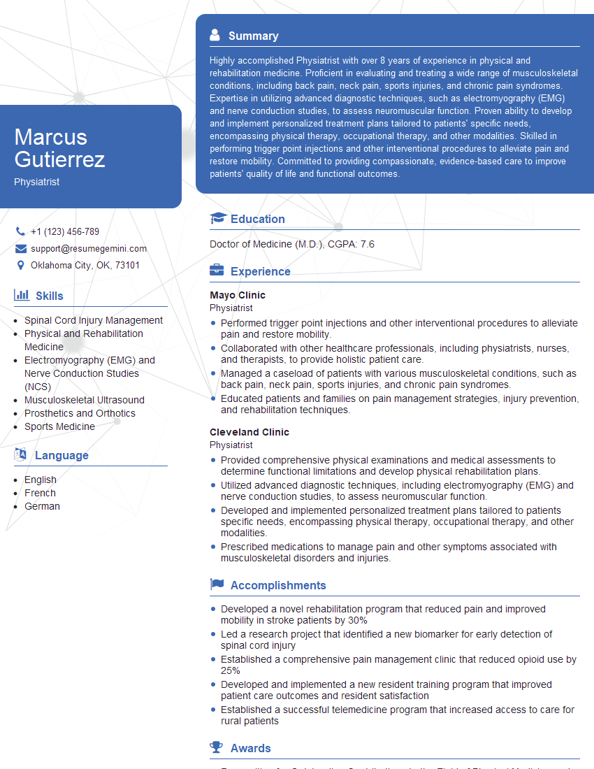
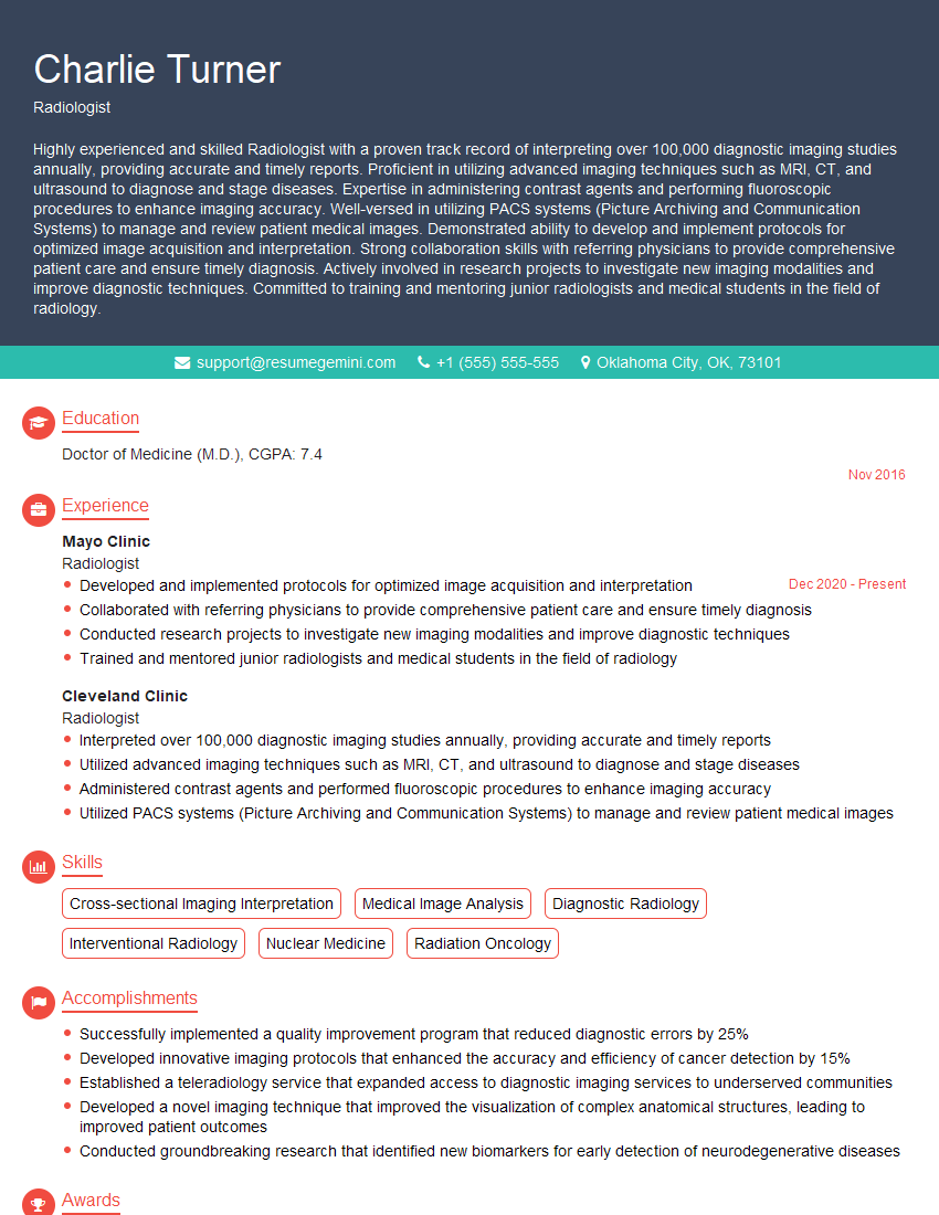
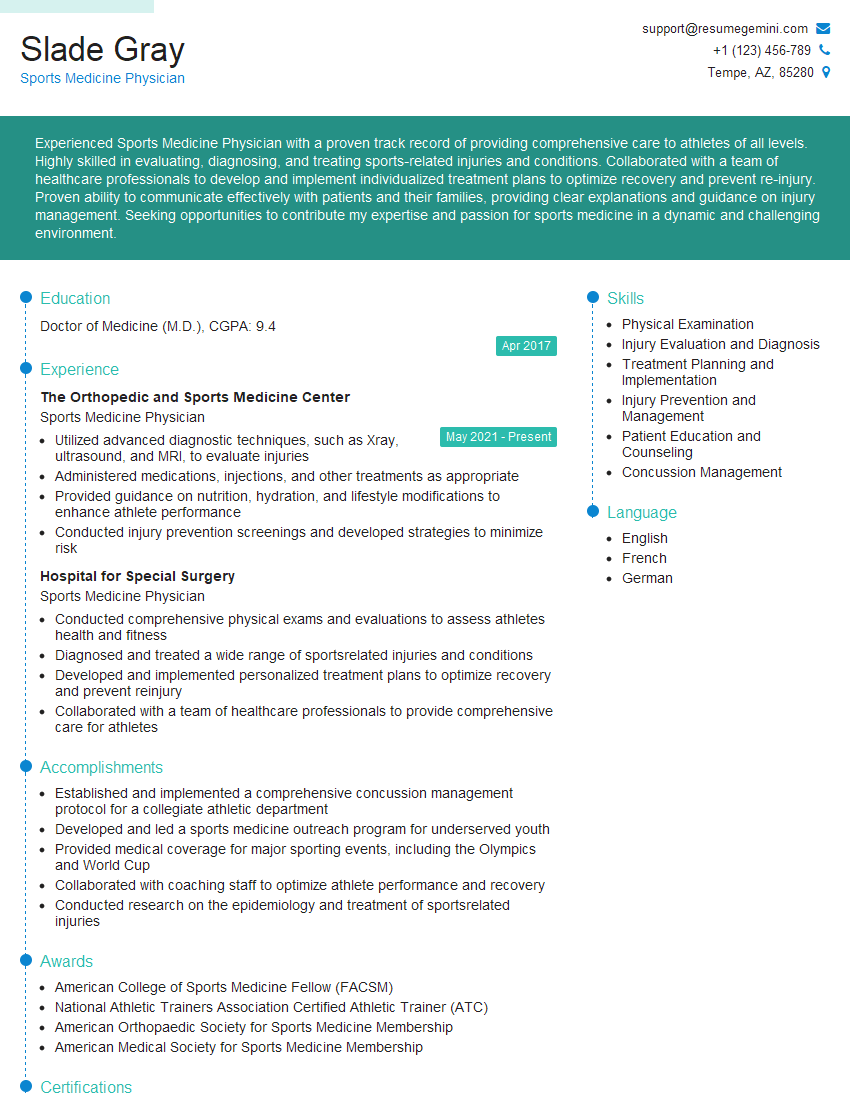
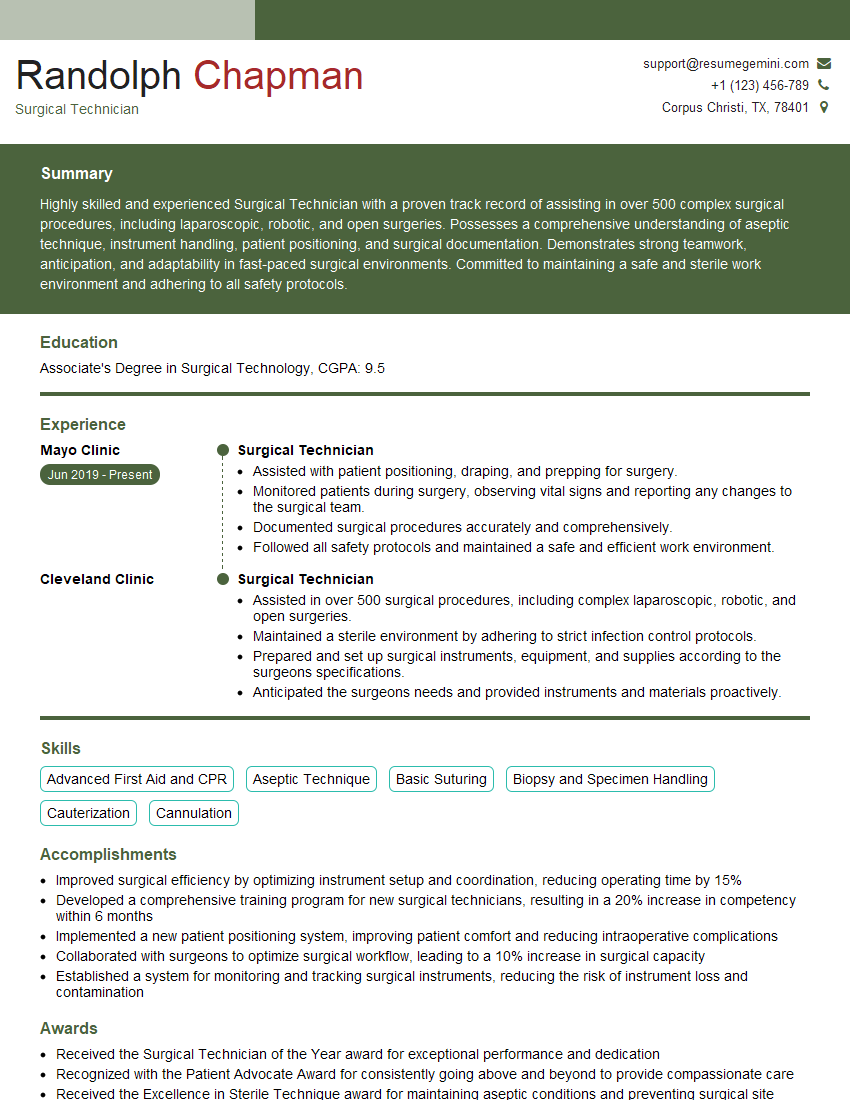
Mastering the complexities of spondylolisthesis is crucial for career advancement in the medical field. A strong understanding of this condition demonstrates expertise and opens doors to specialized roles and increased responsibilities. To maximize your job prospects, it’s vital to present your skills effectively. Creating an ATS-friendly resume is key to getting your application noticed. We recommend using ResumeGemini to build a professional and impactful resume tailored to highlight your spondylolisthesis expertise. ResumeGemini provides examples of resumes specifically designed for this field to help you showcase your skills and experience effectively.
Explore more articles
Users Rating of Our Blogs
Share Your Experience
We value your feedback! Please rate our content and share your thoughts (optional).
What Readers Say About Our Blog
This was kind of a unique content I found around the specialized skills. Very helpful questions and good detailed answers.
Very Helpful blog, thank you Interviewgemini team.
