Interviews are more than just a Q&A session—they’re a chance to prove your worth. This blog dives into essential Ultrasound-Guided Vascular Interventions interview questions and expert tips to help you align your answers with what hiring managers are looking for. Start preparing to shine!
Questions Asked in Ultrasound-Guided Vascular Interventions Interview
Q 1. Describe the different types of ultrasound transducers used in vascular interventions.
Ultrasound transducers used in vascular interventions are categorized primarily by their frequency and imaging capabilities. Higher frequency transducers (7-15 MHz) provide superior image resolution for superficial vessels like those in the neck or wrist, allowing for precise needle placement. Lower frequency transducers (2-5 MHz) penetrate deeper, ideal for visualizing larger, deeper vessels such as the femoral artery or internal jugular vein. The choice depends on the target vessel’s depth and size.
- Linear transducers: Provide a rectangular field of view with high resolution, excellent for superficial structures.
- Curvilinear transducers: Offer a broader, sector-shaped field of view, useful for visualizing deeper vessels and surrounding anatomy.
- Phased array transducers: Allow for electronic steering of the ultrasound beam, offering versatility and often used for cardiac and transthoracic imaging, though they may also be used for vascular access in some cases.
For example, when inserting a central line into the internal jugular vein, a high-frequency linear transducer would typically be preferred due to the vein’s superficial location. Conversely, a lower frequency curvilinear transducer may be necessary for accessing the femoral artery in a larger patient.
Q 2. Explain the principles of Doppler ultrasound in vascular imaging.
Doppler ultrasound utilizes the Doppler effect – the change in frequency of a wave (sound in this case) due to relative motion between the source and the receiver. In vascular imaging, a transducer emits ultrasound waves. When these waves encounter moving blood cells, their frequency shifts. This frequency shift (Doppler shift) is proportional to the velocity of the blood flow. The machine processes this shift, displaying it as a waveform or a color-coded image overlaid on the grayscale anatomical image. This allows us to assess blood flow direction (towards or away from the transducer), velocity, and the presence of any flow disturbances like stenosis (narrowing) or occlusion (blockage).
Imagine a police siren: as the siren approaches, the pitch (frequency) is higher, and as it moves away, the pitch is lower. Doppler ultrasound works on a similar principle, using the change in frequency of the ultrasound waves reflected by moving blood cells to measure blood flow.
Q 3. How do you identify arterial vs. venous structures using ultrasound?
Differentiating arteries from veins on ultrasound relies on several key characteristics:
- Vessel wall thickness: Arterial walls appear thicker and more echogenic (brighter) than venous walls.
- Blood flow pattern: Arterial blood flow is typically pulsatile (showing a waveform mirroring the heartbeat), while venous flow is more continuous, although it may show phasic changes related to respiration.
- Compressibility: Veins are generally more compressible than arteries. Applying gentle pressure with the transducer can collapse a vein, but not an artery.
- Doppler waveform analysis: Arterial waveforms exhibit a characteristic sharp systolic peak followed by a dicrotic notch (a brief dip in velocity during diastole). Venous waveforms tend to be more low-velocity and continuous, although respiratory variations can be observed.
For instance, if a vessel has a thick wall, pulsatile flow and resists compression during transducer pressure, it’s highly likely to be an artery. If the opposite is true – thin wall, continuous flow, and easily compressible – it’s likely a vein. Combining these features allows for reliable identification.
Q 4. What are the safety precautions for ultrasound-guided vascular access?
Safety precautions for ultrasound-guided vascular access are crucial to minimize complications. They include:
- Sterile technique: Maintaining a sterile field is paramount to prevent infection. This includes using sterile gloves, drapes, and appropriate antiseptic solutions.
- Patient positioning and monitoring: Proper patient positioning ensures adequate vessel visualization and minimizes risks. Continuous monitoring of vital signs (heart rate, blood pressure, oxygen saturation) is necessary throughout the procedure.
- Appropriate needle selection: Choosing the correct needle size and type minimizes trauma to the vessel and surrounding tissues.
- Image optimization: Obtaining high-quality ultrasound images is critical for safe and effective needle guidance. Adjustments to transducer position, depth, and gain settings are often necessary.
- Continuous monitoring during needle advancement: Close observation of the ultrasound image throughout needle insertion helps prevent accidental puncture of surrounding structures.
- Hematoma prevention: Applying firm pressure after needle withdrawal is essential to minimize bleeding and hematoma formation.
Remember, understanding the local anatomy and the potential risks is crucial. A comprehensive understanding of the relevant anatomy is always the best preventative measure.
Q 5. Describe the technique for performing ultrasound-guided central venous catheter insertion.
Ultrasound-guided central venous catheter (CVC) insertion, for example into the internal jugular vein, involves the following steps:
- Patient preparation and positioning: The patient is positioned appropriately (typically supine with the head slightly turned away from the side of insertion), and the skin is cleansed using an appropriate antiseptic solution.
- Landmark identification: Using ultrasound, the internal jugular vein is identified. This usually involves visualizing the common carotid artery and then identifying the vein lateral to it.
- Needle insertion: An appropriately sized needle is advanced under ultrasound guidance, aiming to enter the vein at a shallow angle. The needle tip is carefully visualized as it approaches and enters the vein. Real-time guidance ensures a safe and accurate puncture.
- Confirmation of venous access: Once the needle is in the vein, blood return is confirmed, usually by aspiration. Using a guidewire, a catheter is then advanced.
- Catheter placement and securing: The catheter is advanced to the desired location (typically the superior vena cava), and its position is confirmed using fluoroscopy or further ultrasound imaging. Once proper placement is verified, the catheter is secured to prevent dislodgement.
- Dressing application and post-procedure monitoring: A sterile dressing is applied to the insertion site, and the patient is monitored for any signs of complications such as bleeding or infection.
Each step relies heavily on real-time image feedback, allowing for adjustments throughout the procedure. Accurate vessel identification and controlled needle advancement are key to success.
Q 6. How do you manage complications such as hematoma or pneumothorax during vascular access?
Complications during vascular access, such as hematoma or pneumothorax (collapsed lung), require immediate and appropriate management:
- Hematoma: Small hematomas often resolve spontaneously with local pressure. Larger hematomas may necessitate surgical intervention to evacuate the blood. Careful monitoring of the patient’s hematocrit and vital signs is important.
- Pneumothorax: Suspected pneumothorax requires immediate chest x-ray confirmation. Treatment typically involves inserting a chest tube to remove air from the pleural space. This is a serious complication and requires prompt attention to avoid potential respiratory compromise.
- Arterial puncture: Accidental arterial puncture needs immediate pressure applied to the puncture site to control bleeding. If bleeding persists despite pressure, surgical intervention may be necessary.
Careful attention to detail during the procedure is the best way to reduce the risk of these complications, but appropriate preparation for management is also essential. Knowing how to appropriately react to the complications is a crucial skill for all vascular interventionists.
Q 7. What are the indications and contraindications for ultrasound-guided arterial puncture?
Ultrasound-guided arterial puncture has several indications and contraindications:
- Indications:
- Diagnostic angiography: Obtaining arterial blood samples for laboratory analysis.
- Therapeutic interventions: Administering thrombolytic agents or contrast media for clot dissolution or imaging.
- Hemodynamic monitoring: Placing arterial lines for continuous blood pressure monitoring in critically ill patients.
- Difficult access: Accessing arteries in patients with challenging anatomy or previous surgical interventions.
- Contraindications:
- Severe coagulopathy: Patients with significant bleeding disorders are at high risk of hematoma formation.
- Local infection: Infection at the puncture site should be treated before the procedure.
- Severe peripheral artery disease: Severely diseased arteries may be fragile and prone to complications during puncture.
- Patient refusal: Informed consent from the patient is essential before any procedure.
The decision to perform an ultrasound-guided arterial puncture should always be made after careful consideration of the potential benefits and risks, weighing the indications against any contraindications. Proper patient selection and a well-executed procedure are crucial to minimizing complications.
Q 8. Explain the technique for ultrasound-guided peripheral IV cannulation.
Ultrasound-guided peripheral IV cannulation is a technique that uses real-time ultrasound imaging to visualize veins before inserting an intravenous catheter. This significantly improves the success rate and reduces complications compared to the traditional landmark-based approach.
The technique begins with identifying a suitable vein, usually in the forearm or hand, using a high-frequency linear transducer. We look for a vein that appears as a hypoechoic (darker) structure with compressibility. Once a suitable vein is identified, we use the ultrasound to guide needle insertion, ensuring the needle is advanced into the vein lumen. The transducer is kept in place to visualize the needle tip and to confirm successful entry. After the needle is placed, the stylet is removed and a catheter is advanced over the guidewire into the vessel, all while continuously visualized by the ultrasound. We then confirm proper placement by observing blood return and checking for extravasation (leakage). This ensures minimal discomfort and optimal intravenous access.
Step-by-step process:
- Skin Preparation: Cleanse the insertion site with an antiseptic.
- Vein Identification: Use ultrasound to locate a suitable vein and assess its depth, size, and compressibility.
- Needle Insertion: Gently insert the needle into the vein under ultrasound guidance, aiming for the center of the vein lumen.
- Catheter Advancement: Once the needle is in the vein, advance the catheter over a guidewire into the desired position.
- Confirmation of Placement: Observe blood return and check for extravasation. Secure the catheter.
Using ultrasound reduces the risk of multiple needle sticks, nerve damage, and arterial puncture, leading to improved patient comfort and satisfaction. For example, in patients with difficult venous access, such as obese individuals or those with collapsed veins, ultrasound guidance significantly improves success rates.
Q 9. How do you interpret ultrasound images to assess vascular patency and flow?
Interpreting ultrasound images to assess vascular patency and flow involves recognizing specific features indicative of normal and abnormal blood flow. We look for the presence of anechoic (dark) lumen which implies patency and observe for the pulsatile flow within the vessel.
Patency: A patent vessel displays a clear, anechoic lumen (the space within the vessel). A thrombus (blood clot) will appear as an echogenic (bright) filling in the lumen, obstructing the blood flow.
Flow Assessment: Color Doppler ultrasound allows us to visualize the direction and velocity of blood flow. A normal artery will display pulsatile flow, while a vein will show relatively slower flow. Absence or significant reduction in flow can indicate stenosis (narrowing), occlusion (complete blockage), or other vascular pathology. We often use spectral Doppler to get precise velocity measurements, which are important for assessing the degree of stenosis.
For example, in a suspected case of deep vein thrombosis (DVT), the absence of flow within a deep vein along with the presence of filling defects suggests a thrombus. In arterial disease, decreased velocity and disturbed flow patterns can indicate stenosis. The combination of grayscale and Doppler imaging provides a comprehensive assessment of vascular status.
Q 10. Discuss the role of ultrasound in guiding peripheral thrombolysis.
Ultrasound plays a crucial role in guiding peripheral thrombolysis, a procedure that uses medication to dissolve blood clots. Real-time ultrasound monitoring is essential for the safe and effective administration of thrombolytic agents.
Pre-Procedure Assessment: Ultrasound helps in identifying the location, size, and extent of the thrombus. It also allows for the assessment of surrounding vascular anatomy to plan the catheterization approach. We can also assess the collateral circulation to see if there are alternate routes of blood flow.
Catheter Placement: Ultrasound guidance helps to accurately place a catheter within the thrombus. Real-time imaging ensures the catheter remains within the clot and avoids perforation of the vessel wall. We can see the medication being delivered and see if it is causing changes in the thrombus, such as liquefaction.
Monitoring Thrombolysis: During thrombolysis, ultrasound allows for continuous monitoring of the clot lysis. We observe changes in the size and echogenicity of the thrombus, which indicates the effectiveness of the treatment. It also helps in detecting any complications such as vessel perforation or bleeding. For example, we can see a reduction in the size of a clot which is an indicator of thrombolysis.
In summary, ultrasound is indispensable for safe and effective peripheral thrombolysis. Continuous monitoring helps in optimizing the treatment and minimizing potential complications.
Q 11. Describe the use of ultrasound in the placement of dialysis catheters.
Ultrasound guidance is extremely helpful and often essential in the placement of dialysis catheters. It enhances the safety and success rate of this procedure.
Vessel Selection: Ultrasound allows for the visualization of suitable veins, such as the internal jugular, femoral, or subclavian veins, assessing their size, patency, and location relative to surrounding structures. This is crucial for choosing the optimal location for catheter insertion.
Needle Guidance: Real-time imaging guides the placement of the needle into the vein, minimizing the risk of arterial puncture, pneumothorax (collapsed lung), or other complications. We can observe the needle tip as it progresses toward the vessel and confirm its correct positioning.
Catheter Advancement: Ultrasound helps to ensure smooth advancement of the catheter into the vein and confirms proper placement within the vessel, checking for kinking or malposition. We can check for surrounding structures and make sure there are no complications.
Post-Procedure Assessment: Ultrasound also helps in verifying the catheter position and assessing for potential complications such as hematoma (blood collection), thrombosis, or infiltration. The absence of these confirms a successful placement.
In scenarios where traditional landmark-based approaches are challenging, ultrasound guidance is especially valuable. Patients with obesity, prior surgery, or anatomical variations can benefit greatly from this technique. In essence, ultrasound ensures a precise and safe placement of the dialysis catheter, improving the overall outcome and patient care.
Q 12. How do you adjust transducer settings for optimal visualization in different vascular structures?
Adjusting transducer settings for optimal visualization in different vascular structures is critical for achieving accurate diagnostic images and guiding interventions effectively. Different structures require different settings to optimize image quality.
Frequency: Higher-frequency transducers (e.g., 10-18 MHz) provide better resolution for superficial vessels like those in the extremities. Lower-frequency transducers (e.g., 2-5 MHz) are better suited for deeper vessels like the femoral or subclavian veins. A good analogy is that a higher frequency is like a sharper knife, you can see more fine detail but you can’t see as far, while a lower frequency is a blunt knife; you can see further but you can’t see the finer details.
Gain: This adjusts the amplification of the signal. Increasing the gain improves the visibility of weaker signals but also increases noise. We adjust the gain to achieve optimal image brightness and contrast. We avoid excessive gain to prevent noise from interfering with the image interpretation.
Depth: The depth setting determines the field of view. We select an appropriate depth to fully encompass the vessel of interest and surrounding structures. Too shallow a depth might miss important information, too deep adds unnecessary noise and reduces resolution.
Focus: Focusing the ultrasound beam improves resolution. We adjust the focus to ensure optimal visualization of the target vessel. Proper focus ensures that you are evaluating the image with the highest resolution.
Doppler Settings: For flow assessment, we adjust Doppler parameters such as scale, pulse repetition frequency, and wall filter to optimize the visualization of blood flow. These parameters need to be adjusted depending on the specific blood vessel, its size, and the flow rate.
Understanding these settings and adjusting them appropriately for different vascular structures is crucial for the success and safety of ultrasound-guided vascular interventions. Each patient and each vessel is different, and we need to be able to tailor the settings to the specific case.
Q 13. What are the limitations of ultrasound in vascular interventions?
While ultrasound is a powerful tool in vascular interventions, it does have limitations:
- Operator Dependence: Image quality and interpretation are highly dependent on the skill and experience of the sonographer. A poorly performed ultrasound can lead to inaccurate assessment and potentially dangerous interventions.
- Acoustic Shadowing and Enhancement: Bone, air, and calcified plaques can cause acoustic shadowing (reduced signal behind the structure) and enhancement (increased signal in front), obscuring the underlying vascular anatomy.
- Limited Depth Penetration: Ultrasound has limited penetration depth, particularly at higher frequencies. This can make visualizing deeper vessels challenging.
- Obese Patients: Obesity can reduce image quality due to increased attenuation of the ultrasound signal.
- Inability to Visualize All Structures: Ultrasound may not visualize small vessels or structures completely occluded by thrombus.
- Motion Artifacts: Patient movement can introduce artifacts that interfere with image interpretation.
Understanding these limitations is crucial for selecting the appropriate imaging modality and interpreting the results accurately. In some cases, combining ultrasound with other imaging techniques such as fluoroscopy or CT may be necessary to overcome these limitations. For example, fluoroscopy is invaluable in cases where we are placing a catheter in a tortuous vessel.
Q 14. Compare and contrast ultrasound-guided vs. landmark-based vascular access techniques.
Ultrasound-guided vascular access techniques offer significant advantages over landmark-based techniques. Landmark-based approaches rely on anatomical landmarks to guide needle insertion. While simpler, they are less accurate and more prone to complications.
| Feature | Ultrasound-Guided | Landmark-Based |
|---|---|---|
| Accuracy | High; real-time visualization | Lower; relies on anatomical estimations |
| Success Rate | Significantly higher | Lower, especially in challenging patients |
| Complications | Reduced risk of arterial puncture, nerve injury, extravasation | Higher risk of complications |
| Patient Comfort | Generally better, fewer needle sticks | May be more painful, multiple attempts possible |
| Learning Curve | Steeper initial learning curve | Simpler to learn initially |
| Cost | Higher initial cost due to equipment | Lower initial cost |
In summary, while ultrasound-guided vascular access requires specialized training, the higher success rate, reduced complications, and improved patient comfort make it the preferred method in most cases, particularly for challenging patients or complex procedures. Landmark-based techniques remain useful in specific, straightforward situations where ultrasound is not readily available or immediately necessary. But in complex vascular procedures ultrasound guidance is becoming the gold standard.
Q 15. Describe the procedure for ultrasound-guided thrombectomy.
Ultrasound-guided thrombectomy involves removing a blood clot (thrombus) from a blood vessel using ultrasound imaging for guidance. It’s a minimally invasive procedure, reducing the need for large incisions. The procedure typically begins with meticulous skin preparation and draping to maintain sterility. High-frequency ultrasound is used to visualize the thrombus and surrounding vasculature, allowing precise placement of a catheter. Different thrombectomy techniques exist, including aspiration thrombectomy (using a catheter to suction the clot), mechanical thrombectomy (using devices to break up and remove the clot), or a combination of both. The selection of the technique depends on factors such as clot characteristics, vessel location, and patient factors. Post-procedure, close monitoring for bleeding, hematoma formation, and re-thrombosis is crucial.
Example: In a patient with a deep vein thrombosis (DVT) in the femoral vein, ultrasound would guide the insertion of a catheter to the clot’s location. The catheter’s tip would then be used to either aspirate or mechanically fragment the thrombus, with continuous ultrasound monitoring ensuring complete removal and vessel patency.
Career Expert Tips:
- Ace those interviews! Prepare effectively by reviewing the Top 50 Most Common Interview Questions on ResumeGemini.
- Navigate your job search with confidence! Explore a wide range of Career Tips on ResumeGemini. Learn about common challenges and recommendations to overcome them.
- Craft the perfect resume! Master the Art of Resume Writing with ResumeGemini’s guide. Showcase your unique qualifications and achievements effectively.
- Don’t miss out on holiday savings! Build your dream resume with ResumeGemini’s ATS optimized templates.
Q 16. How do you measure vessel diameter and depth using ultrasound?
Ultrasound measures vessel diameter and depth using several principles. Diameter is typically measured directly on the ultrasound image, using the built-in calipers. The image provides a cross-sectional view of the vessel, allowing for direct measurement of its internal lumen. Depth is determined by the ultrasound machine’s ability to calculate the time it takes for the sound wave to travel from the transducer to the vessel and back. This time of flight is directly proportional to the distance – the machine converts this into a depth measurement displayed on the screen. This measurement is highly dependent on the acoustic properties of the tissue being imaged. The accuracy of these measurements depends on the quality of the ultrasound image, the angle of the transducer, and the operator’s skill.
Example: Imagine the vessel as a circle on the screen. The calipers allow you to measure the distance across the circle, giving you the diameter. The depth is displayed numerically beside the image, indicating how far below the skin surface the vessel lies.
Q 17. Explain the concept of in-plane vs. out-of-plane needle insertion.
In-plane and out-of-plane needle insertion refer to the orientation of the needle relative to the ultrasound beam. In in-plane insertion, the needle is advanced within the ultrasound plane; you see the needle’s tip entering the target vessel throughout the procedure. This provides a direct and clear visualization of the needle’s trajectory. Out-of-plane insertion involves advancing the needle perpendicular to the ultrasound plane; only a portion of the needle is visible at a time. While out-of-plane offers greater flexibility for accessing challenging angles, it requires more advanced skill to avoid complications like vessel perforation.
Analogy: Imagine you are looking at a map (ultrasound image). In-plane insertion is like drawing a line directly on the map; you see the entire line as you draw. Out-of-plane is like inserting a pin from above, only seeing the pinhead as it pierces the map.
Q 18. What is the role of contrast agents in ultrasound-guided vascular procedures?
Contrast agents, typically microbubbles of gas encapsulated in a shell, enhance the ultrasound image by increasing the reflectivity of blood flow. This allows for better visualization of the vessel lumen, identification of stenosis (narrowing), and assessment of perfusion (blood flow). Contrast agents are especially helpful in detecting subtle vascular abnormalities or slow flow, which might otherwise be missed. They are commonly used in procedures like angiography (mapping blood vessels) and for assessing the patency (openness) of vessels after an intervention.
Example: During a thrombectomy, a contrast agent can help differentiate the thrombus from the vessel wall, ensuring complete clot removal and confirming the vessel’s patency after the procedure.
Q 19. Describe different types of vascular complications and their management.
Vascular complications during ultrasound-guided interventions can range from minor to life-threatening. Hematoma formation (blood clotting at the puncture site) is common and usually managed conservatively with compression. Arterial puncture can lead to bleeding, hematoma, or even pseudoaneurysm (a false aneurysm). Thrombosis (blood clot formation) can occur at the puncture site or within the vessel. Nerve injury is a possibility, depending on the proximity of nerves to the intervention site. Management varies depending on the severity of the complication. Minor bleeding may require only compression, while more serious complications may necessitate surgical repair, embolization (blocking the bleeding vessel), or blood transfusions.
Example: An arterial puncture might require immediate compression, potentially followed by angiographic embolization to stop the bleeding if compression is unsuccessful. A large hematoma could necessitate surgical evacuation.
Q 20. How would you handle a difficult or compromised vascular anatomy during a procedure?
Dealing with difficult vascular anatomy requires careful planning and adaptability. High-resolution ultrasound with various transducer types (e.g., higher frequency for superficial vessels) becomes crucial. Alternative access sites might be considered. Using smaller-diameter catheters and needles reduces the risk of vessel trauma. Fluoroscopy (X-ray imaging) can provide additional information when ultrasound alone is insufficient. Experienced operators possess the ability to adjust techniques in real time based on the encountered anatomy. In extreme cases, it may be necessary to abandon the procedure and explore alternate treatment strategies.
Example: If a vessel is tortuous (winding) or severely calcified (hardened), a micropuncture technique with smaller catheters and careful needle advancement under ultrasound guidance minimizes the risk of vessel perforation.
Q 21. What are your strategies for minimizing patient discomfort during the procedure?
Minimizing patient discomfort involves a multi-pronged approach. Adequate analgesia (pain relief) is crucial, typically through local anesthesia at the puncture site. Using smaller needles and catheters reduces trauma. Maintaining good communication with the patient throughout the procedure and explaining each step reduces anxiety. A gentle and reassuring demeanor from the operator builds trust and reduces stress. Post-procedure, providing clear instructions for pain management and wound care enhances the overall patient experience. In some cases, sedation may be employed, but only when deemed necessary by the medical team.
Example: Before inserting the needle, topical anesthetic cream or spray can numb the skin, lessening the discomfort of the initial puncture. Explaining the expected sensations (“You might feel a slight pinch or pressure”) helps prepare the patient.
Q 22. How do you maintain a sterile field during ultrasound-guided vascular interventions?
Maintaining a sterile field during ultrasound-guided vascular interventions is paramount to prevent infection. It’s a multi-step process that begins even before the procedure starts. We meticulously prepare the intervention site with antiseptic solutions, following a strict scrub protocol. A sterile drape is then applied, creating a barrier around the puncture site. All instruments and equipment that come into contact with the patient’s skin or vascular system are sterile, including the ultrasound transducer, which is often covered with a sterile sheath or sleeve. Throughout the procedure, we maintain strict sterile technique, ensuring that nothing unsterile touches the sterile field. For example, we’ll use sterile gloves and gowns, and avoid unnecessary movement or reaching over the sterile field. Any breaches in sterility are immediately addressed, such as replacing contaminated drapes or instruments. The entire team is trained to recognize and report potential sources of contamination, creating a culture of safety.
Q 23. Describe your experience with different types of vascular access devices.
My experience encompasses a wide range of vascular access devices, each with its own advantages and disadvantages depending on the clinical scenario. I’m proficient with peripheral intravenous catheters (PIVCs) for short-term access, central venous catheters (CVCs) including subclavian, internal jugular, and femoral approaches for longer-term access and administration of various medications, as well as arterial lines for continuous blood pressure monitoring and blood sampling. I’m also experienced with implantable ports for long-term access. Choosing the appropriate device relies on careful patient assessment considering the duration of therapy needed, the type of medication to be administered, and the patient’s overall clinical condition. For instance, a patient requiring short-term antibiotic administration might receive a PIVC, whereas a patient with cancer needing long-term chemotherapy would benefit from a port-a-cath. In complex cases, such as patients with difficult venous anatomy, I would consider using techniques like ultrasound-guided CVC insertion to increase the success rate and reduce complications. I’m also well-versed in managing complications associated with each device, such as thrombosis or infection.
Q 24. How do you document ultrasound-guided vascular interventions?
Documentation of ultrasound-guided vascular interventions is crucial for maintaining accurate medical records and ensuring patient safety. My documentation method follows a structured format, including a pre-procedure assessment, detailed description of the procedure performed (including the type of access, location, technique used, and imaging findings), any complications encountered and how they were managed, and post-procedure assessment of the patient’s condition. The type of catheter used, insertion depth, and its position confirmed by imaging are also meticulously documented. We include measurements of the vessel’s diameter and depth. Specific details about the ultrasound probe settings, and relevant images taken during the procedure are always recorded. The documentation also specifies the position of the catheter tip, which is critical. This comprehensive approach minimizes the risk of errors and provides a clear and concise record for future reference and auditing purposes. For example, if there was an unexpected bleeding complication, we thoroughly document the cause and our management approach to the situation.
Q 25. What is your understanding of relevant safety regulations and guidelines?
Safety regulations and guidelines are foundational to my practice. I am very familiar with the relevant standards set by organizations such as the Joint Commission and the CDC. These guidelines cover infection control, radiation safety (related to ultrasound use), sterile technique, and appropriate handling of medications. We meticulously follow protocols for safe disposal of sharps and biohazardous materials. I regularly participate in continuing education programs and quality improvement initiatives to stay abreast of updated safety measures. Patient safety is paramount, and our protocols are designed to prevent and manage adverse events, such as hematomas, pneumothorax (during central line placement), and infection. Before each intervention, we perform a thorough risk assessment. Furthermore, incident reports are meticulously completed and reviewed to identify areas for improvement in safety protocols.
Q 26. How do you stay current with the latest advancements in ultrasound-guided vascular techniques?
Keeping up with advancements in ultrasound-guided vascular techniques is an ongoing commitment. I actively participate in professional organizations such as the Society for Vascular Surgery and attend national and international conferences to learn about new technologies and techniques. I regularly review peer-reviewed journals and publications in vascular medicine and ultrasound technology. I also actively seek out opportunities for mentorship and collaboration with colleagues and experts in the field. Online educational platforms and courses are also valuable resources for staying current on the latest techniques and best practices in managing various complications and improving efficiency.
Q 27. Describe a challenging case you encountered and how you overcame the obstacles.
One challenging case involved a patient with severe obesity and a history of multiple failed attempts at central venous catheter placement. Standard landmarks were obscured, making traditional anatomical approaches unsuccessful. Using high-resolution ultrasound, we identified a suitable vessel but its course was tortuous and the vessel itself was quite fragile. We employed a combined technique, using a micropuncture approach guided by ultrasound to minimize trauma. We also used a hydrophilic guidewire to carefully navigate the vessel’s path. Slow and deliberate placement was critical to avoid perforation. Constant real-time ultrasound monitoring allowed us to visualize the catheter tip’s position accurately and avoid complications. Ultimately, we successfully placed the CVC, providing the patient with essential venous access, a result we may not have achieved using traditional methods. The case reinforced the importance of adapting techniques based on individual patient anatomy and using advanced imaging to improve the success rate and patient safety.
Q 28. How would you communicate effectively with the patient and the interdisciplinary team?
Effective communication is essential. Before the procedure, I explain the procedure to the patient in clear, non-technical terms, addressing any concerns or questions they may have. I obtain informed consent and answer any questions from the patient and their family. During the procedure, I communicate regularly with the interdisciplinary team, including nurses, anesthesiologists (if applicable), and other specialists, to ensure a coordinated and efficient intervention. This includes providing regular updates on the procedure’s progress, any challenges encountered, and the plan of action. Post-procedure, we provide the patient and family with clear instructions on post-operative care and potential complications. We use a multidisciplinary approach to care, actively involving all healthcare team members in patient discussions and treatment plans. Open and clear communication minimizes misunderstandings and promotes a positive outcome for the patient.
Key Topics to Learn for Ultrasound-Guided Vascular Interventions Interview
- Image Acquisition and Optimization: Mastering techniques for optimal image acquisition, including probe selection, transducer manipulation, and image adjustments to visualize vascular structures clearly.
- Vascular Anatomy and Pathology: Thorough understanding of normal and abnormal vascular anatomy, common pathologies requiring intervention, and their ultrasound appearances. This includes recognizing stenosis, aneurysms, and thrombi.
- Needle Guidance and Puncture Techniques: Proficiency in various needle puncture techniques, including in-plane and out-of-plane approaches, managing challenges like vessel depth and angulation.
- Safety Considerations and Complications: Understanding potential complications (hematoma, arteriovenous fistula, nerve injury) and implementing safety protocols to minimize risk. Knowing how to handle complications if they arise.
- Specific Intervention Procedures: Detailed knowledge of procedures like thrombolysis, thrombectomy, and angioplasty, including indications, contraindications, and procedural steps.
- Interpretation of Ultrasound Images: Accurate interpretation of Doppler waveforms and color flow imaging to assess blood flow, identify stenosis, and guide intervention.
- Post-Procedure Care and Follow-up: Understanding the post-procedure monitoring and follow-up care necessary for patient safety and recovery.
- Equipment and Technology: Familiarity with different ultrasound machines, catheters, and guidewires used in vascular interventions.
- Radiation Safety Protocols: Understanding and adhering to radiation safety protocols during fluoroscopy-guided procedures.
- Problem-Solving and Troubleshooting: Ability to troubleshoot technical issues during procedures and adapt to unexpected situations.
Next Steps
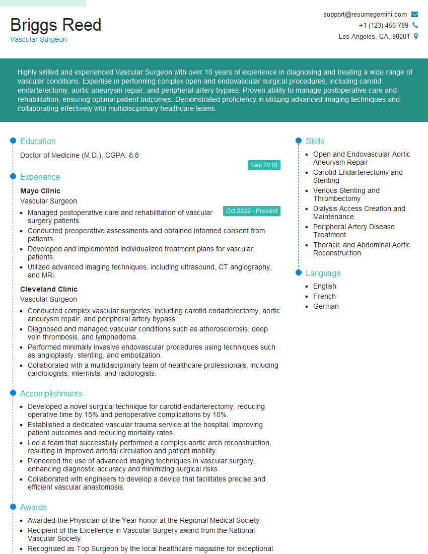
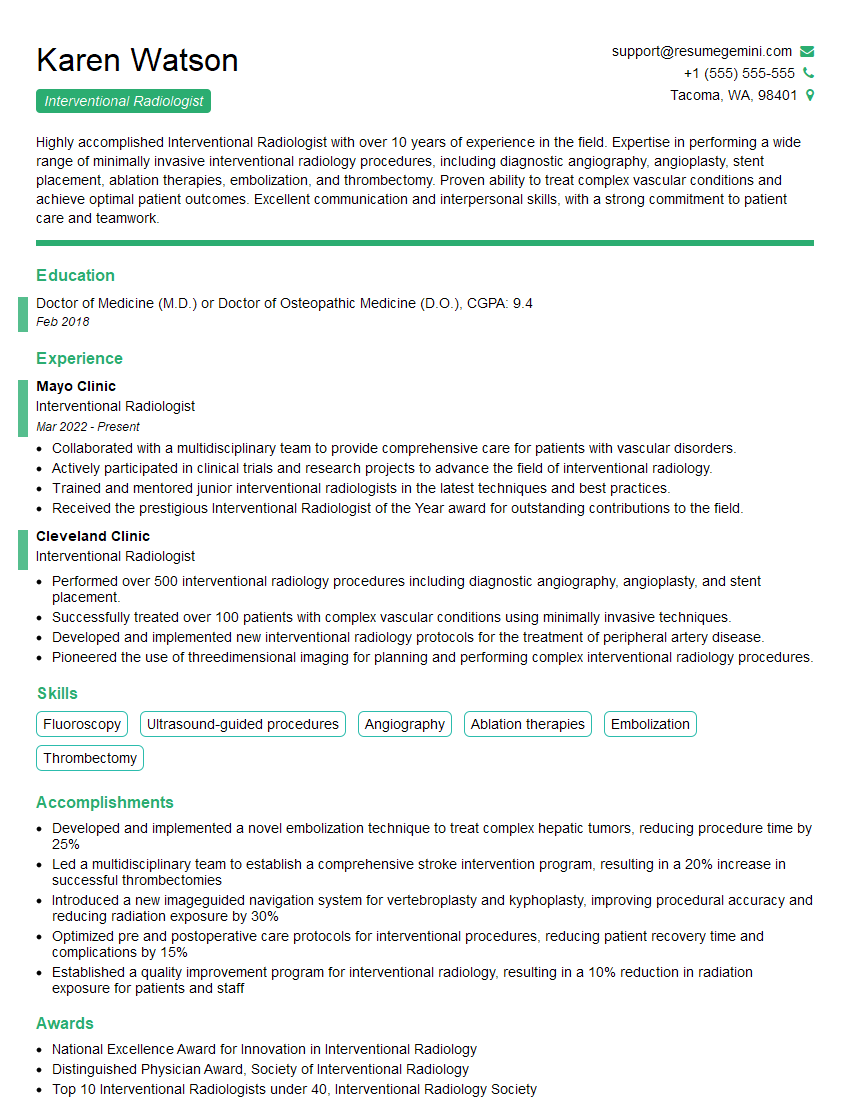
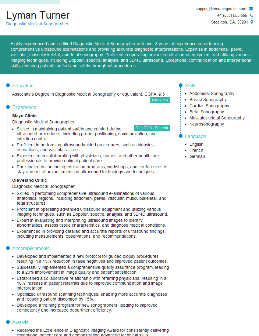
Mastering Ultrasound-Guided Vascular Interventions opens doors to exciting career advancements, offering specialization opportunities and increased earning potential. A strong resume is crucial for showcasing your skills and experience to potential employers. Creating an ATS-friendly resume significantly increases your chances of getting your application noticed. We strongly recommend using ResumeGemini to build a professional and impactful resume that highlights your unique qualifications. ResumeGemini provides examples of resumes tailored specifically to Ultrasound-Guided Vascular Interventions to help you create a document that truly stands out. Invest in your future – invest in your resume.
Explore more articles
Users Rating of Our Blogs
Share Your Experience
We value your feedback! Please rate our content and share your thoughts (optional).
What Readers Say About Our Blog
This was kind of a unique content I found around the specialized skills. Very helpful questions and good detailed answers.
Very Helpful blog, thank you Interviewgemini team.


