Every successful interview starts with knowing what to expect. In this blog, we’ll take you through the top Vascular Assessment of the Lower Extremities interview questions, breaking them down with expert tips to help you deliver impactful answers. Step into your next interview fully prepared and ready to succeed.
Questions Asked in Vascular Assessment of the Lower Extremities Interview
Q 1. Describe the procedure for performing a lower extremity arterial Doppler examination.
A lower extremity arterial Doppler examination is a non-invasive procedure used to assess blood flow in the arteries of the legs and feet. It utilizes a handheld Doppler ultrasound probe that emits high-frequency sound waves. These waves reflect off moving red blood cells, creating an audible signal that is interpreted by the examiner. The signal’s characteristics reveal information about the blood flow’s velocity and direction.
Procedure:
- Patient Preparation: The patient lies supine, and the examiner assesses the patient’s history and performs a thorough physical examination, including palpation of pulses.
- Probe Placement: The Doppler probe is placed over the artery, typically beginning at the brachial artery (in the arm) for comparison, then moving to the arteries of the legs and feet (e.g., femoral, popliteal, posterior tibial, dorsalis pedis arteries). Gel is used to ensure good acoustic coupling between the probe and the skin.
- Signal Acquisition: The examiner adjusts the Doppler settings to optimize the signal, listening to the characteristic sounds of arterial blood flow. The waveform is visualized on a screen and analyzed.
- Measurements: Blood pressure is measured at both arms and ankles using a sphygmomanometer. Pulse wave velocity (PWV) may be calculated in some instances. The waveforms are analyzed to assess their characteristics (see subsequent answers for more on waveform analysis).
- Documentation: All findings, including waveforms, blood pressure readings, and any abnormalities, are meticulously documented.
Example: Imagine a patient with suspected peripheral artery disease (PAD). The Doppler exam would help to determine the location and severity of arterial blockages by analyzing the changes in the waveform and blood pressure readings in the affected limb compared to the unaffected one.
Q 2. Explain the difference between ABI and TBI measurements.
Both ABI and TBI are non-invasive methods used to assess lower extremity arterial disease by comparing blood pressures in the arms and legs. The key difference lies in the specific pressure points used.
- Ankle-Brachial Index (ABI): This is the most common method. It compares the systolic blood pressure at the ankle (using the posterior tibial or dorsalis pedis artery) to the systolic blood pressure at the brachial artery (in the arm). It is calculated as: ABI = Ankle Systolic Pressure / Brachial Systolic Pressure
- Toe-Brachial Index (TBI): This is a more precise method used when the ankle pressures are difficult to obtain accurately (e.g., due to calcification of the arteries). The TBI compares the systolic blood pressure at the toe to the brachial systolic blood pressure: TBI = Toe Systolic Pressure / Brachial Systolic Pressure.
In essence, a lower ABI or TBI suggests arterial narrowing or blockage. While ABI is more common, TBI can provide more detailed information, especially in patients with calcified arteries where ankle pressures might be falsely elevated.
Q 3. What are the normal ranges for ankle-brachial index (ABI)?
Normal ABI values typically range from 0.9 to 1.3. Values below 0.9 suggest peripheral arterial disease (PAD). Values above 1.3 may indicate incompressible arteries, often due to calcification, and require further investigation with alternative methods.
- 0.9-1.3: Normal
- 0.8-0.9: Mild PAD
- 0.4-0.7: Moderate PAD
- <0.4: Severe PAD
- >1.3: Incompressible arteries (likely due to calcification)
It’s crucial to note that these are general guidelines, and interpretation should always consider the patient’s overall clinical picture and other examination findings.
Q 4. How do you identify and interpret a triphasic waveform in a lower extremity arterial Doppler exam?
A triphasic waveform is a characteristic arterial waveform seen in a healthy lower extremity Doppler examination. It is a reflection of the normal pulsatile blood flow.
Characteristics:
- Sharp systolic upstroke: A rapid increase in blood flow velocity at the beginning of systole.
- Early diastolic flow reversal: A brief period of reversed flow at the beginning of diastole due to the elastic recoil of the arterial walls.
- Late diastolic forward flow: A resumption of forward flow in the later part of diastole representing the sustained pressure driving blood flow.
Interpretation: The presence of a triphasic waveform suggests normal arterial compliance and efficient blood flow. The three phases clearly reflect the normal pulsatile nature of the arterial system. Seeing triphasic waves in multiple arterial sites strengthens the interpretation of normal flow.
Example: A patient with no history of vascular disease would be expected to have triphasic waveforms in the femoral, popliteal, posterior tibial and dorsalis pedis arteries.
Q 5. Describe the characteristics of a monophasic waveform and its clinical significance.
A monophasic waveform represents an abnormal arterial waveform where there is only one phase of forward blood flow in the arterial cycle, with no diastolic flow reversal.
Characteristics: The waveform shows a sustained forward flow throughout systole and diastole. It lacks the sharp systolic upstroke and diastolic flow reversal typical of a triphasic waveform. The absence of the distinct components suggests significant impairment of arterial flow.
Clinical Significance: A monophasic waveform strongly suggests the presence of significant arterial stenosis (narrowing) or occlusion (blockage) distal to the point of Doppler probe placement. The loss of diastolic flow reversal is indicative of reduced arterial compliance and suggests decreased blood flow to the distal tissues.
Example: In a patient with critical limb ischemia, a monophasic waveform in the posterior tibial artery might indicate severe blockage in the distal arterial circulation, potentially leading to significant tissue damage.
Q 6. What are the indications for performing a lower extremity venous Doppler examination?
Lower extremity venous Doppler examinations are indicated for a variety of conditions related to venous insufficiency and thrombosis.
- Suspected Deep Vein Thrombosis (DVT): This is a major indication. DVT is a dangerous condition characterized by blood clots forming in the deep veins of the legs.
- Venous Insufficiency: This involves problems with the venous valves or veins themselves, resulting in inadequate return of blood to the heart.
- Chronic Venous Disease: This encompasses various manifestations of venous insufficiency such as varicose veins, leg ulcers, and edema (swelling).
- Post-thrombotic syndrome: This is a condition that develops after a DVT, causing long-term leg problems.
- Pre-operative assessment: Venous assessment can be performed to determine suitability for surgical procedures.
A venous Doppler study helps in identifying the location and extent of venous abnormalities and guiding appropriate management. In essence, it aids in the diagnosis and management of a range of venous conditions.
Q 7. Explain the process of performing a lower extremity venous ultrasound exam and how you identify venous reflux.
A lower extremity venous ultrasound exam uses a handheld ultrasound transducer to visualize the veins of the legs and assess blood flow. This is usually combined with Doppler techniques to evaluate blood flow direction and velocity.
Process:
- Patient Positioning: The patient usually lies supine, with the legs slightly elevated.
- Transducer Placement: The transducer is moved along the course of the veins (e.g., common femoral, popliteal, and calf veins).
- Image Acquisition: The ultrasound machine produces images of the veins, showing their structure and any abnormalities.
- Doppler Evaluation: Doppler is used to assess blood flow direction. Normal venous flow is towards the heart. The presence of reversed flow (reflux) indicates valvular incompetence.
Identifying Venous Reflux: Venous reflux is identified by applying compression to the vein proximal to the transducer. In a normal vein, this compression stops blood flow momentarily; upon release, flow resumes in the direction of the heart. Venous reflux is indicated by retrograde flow (flow away from the heart) after the release of compression.
Example: When evaluating a patient with suspected varicose veins, a venous ultrasound with Doppler might reveal reflux in the greater saphenous vein, signifying valvular insufficiency, a common cause of varicose veins.
Q 8. What are the different types of venous insufficiency, and how are they identified during ultrasound?
Venous insufficiency describes inadequate venous return from the lower extremities, leading to symptoms like swelling, pain, and skin changes. Several types exist, primarily categorized by their location and cause. Ultrasound plays a crucial role in identifying these:
- Chronic Venous Insufficiency (CVI): This is the most common type, often resulting from valve dysfunction within the superficial or deep veins. Ultrasound reveals dilated, tortuous veins, often with incompetent valves demonstrated by reflux during Doppler assessment. We look for signs of venous hypertension such as edema, skin changes (lipodermatosclerosis, stasis dermatitis, hyperpigmentation), and possibly ulcers.
- Superficial Venous Insufficiency: This involves problems with the superficial veins (great saphenous vein, small saphenous vein, and their tributaries). Ultrasound shows dilated and refluxing superficial veins. This often leads to varicose veins.
- Deep Venous Insufficiency: This involves the deep venous system, often following a previous DVT or congenital anomalies. Ultrasound demonstrates venous dilation, incompetence of deep vein valves and collateral vein formation. This can lead to more severe symptoms and complications than superficial venous insufficiency.
- Post-thrombotic Syndrome: This is a long-term complication of DVT, where damage to the venous valves leads to chronic venous insufficiency. Ultrasound reveals residual thrombus, venous dilation, and valve incompetence.
During ultrasound, we use both B-mode imaging (grayscale) to visualize the vein anatomy and Doppler to assess blood flow and valve function. Identifying reflux (backward flow) is key to diagnosing venous insufficiency. We would also look for signs of thrombosis (blood clot formation).
Q 9. How do you differentiate between acute and chronic deep vein thrombosis (DVT) using ultrasound?
Differentiating acute from chronic DVT using ultrasound relies on subtle yet crucial differences in the thrombus appearance and surrounding vascular structures.
- Acute DVT: Shows a hypoechoic (darker on ultrasound) thrombus that is poorly compressible (doesn’t flatten when pressure is applied with the ultrasound probe). The vein may be distended and the surrounding tissues can be edematous (swollen). The thrombus may appear heterogeneous (uneven texture).
- Chronic DVT: The thrombus often appears more echogenic (brighter), and may be partially or completely organized (becoming fibrous). The vein may be contracted or recanalized (partially reopened), and the thrombus may be more compressible than in an acute DVT. The surrounding tissues are less likely to be edematous as in the acute phase. Often there’s evidence of collateral venous circulation.
In essence, the acute DVT is a fresh clot, appearing dark, unorganized, and filling the vein, while a chronic DVT has been present for a longer period, causing changes in the clot’s appearance and vein structure.
Q 10. Explain the use of compression ultrasonography in the diagnosis of DVT.
Compression ultrasonography is the cornerstone of DVT diagnosis. It leverages the fact that normal veins compress easily with the ultrasound probe, while veins containing thrombi do not.
The technique involves applying gentle pressure with the ultrasound transducer to the vein in question. A compressible vein suggests normal patency (no clot), while an incompressible vein strongly indicates the presence of a thrombus. This is the gold standard because it directly assesses the compressibility, providing a clear visual differentiation.
However, it’s important to note that compression isn’t always possible in all venous segments (e.g., popliteal vein in some body habits) or when dealing with a very large thrombus that entirely occludes the vein. In these cases, indirect signs must be used.
Q 11. Describe the physiological basis for performing arterial and venous Doppler ultrasound.
Doppler ultrasound relies on the Doppler effect, which describes the change in frequency of a wave (in this case, ultrasound) due to the relative motion between the source (transducer) and the receiver (blood cells).
- Arterial Doppler: We use this to assess arterial blood flow, looking for characteristics like velocity, flow patterns (triphasic flow is normal in the lower extremities), and presence of stenosis (narrowing). A high-pitched, pulsatile sound reflects the nature of arterial flow.
- Venous Doppler: This assesses venous flow. We look for spontaneous flow, augmentation with distal compression (manual compression below the probe), and absence of reflux (backward flow). A lower-pitched, continuous sound (or phasic flow in the calf veins) characterizes venous blood flow.
In both cases, the Doppler shift frequency is processed to create an audible signal and a velocity profile, which provides quantitative information about blood flow. For example, a high resistance pattern in an artery suggests possible stenosis, while the presence of venous reflux signals incompetence of venous valves.
Q 12. What are the limitations of Doppler ultrasound in assessing lower extremity vasculature?
While Doppler ultrasound is a valuable tool, it has limitations:
- Operator Dependence: The quality of the exam heavily relies on the examiner’s skill and experience. A less experienced sonographer may miss subtle findings or misinterpret the images.
- Limited Visualization of Small Vessels: Doppler may struggle to visualize very small vessels or those buried deep within tissues, leading to potential missed diagnoses.
- Artifacts: Several artifacts (e.g., air bubbles, calcifications, etc.) can mimic or obscure true pathology.
- Inability to assess distal perfusion: Doppler alone cannot provide information about the distal perfusion at the level of the capillaries. Other imaging techniques like contrast angiography might be more informative.
- Limited detection of some types of stenosis: Doppler might not be sensitive enough to detect very early or non-obstructive stenosis.
Therefore, it is essential to consider the limitations and to correlate the findings with the clinical presentation and other imaging modalities if necessary.
Q 13. How do you document your findings from a vascular ultrasound examination?
Documentation of a vascular ultrasound examination is critical for accurate medical record-keeping and communication. It should include:
- Patient demographics: Name, age, date of birth, date of exam.
- Clinical history: Reason for the exam, relevant symptoms (pain, swelling, etc.).
- Technique: The ultrasound techniques used (e.g., B-mode, Doppler).
- Findings: A detailed description of the findings for each vessel examined (e.g., ‘Right common femoral artery shows normal triphasic waveform’ or ‘Left great saphenous vein demonstrates significant reflux’).
- Measurements: Quantitative data such as vessel diameter, flow velocities, etc., where applicable.
- Images: Relevant ultrasound images should be included.
- Impression/Conclusion: A concise summary of the findings and their clinical implications.
A standardized reporting format ensures consistency and completeness, facilitating accurate interpretation by other healthcare providers.
Q 14. What are the potential artifacts encountered during vascular ultrasound examinations, and how do you mitigate them?
Several artifacts can complicate vascular ultrasound interpretations. Understanding these artifacts is crucial for accurate diagnosis:
- Aliasing: Occurs when the Doppler shift frequency exceeds the pulse repetition frequency, causing a wrap-around effect in the velocity waveform. This can be mitigated by increasing the pulse repetition frequency or using lower frequencies.
- Reverberation: Multiple echoes from a strong reflector (e.g., air bubbles, calcifications) create multiple lines. This can be reduced by adjusting gain or selecting another ultrasound window.
- Shadowing: Occurs behind highly attenuating structures (e.g., calcifications, thrombi) where sound waves are blocked. This can obscure underlying structures. This is a characteristic feature for calcification and not necessarily for thrombi which usually have lower echogenicity.
- Enhancement: Increased brightness behind fluid-filled structures (e.g., blood vessels), due to less attenuation. It occurs usually next to anechoic (black) structures.
- Acoustic Noise: Random speckles in the image that doesn’t correspond to any anatomical structure. It’s important to differentiate it from small thrombi that can be heterogeneous.
Proper technique, meticulous attention to detail, and using different imaging settings (frequency, gain, depth) can help minimize or identify artifacts, ensuring reliable interpretation of the results.
Q 15. How do you determine the appropriate transducer frequency for different vascular assessments?
Transducer frequency selection in vascular assessment is crucial for optimal image quality. Higher frequencies (e.g., 7-10 MHz) provide superior resolution, ideal for visualizing superficial vessels like the veins in the lower leg. The higher frequency sound waves penetrate less deeply, meaning we get a clearer picture of the structures closer to the skin’s surface. However, penetration depth is sacrificed. Conversely, lower frequencies (e.g., 2-5 MHz) offer better penetration depth, necessary for visualizing deeper structures such as the common femoral artery or iliac arteries. The trade-off here is that resolution decreases.
In practice, I often start with a higher frequency transducer to assess superficial veins. If I need to visualize deeper structures or there’s significant body habitus, I switch to a lower frequency transducer. Think of it like choosing the right lens for a camera; a telephoto lens (high frequency) is great for close-ups, while a wide-angle lens (low frequency) captures a broader view.
Career Expert Tips:
- Ace those interviews! Prepare effectively by reviewing the Top 50 Most Common Interview Questions on ResumeGemini.
- Navigate your job search with confidence! Explore a wide range of Career Tips on ResumeGemini. Learn about common challenges and recommendations to overcome them.
- Craft the perfect resume! Master the Art of Resume Writing with ResumeGemini’s guide. Showcase your unique qualifications and achievements effectively.
- Don’t miss out on holiday savings! Build your dream resume with ResumeGemini’s ATS optimized templates.
Q 16. Explain the role of color Doppler in assessing lower extremity blood flow.
Color Doppler ultrasound is an invaluable tool in lower extremity vascular assessment because it allows us to visualize and quantify blood flow. It overlays color information onto the grayscale anatomical image, indicating the direction and velocity of blood flow. Red typically represents flow towards the transducer, while blue represents flow away. The intensity or brightness of the color is related to the speed of blood flow. This helps us assess for stenosis (narrowing), occlusion (blockage), and other abnormalities. For example, a significantly reduced or absent color signal within a vessel suggests a significant stenosis or occlusion.
In a patient with suspected deep vein thrombosis (DVT), color Doppler helps identify the absence of blood flow within a vein, a key characteristic of a thrombus. Similarly, in a patient with peripheral artery disease (PAD), color Doppler allows us to identify areas of reduced or absent blood flow in the arteries, indicating areas of stenosis or occlusion.
Q 17. How do you interpret different color Doppler patterns?
Interpreting color Doppler patterns requires experience and a systematic approach. A homogenous, bright red or blue signal within a vessel indicates laminar (smooth) flow. A mosaic pattern, characterized by a mixture of red and blue, usually reflects turbulent flow often associated with stenosis or post-stenotic dilation. Absence of color flow suggests occlusion.
For example, seeing a triphasic waveform in a lower extremity artery is usually normal, characterized by a sharp systolic peak, early diastolic flow reversal, and a late diastolic forward flow. However, a monophasic or biphasic waveform might indicate disease. We also need to consider the vessel’s size, location, and the patient’s overall clinical presentation. A lack of color fill in a vein in combination with other findings could suggest a thrombus. Each case needs to be assessed holistically to reach accurate conclusions.
Q 18. Describe your experience with different types of lower extremity vascular pathologies.
Throughout my career, I’ve encountered a wide range of lower extremity vascular pathologies. This includes various types of arterial disease, such as peripheral artery disease (PAD) encompassing atherosclerosis, acute arterial occlusion and thromboembolism; venous conditions like deep vein thrombosis (DVT), superficial venous insufficiency, and varicose veins; and also more complex situations such as arteriovenous malformations (AVMs) and pseudoaneurysms.
I’ve been involved in the diagnosis and management of patients with critical limb ischemia, requiring complex assessments and often multidisciplinary consultations. I have experience with post-surgical assessment, such as evaluating patency of bypass grafts or endovascular interventions. Each case presents unique challenges, necessitating a thorough understanding of the patient’s history, physical examination findings, and the appropriate use of imaging modalities.
Q 19. What is your experience with patient communication and education related to vascular ultrasound findings?
Patient communication and education are paramount in vascular ultrasound. I believe in explaining findings in a clear, concise, and understandable manner, avoiding medical jargon as much as possible. I always correlate the ultrasound findings with the patient’s clinical symptoms. For instance, if a patient presents with leg pain and swelling, and the ultrasound shows a DVT, I explain the implications of the diagnosis, including the risks and treatment options.
I provide patients with visual aids, such as anatomical diagrams, to help them understand the location and nature of the vascular problem. I ensure patients understand the next steps, such as follow-up appointments or referrals to specialists. I always encourage patients to ask questions, and I answer them patiently and thoroughly, empowering them to actively participate in their healthcare.
Q 20. How do you handle challenging or complex vascular cases?
Challenging vascular cases require a structured approach. This involves carefully reviewing the patient’s history, conducting a thorough physical examination, and obtaining additional imaging if necessary, such as CT or MRI. I would collaborate with other specialists, such as vascular surgeons or interventional radiologists, to develop a comprehensive management plan.
For example, in a case of an ambiguous lesion, I would discuss my findings with a vascular surgeon or radiologist to determine the best approach for further evaluation. In situations of difficult-to-visualize vessels due to obesity or severe scarring, I will employ different techniques such as using lower frequency probes or changing patient positioning to optimize image quality. The key is a systematic approach, meticulous image acquisition, and effective team communication.
Q 21. What is your experience with quality control and quality assurance in a vascular laboratory?
Quality control and quality assurance (QA) are integral to a well-functioning vascular laboratory. This includes regular equipment maintenance and calibration, ensuring optimal performance and image quality. We participate in regular quality control programs with phantoms to assess the accuracy and precision of our measurements. We maintain detailed records of all examinations, including images and reports.
Our QA program also includes adherence to established protocols and guidelines for vascular ultrasound examinations. We regularly review our processes, looking for areas of improvement. This may include internal audits or external proficiency testing to ensure we are providing accurate and reliable results, maintaining the highest level of patient care and diagnostic confidence.
Q 22. Describe your knowledge of relevant safety protocols for vascular ultrasound examinations.
Safety protocols in vascular ultrasound are paramount to patient and operator well-being. They encompass several key areas:
- Infection Control: Strict adherence to hand hygiene, using sterile or disinfected probes and gel, and proper disposal of used materials is crucial. We follow our facility’s guidelines, which typically align with CDC recommendations. For instance, using probe covers and changing them between patients is standard practice.
- Electrical Safety: Ensuring the ultrasound machine is properly grounded and functioning correctly is essential to prevent electrical shocks. Regular equipment maintenance and safety checks are vital. If I notice any malfunction, such as a frayed cord or unusual electrical noise, I immediately report it to the biomedical engineering department.
- Patient Safety: Careful attention is paid to patient positioning and comfort to prevent falls or injuries. We continuously monitor vital signs, especially during prolonged examinations. Patients are informed of the procedure, potential risks, and what to expect. A detailed explanation of the study and any necessary pre-test instructions helps patient anxiety and helps ensure study compliance.
- Radiation Safety (if applicable): While vascular ultrasound doesn’t utilize ionizing radiation, if using a combined modality system with features such as Doppler capabilities, understanding any associated safety protocols is still essential.
Regular training and competency assessments ensure our adherence to these protocols. We participate in regular safety training sessions covering topics such as infection control and electrical safety.
Q 23. How do you maintain patient confidentiality during vascular assessments?
Maintaining patient confidentiality is a core value and a legal obligation. We adhere to HIPAA regulations and our institution’s privacy policies diligently. This includes:
- Secure Data Storage: All patient information, including images and reports, are stored on password-protected computers and servers with restricted access. Only authorized personnel have access to patient data. Access is carefully documented and monitored.
- Confidential Communication: Discussions regarding patient cases occur only in private settings or via secure electronic communication channels. Patient information is never discussed in public areas or with unauthorized individuals.
- Patient Identification: Patients are always identified using appropriate methods, such as a combination of name and date of birth, to avoid any errors or misidentification. We always verify patient identification and compare it to the requisition prior to starting the exam.
- Data Disposal: Appropriate disposal methods are followed for any physical or electronic records when they are no longer needed, following both hospital and departmental policies.
We treat every piece of patient information with the utmost respect and care, recognizing the sensitive nature of medical information. This commitment extends to every aspect of our work, from obtaining consent to managing patient data.
Q 24. What is your experience with using vascular imaging software and reporting systems?
I have extensive experience using various vascular imaging software and reporting systems, including GE PACS, Siemens syngo, and Philips IntelliSpace Portal. My expertise encompasses image acquisition, processing, measurement, and reporting.
I’m proficient in using these systems to:
- Acquire high-quality images: Adjusting parameters such as gain, depth, frequency, and TGC to optimize image visualization for various vascular structures in the lower extremities.
- Perform precise measurements: Accurately measuring vessel diameters, wall thickness, and blood flow velocities using automated and manual measurement tools. These measurements are critical for diagnosis and treatment planning.
- Generate comprehensive reports: Creating detailed reports that include images, measurements, and interpretations, tailored to the referring physician’s needs and following established formatting standards.
- Manage patient data: Efficiently managing patient data, including demographic information, examination parameters, and reporting findings, within the confines of the software interface.
I am adept at troubleshooting minor technical issues within these systems, ensuring minimal disruption to patient care. For example, recently I resolved a problem with image freezing by adjusting the system’s settings and communicating the issue to the biomedical engineering department.
Q 25. Describe your knowledge of relevant anatomy and physiology of the lower extremities vascular system.
A thorough understanding of the lower extremity vascular anatomy and physiology is fundamental to performing accurate vascular assessments. This includes knowledge of:
- Arteries: The abdominal aorta, common iliac arteries, external and internal iliac arteries, common femoral arteries, superficial femoral arteries, profunda femoris arteries, popliteal arteries, anterior and posterior tibial arteries, peroneal arteries, and their branches. Understanding their branching patterns, locations, and typical sizes is crucial for identifying stenosis or occlusions.
- Veins: The deep and superficial venous systems, including the femoral veins, popliteal veins, tibial veins, peroneal veins, great saphenous vein, small saphenous vein, and their tributaries. Knowing the venous valves and their function is also critical in evaluating venous reflux.
- Physiology: Understanding blood flow dynamics, including laminar and turbulent flow, pressure gradients, and the impact of various factors such as age, disease, and medications on vascular function. This is essential for interpreting Doppler waveforms.
This detailed anatomical knowledge allows me to identify variations from the norm and correlate findings with clinical presentation, leading to accurate diagnoses.
For example, understanding the relationship between the superficial femoral artery and the profunda femoris artery is vital for interpreting flow patterns and identifying potential collateral circulation pathways. Similarly, understanding the location of the perforating veins is crucial for evaluating venous insufficiency and varicose veins.
Q 26. How do you stay updated with the latest advancements in vascular ultrasound technology and techniques?
Staying current in vascular ultrasound requires a multi-pronged approach:
- Continuing Medical Education (CME): I actively participate in CME courses, webinars, and conferences focusing on advancements in ultrasound technology, techniques, and interpretation. This ensures my skills remain sharp and my knowledge base is updated on the latest research and guidelines.
- Professional Organizations: Membership in professional organizations like the Society for Vascular Ultrasound (SVU) and the American Institute of Ultrasound in Medicine (AIUM) provides access to educational resources, journals, and networking opportunities. Participation in these organizations helps keep me abreast of the latest guidelines and research.
- Peer Review and Collaboration: Engaging in peer review of studies and collaborating with colleagues allows for the exchange of knowledge and experiences. This is particularly beneficial for discussing challenging cases and learning from the expertise of others. Sharing complex cases and learning how other specialists approached the diagnosis is very helpful.
- Journal Articles and Literature: Regularly reading peer-reviewed journals and publications on vascular ultrasound keeps me up-to-date on the latest research findings and technological advancements in the field.
Continuous learning is not just a professional obligation but a personal commitment to providing the highest quality patient care. I’m constantly striving to enhance my skills and remain at the forefront of vascular ultrasound expertise.
Q 27. Describe a situation where you had to troubleshoot a technical issue during a vascular ultrasound exam.
During a lower extremity venous ultrasound, I encountered a situation where the image quality was significantly degraded due to excessive air in the coupling gel. The images were blurry and artifacts were obscuring the venous structures. Initially, I tried adjusting the TGC and gain settings to improve the image, but the problem persisted.
My troubleshooting steps included:
- Re-application of Gel: I carefully removed the excess air bubbles from the coupling gel by applying a thin, even layer and using gentle pressure to ensure good contact between the transducer and the skin. This eliminated most of the air pockets.
- Transducer Angle: I adjusted the angle of the transducer to optimize the acoustic window, further minimizing the air artifact influence. A slight change in angle optimized the image quality.
- Frequency Adjustment: I then tried changing the transducer frequency, using a higher frequency to get a clearer picture of the superficial veins while using a lower frequency for the deeper veins.
- Alternative Gel: Since the issue didn’t completely resolve, I switched to a different brand of ultrasound gel known for its better viscosity and reduced air bubble formation. This brand produces less air bubbles and is easier to apply, resulting in higher image quality.
By systematically addressing potential causes, I was able to restore optimal image quality and complete the examination. This experience reinforced the importance of meticulous technique and troubleshooting skills in vascular ultrasound.
Key Topics to Learn for Vascular Assessment of the Lower Extremities Interview
- History Taking & Patient Presentation: Understanding the patient’s medical history, symptoms (pain, swelling, discoloration), and risk factors for peripheral arterial disease (PAD) or venous insufficiency.
- Physical Examination Techniques: Mastering palpation of peripheral pulses (e.g., femoral, popliteal, posterior tibial, dorsalis pedis), inspection for signs of arterial or venous disease (e.g., skin changes, edema, ulceration), and assessment of capillary refill time.
- Non-Invasive Diagnostic Tests: Understanding the principles and interpretation of ankle-brachial index (ABI), Doppler ultrasound, and segmental pressure measurements. Knowing the limitations of each test.
- Differential Diagnosis: Differentiating between arterial, venous, and other causes of lower extremity symptoms. Being able to explain your reasoning process.
- Clinical Reasoning & Problem Solving: Applying your knowledge to interpret findings from history, physical exam, and diagnostic tests to formulate a diagnosis and treatment plan. Practice explaining your thought process clearly.
- Specific Vascular Conditions: Deep understanding of PAD, chronic venous insufficiency, deep vein thrombosis (DVT), and other relevant vascular conditions of the lower extremities. Be ready to discuss their pathophysiology, clinical presentation, and management.
- Case Studies & Scenario-Based Questions: Prepare to analyze hypothetical cases, applying your knowledge to diagnose and manage patient scenarios. Practice explaining your reasoning behind your decisions.
Next Steps
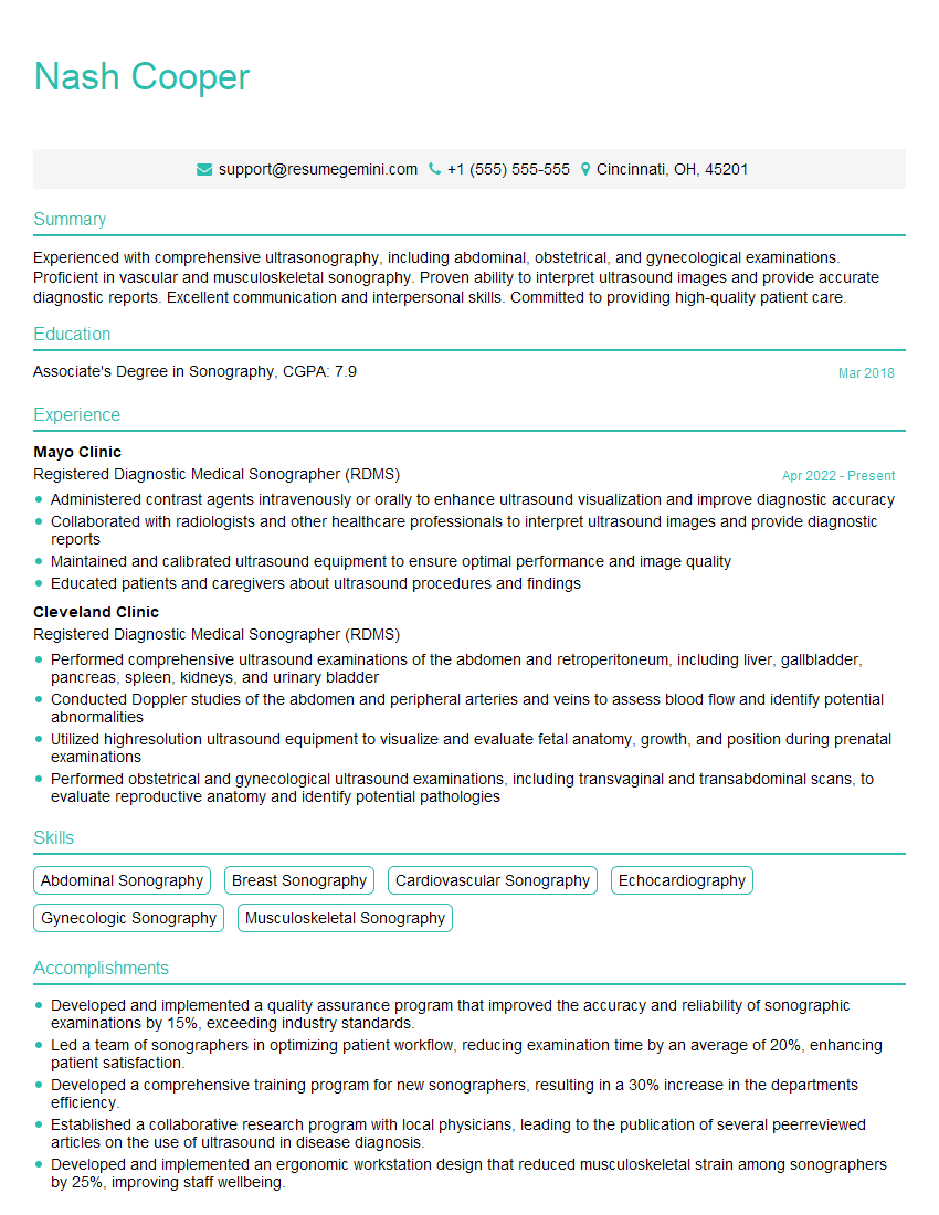
Mastering vascular assessment of the lower extremities is crucial for career advancement in many healthcare fields. A strong understanding of these concepts will significantly enhance your clinical skills and make you a more valuable asset to any healthcare team. To maximize your job prospects, it’s essential to present your skills effectively. Creating an ATS-friendly resume is key to getting your application noticed by recruiters. ResumeGemini is a trusted resource that can help you build a professional and impactful resume tailored to highlight your expertise in vascular assessment. Examples of resumes specifically tailored to this area are available to help you get started.
Explore more articles
Users Rating of Our Blogs
Share Your Experience
We value your feedback! Please rate our content and share your thoughts (optional).
What Readers Say About Our Blog
This was kind of a unique content I found around the specialized skills. Very helpful questions and good detailed answers.
Very Helpful blog, thank you Interviewgemini team.


