Feeling uncertain about what to expect in your upcoming interview? We’ve got you covered! This blog highlights the most important Pleural Biopsy interview questions and provides actionable advice to help you stand out as the ideal candidate. Let’s pave the way for your success.
Questions Asked in Pleural Biopsy Interview
Q 1. Describe the indications for performing a pleural biopsy.
A pleural biopsy is indicated when we need to determine the cause of a pleural effusion (fluid buildup in the pleural space) that cannot be diagnosed with less invasive methods like thoracentesis (fluid removal). This is crucial because pleural effusions can stem from a wide range of conditions, from infections like tuberculosis or pneumonia to cancers such as lung cancer or lymphoma, and even autoimmune diseases.
- Suspected malignancy: When imaging suggests a cancerous mass or concerning features within the pleura, a biopsy is essential for definitive diagnosis and staging.
- Infectious causes: If cultures from thoracentesis are negative, but clinical suspicion for an infection like tuberculosis remains high, a biopsy helps confirm the diagnosis and guide treatment.
- Diagnostic uncertainty: When the nature of the pleural effusion remains unclear after thorough clinical evaluation and basic laboratory tests, a biopsy provides a tissue sample for microscopic examination.
- Autoimmune disorders: Pleural effusions associated with autoimmune diseases like lupus or rheumatoid arthritis may require biopsy for confirmation and to guide treatment decisions.
In essence, a pleural biopsy is a valuable diagnostic tool when non-invasive methods are insufficient to determine the underlying cause of a pleural effusion.
Q 2. What are the contraindications for a pleural biopsy?
Contraindications to pleural biopsy aren’t absolute, but rather situations where the risks significantly outweigh the benefits. These include:
- Severe coagulopathy: Patients with severely impaired blood clotting have an increased risk of bleeding complications. We usually correct this before proceeding.
- Uncontrolled respiratory compromise: If the patient already has severe respiratory distress, the procedure could exacerbate their condition.
- Lack of pleural fluid: A biopsy is pointless without enough fluid to target.
- Patient refusal or inability to cooperate: The procedure requires patient cooperation for accurate sampling and to minimise movement.
- Severe underlying cardiac disease: In these cases, the procedure may pose significant cardiovascular risk.
Careful evaluation of the patient’s overall health and the specific clinical scenario is vital in deciding whether to proceed with a pleural biopsy.
Q 3. Explain the different techniques used for pleural biopsy (e.g., blind needle, image-guided).
Several techniques exist for pleural biopsy, each with its own advantages and disadvantages:
- Blind Needle Biopsy: This is a less invasive approach, using a needle guided only by anatomical landmarks. It is quicker and simpler than image-guided methods, but has a higher risk of missing the target and causing complications.
- Image-Guided Biopsy (Ultrasound or CT): These techniques provide real-time visualization of the needle and the pleural space, allowing for precise targeting and minimizing complications. Ultrasound is often preferred for its lower radiation dose and portability, while CT scans offer greater anatomical detail.
- Video-Assisted Thoracoscopic Surgery (VATS): For more complex cases or when larger tissue samples are needed, VATS provides a minimally invasive approach with excellent visualization and maneuverability. It is more invasive than needle biopsies and requires general anesthesia.
The choice of technique depends on factors such as the location and extent of the pleural abnormality, the availability of imaging, patient characteristics, and physician expertise. For example, a suspected peripheral lesion might be readily accessible via ultrasound-guided biopsy, whereas a centrally located lesion might necessitate CT guidance or even VATS.
Q 4. What are the potential complications of a pleural biopsy?
Pleural biopsy, like any invasive procedure, carries potential complications. These include:
- Pneumothorax: The most common complication, involving air leaking into the pleural space, causing lung collapse. This can range from mild to life-threatening.
- Bleeding: Can occur at the biopsy site and range from minor to significant hemorrhage requiring intervention.
- Infection: Introduction of bacteria at the puncture site can lead to local or systemic infection.
- Injury to adjacent structures: Although rare, the needle can potentially damage adjacent organs such as the lung, diaphragm or intercostal vessels.
- Pain: Post-procedural pain is common and can be managed with analgesics.
The risk of complications varies depending on the technique used, the operator’s experience, and the patient’s overall health. Image-guided biopsies generally have a lower complication rate than blind needle biopsies.
Q 5. How do you manage complications like pneumothorax after a pleural biopsy?
Management of pneumothorax after a pleural biopsy depends on its severity. Most small pneumothoraces resolve spontaneously with observation and supportive care (oxygen therapy, monitoring). Larger pneumothoraces, which cause significant respiratory distress, may require:
- Chest tube insertion: This allows for evacuation of air from the pleural space and re-expansion of the lung. This is the most common intervention for significant pneumothorax.
- Supplemental oxygen: To improve oxygenation.
- Pain management: Analgesics to manage chest pain.
Continuous monitoring of respiratory status is crucial, including monitoring oxygen saturation and respiratory rate. In rare cases, surgical intervention may be necessary.
It’s crucial to emphasize the importance of pre-procedural counseling, where the patient is informed about the possibility of this complication and the subsequent management strategy.
Q 6. Discuss the role of imaging (e.g., ultrasound, CT) in pleural biopsy.
Imaging plays a crucial role in pleural biopsy, primarily in guiding the procedure and assessing the extent of pleural disease. Ultrasound and CT scans are the most commonly used imaging modalities:
- Ultrasound: Provides real-time visualization of the pleural fluid collection, guiding the needle to the target area. It is particularly useful for identifying areas of pleural thickening or masses that may be suitable for biopsy.
- CT scan: Offers detailed anatomical information, useful for identifying the exact location of the lesion and guiding the needle for difficult-to-reach areas. It’s also invaluable in assessing potential complications like pneumothorax.
The use of these imaging techniques improves the accuracy and safety of the procedure. It helps reduce the risk of complications by ensuring that the needle is placed precisely at the target site and avoids injury to surrounding structures. Pre-procedural imaging also helps us assess the feasibility and safety of the procedure in each individual patient. For example, ultrasound can guide a needle biopsy of a pleural effusion, while CT might be better suited to guide a biopsy of a small, subtle pleural nodule.
Q 7. Describe the pre-procedural patient preparation for a pleural biopsy.
Pre-procedural preparation for a pleural biopsy involves several steps to ensure patient safety and comfort and to optimize the procedure’s success:
- Informed consent: The patient must be fully informed about the procedure, its benefits, risks, and potential complications. A well-documented informed consent process is essential.
- Assessment of coagulation status: Blood tests are performed to check the patient’s clotting ability. Patients with coagulopathy require correction before the procedure.
- Review of medications: Medications that may increase bleeding risk, such as anticoagulants, are often temporarily withheld or adjusted. This needs careful individual assessment.
- NPO status: Patients are usually instructed to refrain from eating or drinking for several hours before the procedure to minimize the risk of aspiration.
- Antibiotics: Prophylactic antibiotics are sometimes given to prevent infection.
- Pain control: An appropriate pain management plan should be discussed and implemented pre-procedure and post-procedure.
These preparations help minimize risks, ensure patient cooperation, and improve the overall outcome of the procedure.
Q 8. What are the post-procedural care instructions for a patient after a pleural biopsy?
Post-procedural care after a pleural biopsy focuses on monitoring for complications and ensuring patient comfort. Patients are typically monitored for several hours after the procedure for signs of bleeding, pneumothorax (collapsed lung), or infection. This involves checking vital signs (heart rate, blood pressure, respiratory rate, oxygen saturation), auscultating the lungs to detect breath sounds, and monitoring chest tube drainage if one was placed.
- Pain Management: Analgesics are prescribed to manage pain, which can range from mild discomfort to moderate pain depending on the technique used.
- Activity Restrictions: Patients are advised to restrict strenuous activity for a few days to allow the puncture site to heal.
- Wound Care: The puncture site is monitored for signs of infection, such as redness, swelling, or drainage. Patients are instructed on proper wound care to keep the area clean and dry.
- Follow-up: A follow-up appointment is scheduled to review the biopsy results and assess the patient’s recovery. Chest X-rays are often taken to check for pneumothorax or other complications.
- Signs and Symptoms to Report: Patients are instructed to report any concerning symptoms, such as shortness of breath, chest pain, fever, or excessive bleeding from the puncture site, to their healthcare provider immediately.
For instance, a patient might be discharged with a prescription for pain medication and instructions to rest for 24-48 hours. They would also be given specific instructions on what to watch for and when to seek immediate medical attention.
Q 9. How do you interpret the results of a pleural biopsy?
Interpreting pleural biopsy results involves a multidisciplinary approach, typically involving a pathologist and the treating physician. The pathologist examines the tissue sample microscopically to identify the cells present and their characteristics. This assessment helps to determine the cause of the pleural effusion (fluid buildup in the pleural space). Key features examined include:
- Cytology: Examines individual cells for malignancy. Malignant cells typically show abnormal morphology and growth patterns.
- Histopathology: Examines the tissue architecture, identifying specific cell types and their arrangement. This is critical in determining the type of tumor (e.g., adenocarcinoma, mesothelioma) if cancer is present.
- Special stains and immunohistochemistry: These techniques help to further characterize the cells and assist in diagnosis, especially in differentiating various types of cancers.
- Culture and Sensitivity: If an infection is suspected, cultures are performed to identify the specific pathogen and determine the appropriate antibiotic therapy.
For example, finding malignant cells with characteristic features of adenocarcinoma on cytology and confirmed with histopathology would indicate a metastatic adenocarcinoma involving the pleura.
Q 10. What are the diagnostic yield and limitations of a pleural biopsy?
The diagnostic yield of a pleural biopsy, meaning the percentage of procedures resulting in a definitive diagnosis, is generally high, ranging from 70-90% depending on the technique and the underlying disease process. However, it has limitations:
- Sampling Error: The biopsy may not obtain a representative sample of the pleural tissue. This is more common with small or localized lesions.
- Non-diagnostic results: Sometimes the sample is too small or insufficient for conclusive diagnosis, leading to inconclusive results.
- Complications: The procedure carries risks, including bleeding, pneumothorax, and infection. These complications can affect the diagnostic yield by delaying the procedure or necessitating further interventions.
- Inability to diagnose certain conditions: Pleural biopsy may not be helpful in diagnosing certain conditions, such as early-stage tuberculosis or certain viral infections.
For instance, a pleural biopsy might not be able to differentiate between different types of lymphoma based on a small sample. In such cases, additional diagnostic tests might be required.
Q 11. Compare and contrast the different biopsy techniques in terms of safety and diagnostic accuracy.
Several biopsy techniques exist, each with its strengths and weaknesses:
- Image-guided needle biopsy (CT or ultrasound guided): This approach offers excellent visualization of the pleural lesion and allows for precise sampling. It is generally well-tolerated with minimal complications. The accuracy is higher than blind techniques.
- Blind needle biopsy: This older technique is less precise and carries a higher risk of complications such as pneumothorax. However, it is a less invasive procedure than thoracoscopy.
- Video-assisted thoracoscopic surgery (VATS): This minimally invasive surgical technique allows for direct visualization of the pleura and collection of multiple tissue samples. It offers superior diagnostic accuracy, particularly when dealing with localized or small lesions. However, it is more invasive and has a higher risk of complications than needle biopsy techniques.
- Open surgical biopsy: This is a rarely used procedure reserved for cases where other techniques fail to yield a diagnosis. It is the most invasive with a higher risk of significant complications.
In terms of safety, image-guided needle biopsy is generally the safest, followed by VATS, blind needle biopsy, and lastly open surgical biopsy. Regarding diagnostic accuracy, VATS generally provides the highest diagnostic yield, followed by image-guided needle biopsy, and then blind needle biopsy.
Q 12. How do you select the optimal biopsy technique for a particular patient?
Selecting the optimal biopsy technique is a crucial decision. Several factors influence this choice:
- Clinical presentation: The patient’s symptoms, imaging findings, and overall health status help guide the decision.
- Location and size of the pleural lesion: Accessibility and size significantly impact technique selection. Small or hard-to-reach lesions might require VATS.
- Prior procedures: Previous chest surgeries or lung conditions may influence the choice of technique.
- Risk-benefit assessment: The physician weighs the potential benefits of a more accurate diagnosis against the risks of more invasive procedures.
- Patient preference: Whenever possible, the patient’s preferences and understanding of the risks and benefits are considered.
For instance, a patient with a large, easily accessible pleural mass on imaging might undergo an image-guided needle biopsy. A patient with a suspicious small lesion difficult to access through needle biopsy might require VATS for better sampling.
Q 13. Describe your experience with managing bleeding complications during a pleural biopsy.
Managing bleeding complications during a pleural biopsy involves prompt recognition and intervention. Minor bleeding is usually self-limiting and managed conservatively with close monitoring. However, significant bleeding requires immediate intervention.
- Close monitoring of vital signs: Continuous monitoring of heart rate, blood pressure, and respiratory rate is crucial to detect early signs of blood loss.
- Chest tube insertion: In cases of significant bleeding, a chest tube is inserted to evacuate blood and prevent hemothorax (blood in the pleural space).
- Surgical intervention: If bleeding continues despite chest tube placement, surgical intervention may be necessary to control the bleeding source. This might involve thoracotomy (open chest surgery).
- Blood transfusion: If the patient develops significant blood loss, blood transfusions may be required to maintain hemodynamic stability.
In my experience, most bleeding episodes are minor and managed conservatively. However, having a plan for managing more significant bleeding, including access to appropriate resources and expertise, is essential.
Q 14. How do you differentiate between malignant and benign pleural effusions based on biopsy results?
Differentiating between malignant and benign pleural effusions relies on a thorough evaluation of the biopsy results, in conjunction with clinical data and imaging findings. Key features differentiating malignant from benign effusions are:
- Cytology: The presence of malignant cells, with features such as nuclear atypia (abnormal cell nuclei), increased nuclear-to-cytoplasmic ratio, and abnormal mitotic figures (cell division), strongly indicates malignancy.
- Histopathology: Tissue architecture, presence of specific cell types, and immunohistochemical staining patterns can help in determining the type of malignancy and its origin.
- Clinical presentation: Symptoms such as unexplained weight loss, fever, night sweats, and persistent cough are more indicative of malignancy.
- Imaging: Chest X-rays and CT scans can reveal the presence of a mass, pleural thickening, or other suspicious findings.
For example, the finding of mesothelial cells with atypical features in a pleural biopsy, combined with clinical features of weight loss and a pleural mass on CT scan, would strongly suggest malignant mesothelioma.
Q 15. What are the specific cytological and histological findings indicative of different pleural diseases?
Pleural biopsy cytology and histology are crucial for diagnosing various pleural diseases. The findings can be broadly categorized, but interpreting them requires expertise and correlation with clinical findings.
- Malignancy: Cytology might reveal malignant cells (e.g., adenocarcinoma, squamous cell carcinoma, mesothelioma). Histology provides more detail, identifying specific tumor types and grading. For example, finding numerous atypical mesothelial cells with nuclear atypia on histology is highly suggestive of mesothelioma.
- Infection: Cytology might show numerous neutrophils and bacteria (in bacterial infections). Histology might demonstrate granulomas in tuberculosis or fungal infections. Finding acid-fast bacilli on Ziehl-Neelsen stain is diagnostic of tuberculosis.
- Autoimmune Diseases: In cases like rheumatoid pleuritis, histology might show lymphocytic inflammation and fibrin deposition. There might be an absence of specific infectious or malignant cytological findings.
- Other Causes: Conditions like pulmonary embolism or drug-induced lung injury may present with non-specific findings, often requiring correlation with clinical presentation and other investigations.
It’s essential to remember that negative results don’t always rule out disease. For example, a negative cytology doesn’t definitively exclude malignancy, especially in suspicious cases where further investigation like a repeat biopsy or other imaging may be indicated.
Career Expert Tips:
- Ace those interviews! Prepare effectively by reviewing the Top 50 Most Common Interview Questions on ResumeGemini.
- Navigate your job search with confidence! Explore a wide range of Career Tips on ResumeGemini. Learn about common challenges and recommendations to overcome them.
- Craft the perfect resume! Master the Art of Resume Writing with ResumeGemini’s guide. Showcase your unique qualifications and achievements effectively.
- Don’t miss out on holiday savings! Build your dream resume with ResumeGemini’s ATS optimized templates.
Q 16. Discuss the role of microbiology in pleural biopsy analysis.
Microbiology plays a vital role when infection is suspected. Cultures from pleural fluid and biopsy specimens (aerobic, anaerobic, fungal, and AFB) are performed to identify the causative organism. This guides targeted antibiotic therapy. For example, if a culture grows Streptococcus pneumoniae, treatment with penicillin would be appropriate. Direct microscopy (Gram stain) may offer preliminary information but definitive diagnosis relies on culture. In cases of suspected tuberculosis, acid-fast bacilli staining is crucial, followed by culture confirmation, molecular tests (PCR), and even histological examination for granulomas. Molecular testing also offers rapid identification of various bacterial and fungal pathogens in pleural specimens.
Q 17. How do you counsel patients about the risks and benefits of a pleural biopsy?
Counseling patients about pleural biopsy involves a balanced approach, clearly outlining both the potential benefits and risks. I always start by explaining why the procedure is necessary, emphasizing how it can help diagnose their condition and guide treatment.
- Benefits: I explain that a definitive diagnosis can be achieved, enabling appropriate treatment and potentially improving prognosis. I tailor this explanation to the individual patient’s specific situation, making it relevant to their concerns.
- Risks: I detail the potential complications, including pneumothorax (collapsed lung), bleeding, infection, and pain. I use clear and simple language, avoiding medical jargon. I discuss the likelihood of these complications, explaining that while they are possible, they are relatively uncommon with careful technique. For example, I explain that pneumothorax occurs in about 5-10% of cases and is usually manageable with observation or simple chest tube drainage.
I encourage patients to ask questions and address any concerns they may have. I offer them time to process information and make an informed decision. I find that taking a patient-centered approach leads to increased patient satisfaction and cooperation.
Q 18. What are the ethical considerations related to pleural biopsy?
Ethical considerations are paramount in pleural biopsy.
- Informed Consent: Obtaining fully informed consent is essential, ensuring the patient understands the procedure, its risks, benefits, and alternatives. This involves using plain language and addressing all patient questions thoroughly.
- Patient Autonomy: Respecting the patient’s autonomy and right to refuse the procedure is crucial. Coercion should never be used. If a patient chooses not to proceed, their decision must be respected.
- Minimizing Risk: The procedure should be performed using appropriate techniques and by skilled professionals to minimize the risk of complications. The benefits should always outweigh the risks.
- Resource Allocation: Ethical considerations related to the allocation of healthcare resources should be considered. The decision to perform a pleural biopsy needs to be justified in relation to the patient’s clinical presentation and the potential benefit compared to other investigations.
Q 19. Describe your experience with image-guided pleural biopsy using ultrasound or CT guidance.
I have extensive experience with image-guided pleural biopsies, primarily using ultrasound and CT guidance. Ultrasound-guided biopsy is often the preferred method for superficial lesions due to its portability, real-time imaging, and avoidance of ionizing radiation. CT guidance is usually reserved for deeper lesions or those in challenging anatomical locations.
The procedure typically involves local anesthesia, followed by insertion of a needle under real-time image guidance, targeting the suspicious area. Biopsy samples are obtained using either a cutting needle or a vacuum-assisted device. The process requires meticulous attention to detail and a deep understanding of both pleural anatomy and imaging techniques.
For example, a recent case involved a patient with a suspicious pleural mass detected on CT scan. The lesion was located deep within the chest. Using CT guidance, we were able to safely obtain tissue samples for pathological analysis, which confirmed the diagnosis of adenocarcinoma. In another case, a smaller peripheral lesion was effectively biopsied using ultrasound guidance, avoiding the need for CT and minimizing radiation exposure.
Q 20. How do you manage a patient with a persistent air leak after a pleural biopsy?
Persistent air leak after pleural biopsy is a concerning complication. Management depends on the severity.
- Observation: Small air leaks often resolve spontaneously with conservative management, including close monitoring of respiratory status, oxygen supplementation if needed, and chest X-rays to track the size of the pneumothorax.
- Chest Tube Placement: For larger or persistent air leaks, chest tube insertion is necessary to evacuate the air and allow the lung to re-expand. The chest tube remains in place until the air leak resolves.
- Surgical Intervention: In rare cases, surgical intervention might be necessary if conservative measures fail. This could involve surgical repair of the lung defect or pleurodesis (scarring of the pleural space).
Regular monitoring of respiratory parameters, chest X-rays, and the patient’s clinical status are crucial throughout the management process. If signs of tension pneumothorax develop (acute respiratory distress, severe hypotension), immediate intervention is required.
Q 21. How do you differentiate between a pneumothorax and a pleural effusion on imaging?
Differentiating pneumothorax from pleural effusion on imaging requires careful analysis of the radiographic findings.
- Pneumothorax: A pneumothorax demonstrates a visceral pleural line (the edge of the collapsed lung) separated from the parietal pleura, creating a lucent (air-filled) space. The lung volume is reduced on the affected side.
- Pleural Effusion: A pleural effusion shows a homogenous opacity obscuring the costophrenic angle and potentially the lung margins. There is no visceral pleural line visible.
However, subtle or complicated cases can be challenging. In some instances, both pneumothorax and pleural effusion may coexist (hydropneumothorax). In these scenarios, additional imaging techniques, like ultrasound, might be beneficial. Ultrasound is particularly helpful in differentiating fluid from air at the pleural interface.
Q 22. What are the common causes of pleural effusion?
Pleural effusion, the accumulation of fluid in the pleural space (the area between the lungs and the chest wall), has a wide range of causes. It’s essentially a symptom, not a disease itself, and understanding the underlying cause is crucial for effective treatment.
- Congestive Heart Failure (CHF): This is a very common cause. The heart’s inability to pump efficiently leads to fluid buildup throughout the body, including the pleural space.
- Pneumonia: Infection of the lung can trigger inflammation and fluid leakage into the pleura.
- Cancer: Lung cancer, other cancers metastasizing to the pleura, or lymphoma can all lead to pleural effusion, often through blockage of lymphatic drainage.
- Tuberculosis (TB): TB infection can cause significant pleural inflammation and effusion.
- Pulmonary Embolism (PE): A blood clot in the lung’s blood vessels can lead to pleural effusion.
- Autoimmune Diseases: Conditions like rheumatoid arthritis and lupus can cause inflammation affecting the pleura.
- Trauma: Chest injury can result in pleural fluid accumulation.
- Cirrhosis of the liver: Advanced liver disease can contribute to fluid buildup in various body cavities, including the pleura.
Diagnosing the underlying cause often requires a combination of tests, including a thorough history, physical exam, chest X-ray, and sometimes a pleural fluid analysis and biopsy.
Q 23. Discuss the role of thoracoscopy in the evaluation of pleural disease.
Thoracoscopy plays a vital role in the evaluation of pleural disease. It’s a minimally invasive procedure where a small scope is inserted into the pleural space, allowing direct visualization of the pleura and lung surface. This offers significant advantages over blind needle biopsies.
- Direct Visualization: Thoracoscopy provides a direct view of the pleural lining, enabling precise identification of lesions, adhesions, and the extent of the disease process. Imagine trying to find a small spot on a large, dark surface; thoracoscopy provides the ‘light’ and magnification needed.
- Targeted Biopsy: Instead of a blind needle biopsy, we can obtain targeted biopsies from suspicious areas, improving the diagnostic yield significantly. This is particularly important in cases of malignancy or when trying to differentiate between various causes of pleural effusion.
- Pleural Fluid Sampling: Thoracoscopy allows for more effective collection of pleural fluid for cytology and microbiology analysis.
- Therapeutic Procedures: Beyond diagnosis, thoracoscopy can also be used therapeutically to perform pleurodesis (to prevent recurrent effusions), remove adhesions, or place chest tubes.
However, it’s important to note that thoracoscopy is a more invasive procedure than a needle biopsy and carries slightly higher risks, though usually minimal.
Q 24. What are the alternative diagnostic procedures if pleural biopsy is contraindicated?
If a pleural biopsy is contraindicated due to factors like severe coagulopathy or patient inability to tolerate the procedure, alternative diagnostic methods exist.
- Thoracentesis with comprehensive fluid analysis: This involves removing pleural fluid for cytological examination, biochemical analysis (looking for things like elevated LDH or protein levels indicative of certain conditions), microbiological cultures (to identify infection), and possibly other tests to guide diagnosis.
- Imaging studies: High-resolution CT scans of the chest can reveal characteristics suggestive of certain diseases, although it doesn’t replace a tissue diagnosis.
- Bronchoscopy: This procedure allows visualization of the airways and can help to obtain samples for diagnosis if a central lung lesion is suspected as the cause of pleural effusion.
- Mediastinoscopy: If mediastinal lymph node involvement is suspected, this procedure may be necessary to obtain a tissue diagnosis.
The choice of alternative depends heavily on the clinical suspicion and the patient’s specific circumstances. It often involves a close collaboration between the pulmonologist, radiologist, and other relevant specialists.
Q 25. Explain the importance of obtaining informed consent before a pleural biopsy.
Obtaining informed consent before a pleural biopsy is paramount. It’s not simply a matter of a signature on a form; it’s about ensuring the patient fully understands the procedure, its potential benefits, risks, and alternatives.
The process involves:
- Explaining the procedure: Clearly describing the process, including how the sample is obtained and what will be done with it.
- Discussing the benefits: Highlighting how the results will help in diagnosing the condition and guiding management.
- Outlining the risks: Openly discussing potential complications, such as bleeding, pneumothorax (collapsed lung), infection, and pain. I always use concrete examples to illustrate the likelihood of these risks, tailoring the explanation to the patient’s level of understanding.
- Presenting alternatives: Clearly explaining alternative diagnostic procedures and their respective risks and benefits.
- Answering questions: Thoroughly answering all the patient’s questions in a manner they can easily understand. I find using visual aids like diagrams can greatly improve understanding.
- Documenting consent: Maintaining a comprehensive record of the consent discussion and the patient’s understanding.
This process ensures patient autonomy and promotes a trusting physician-patient relationship. A poorly explained consent process can lead to legal ramifications and negatively affect patient trust. I always make sure the patient feels comfortable asking questions and that their concerns are addressed fully.
Q 26. Describe your experience with different types of biopsy needles and their applications.
My experience encompasses a range of biopsy needles, each with specific applications.
- Conventional Cutting Needles: These needles have a sharp cutting edge, allowing for relatively large tissue samples to be obtained. They are suitable for most pleural biopsies and are generally my first choice when the pleural lesion is easily accessible and well-visualized.
- Tru-Cut Needles: These needles provide larger tissue cores than cutting needles. They’re particularly useful when the clinician needs a larger tissue sample, for example when trying to obtain sufficient tissue for immunohistochemistry.
- Franseen Needles: These needles have a side-cutting mechanism and are often used for smaller pleural lesions or when the pleura is thickened or adherent to the lung.
- Fine-Needle Aspiration (FNA) Needles: These smaller needles are used mainly for obtaining cells for cytology. While not providing a tissue sample, they can be diagnostically helpful, especially when a large tissue sample is not required.
The selection of the needle depends on various factors including the size and location of the lesion, the thickness of the pleura, and the type of pathology suspected. For example, in a case where I’m suspicious of malignancy, a Tru-Cut needle might be preferred for obtaining a larger and more representative tissue sample.
Q 27. How do you determine the optimal biopsy site?
Determining the optimal biopsy site is crucial for maximizing diagnostic yield and minimizing complications. It’s a clinical judgment based on a combination of factors.
- Imaging Guidance: Chest X-ray, CT scan, or ultrasound are used to identify the most suspicious area of pleural thickening or abnormality. This allows for targeted sampling.
- Palpable Lesion: If a lesion is palpable on physical examination, this can obviously be a guide to biopsy site selection.
- Avoiding Vascular Structures: We carefully avoid major blood vessels to minimize the risk of bleeding. Imaging studies are essential in identifying and avoiding these structures.
- Accessibility: The site must be easily accessible through the chest wall, avoiding anatomical barriers.
- Minimizing Lung Injury: We aim to choose a site that minimizes the risk of lung injury, particularly pneumothorax.
In practice, this involves a careful review of the imaging studies, correlating the findings with the clinical picture, and then planning the biopsy accordingly. This is usually done in discussion with the radiologist to get their perspective on accessibility and safety.
Q 28. How do you manage patients with coagulopathies undergoing pleural biopsy?
Managing patients with coagulopathies undergoing pleural biopsy requires careful consideration and often involves a multidisciplinary approach.
- Preoperative Assessment: A thorough assessment of the patient’s coagulation profile is essential. This includes measuring prothrombin time (PT), activated partial thromboplastin time (aPTT), and platelet count. A consultation with a hematologist might be needed to optimize coagulation status.
- Correction of Coagulopathy: If possible, the coagulopathy should be corrected prior to the biopsy. This might involve administering vitamin K, fresh frozen plasma, or platelets, depending on the specific deficiency.
- Minimally Invasive Technique: A smaller gauge needle, such as a fine-needle aspiration (FNA) needle, may be preferred to minimize bleeding.
- Close Monitoring: Post-procedure monitoring for bleeding is crucial. Vital signs, especially blood pressure and heart rate, are monitored carefully. Serial hematocrits may be ordered.
- Pressure Dressing: A firm pressure dressing is applied to the biopsy site to help prevent hematoma formation.
- Post-Biopsy Management: The patient is advised on what to look for regarding post-biopsy bleeding or pneumothorax. They are usually admitted for observation in cases of significant coagulopathy.
The decision to proceed with a pleural biopsy in a patient with coagulopathy requires careful risk-benefit assessment. In some cases, the diagnostic information gained from the biopsy may outweigh the risks, but other times, alternative diagnostic methods might be preferred.
Key Topics to Learn for Pleural Biopsy Interview
- Indications and Contraindications: Understanding the appropriate scenarios for performing a pleural biopsy and recognizing situations where it’s contraindicated. Consider the patient’s overall health and potential risks.
- Procedure Techniques: Mastering the different techniques used in pleural biopsy (e.g., ultrasound-guided, CT-guided), including their advantages and disadvantages. Be prepared to discuss the specifics of each approach.
- Pre-procedure Preparation: Thoroughly understanding patient preparation, including informed consent, appropriate fasting guidelines, and necessary laboratory investigations. This demonstrates attention to detail and patient safety.
- Intra-procedure Management: Discuss potential complications during the procedure and how to address them, such as pneumothorax, bleeding, and infection. Highlight your problem-solving skills in these challenging scenarios.
- Post-procedure Care: Explain the importance of post-procedure monitoring, pain management, and patient education. This shows you understand the complete patient journey.
- Interpretation of Results: Discuss how to interpret the cytology and histology results, and how this information informs further patient management. Demonstrate your understanding of the diagnostic process.
- Image Guidance and Interpretation: Explain the role of imaging (ultrasound, CT) in guiding the procedure and interpreting the findings. Show your understanding of medical imaging techniques.
- Complications and their Management: Be prepared to discuss common and rare complications, including their prevention and management. This showcases your clinical judgment and problem-solving capabilities.
Next Steps
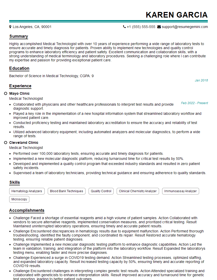
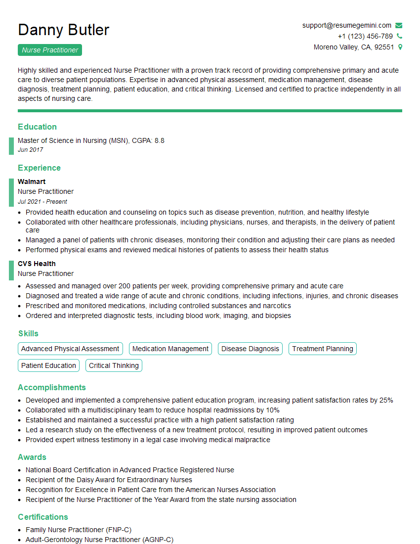
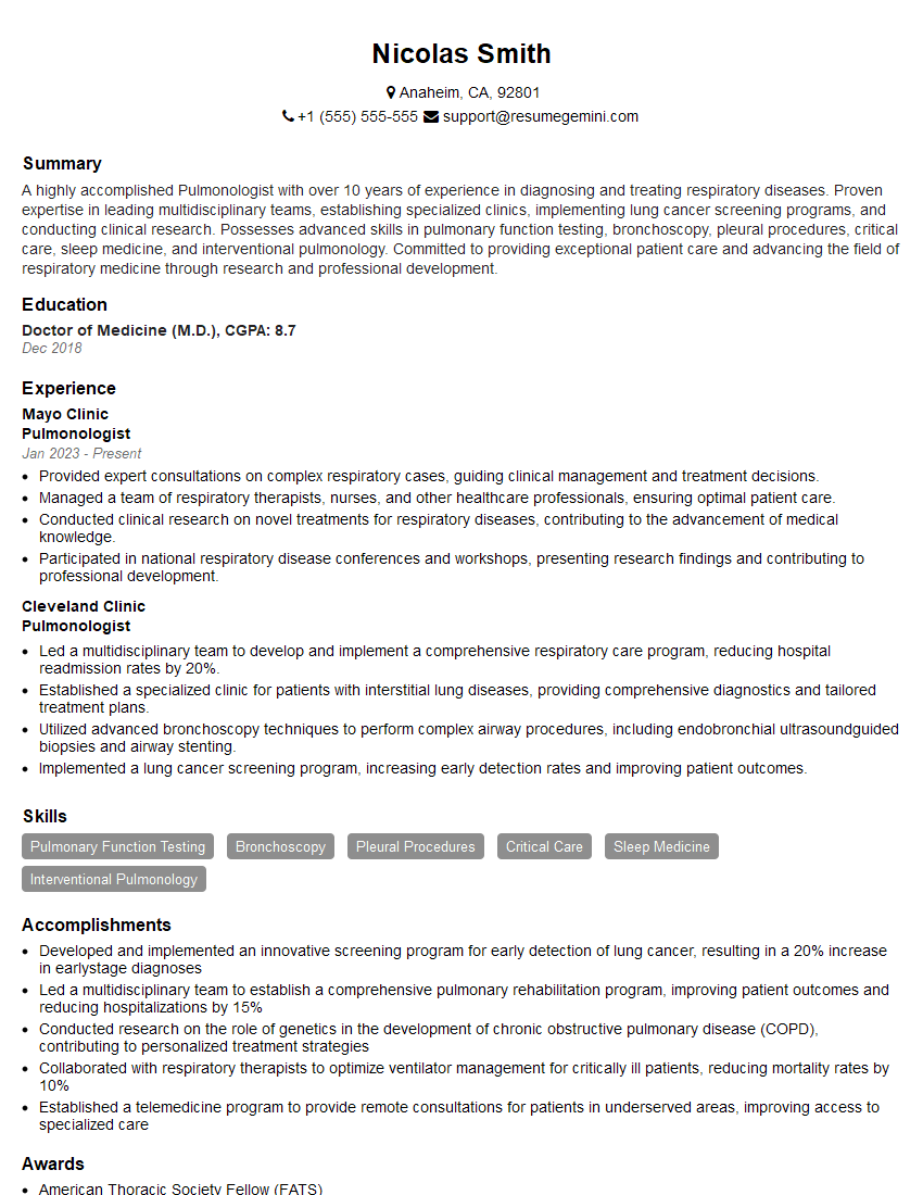
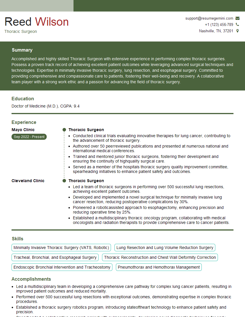
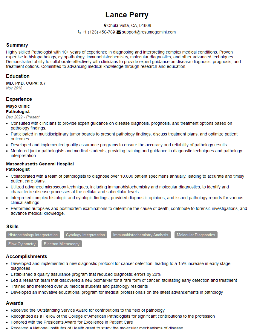
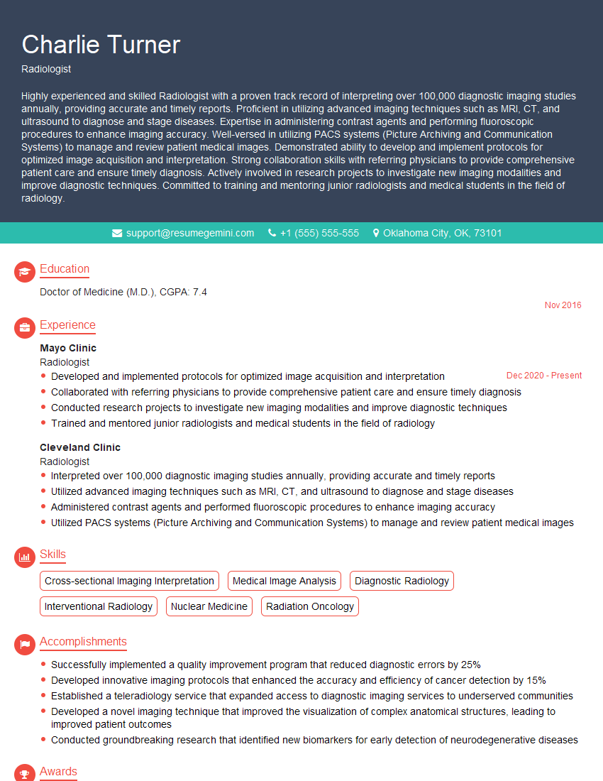
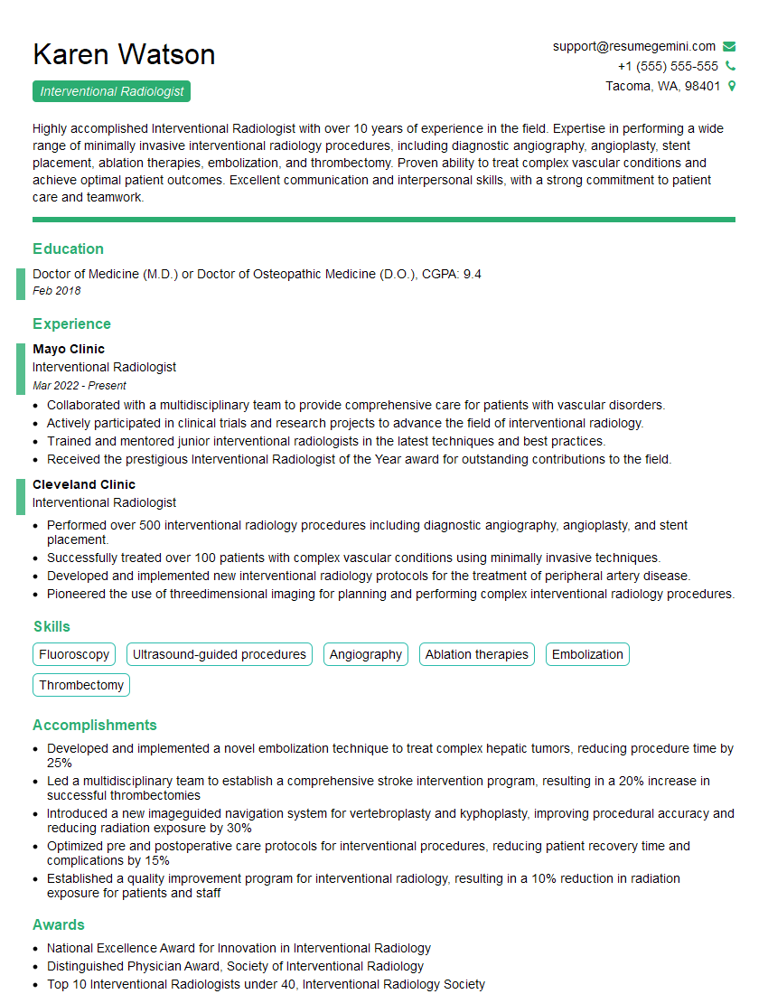
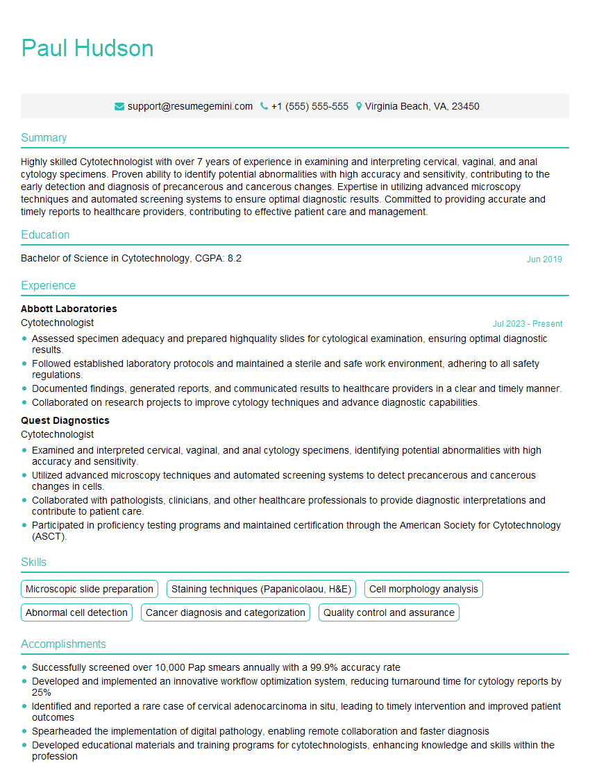
Mastering the intricacies of pleural biopsy significantly enhances your value as a healthcare professional, opening doors to specialized roles and career advancement. To maximize your job prospects, crafting a compelling and ATS-friendly resume is crucial. ResumeGemini is a trusted resource to help you build a professional resume that highlights your skills and experience effectively. Examples of resumes tailored to pleural biopsy expertise are available to guide you, ensuring your application stands out.
Explore more articles
Users Rating of Our Blogs
Share Your Experience
We value your feedback! Please rate our content and share your thoughts (optional).
What Readers Say About Our Blog
Hi, I have something for you and recorded a quick Loom video to show the kind of value I can bring to you.
Even if we don’t work together, I’m confident you’ll take away something valuable and learn a few new ideas.
Here’s the link: https://bit.ly/loom-video-daniel
Would love your thoughts after watching!
– Daniel
This was kind of a unique content I found around the specialized skills. Very helpful questions and good detailed answers.
Very Helpful blog, thank you Interviewgemini team.
