Preparation is the key to success in any interview. In this post, we’ll explore crucial Electromyography and Nerve Conduction Studies interview questions and equip you with strategies to craft impactful answers. Whether you’re a beginner or a pro, these tips will elevate your preparation.
Questions Asked in Electromyography and Nerve Conduction Studies Interview
Q 1. Explain the difference between sensory and motor nerve conduction studies.
Sensory nerve conduction studies (NCS) assess the function of sensory nerves, while motor NCS evaluate the function of motor nerves. Think of it like this: sensory nerves are responsible for feeling (touch, temperature, pain), while motor nerves control movement. Sensory NCS measure how quickly a sensory nerve impulse travels from a stimulated point to a recording point. This is done by stimulating a sensory nerve at a specific location and recording the response at another location. The speed of the signal is the nerve conduction velocity (NCV). Motor NCS, on the other hand, stimulate a motor nerve and record the resulting muscle response (compound muscle action potential or CMAP). Again, the speed of the signal reflects the NCV. In essence, sensory NCS tell us how well you feel, while motor NCS tell us how well you move.
For example, in a sensory study we might stimulate the median nerve at the wrist and record the response at the elbow. In a motor study, we might stimulate the median nerve at the wrist and record the CMAP from the abductor pollicis brevis muscle in the thumb.
Q 2. Describe the procedure for performing a needle EMG.
A needle EMG involves inserting a fine needle electrode into a muscle to record the electrical activity of individual muscle fibers. It’s a bit like using a tiny microphone to listen to the whispers of your muscles. First, the skin is cleaned and disinfected. Then, using a sterile needle electrode, we carefully insert it into the target muscle. This can sometimes cause a slight pinch or discomfort. Once the needle is in place, we record the electrical activity, both spontaneously and during voluntary muscle contractions. We listen for spontaneous activity, such as fibrillation potentials or positive sharp waves, indicative of muscle fiber damage. During contraction, we analyze the motor unit action potentials (MUAPs) to assess the health of the motor neurons supplying the muscle. The size, shape, and duration of the MUAPs provide valuable information about the underlying pathology.
The procedure is usually well-tolerated but can be slightly uncomfortable. The patient’s comfort is paramount, and appropriate measures are taken to minimize discomfort.
Q 3. What are the common artifacts encountered during EMG/NCS studies and how are they addressed?
Several artifacts can interfere with EMG/NCS studies, leading to inaccurate results. Common artifacts include movement artifacts (from patient movement), electrical interference (from nearby electrical equipment), and electrode problems (poor contact or displacement). Movement artifacts appear as erratic, high-amplitude signals obscuring the true nerve signal. Electrical interference creates a 60Hz hum or other regular waveforms. Electrode problems result in inconsistent or absent signals.
Addressing these artifacts requires a systematic approach. Patient education and relaxation techniques can minimize movement. Proper grounding and shielding of equipment reduce electrical interference. Careful electrode placement and use of conductive gel ensure optimal signal quality. If the artifact is significant and cannot be corrected, the study might need to be repeated.
Q 4. Interpret a simple nerve conduction study tracing, identifying abnormalities.
Interpreting a nerve conduction study tracing requires careful analysis of several parameters, including amplitude, latency, and velocity. Let’s say we’re looking at a median nerve motor study. A normal tracing would show a CMAP of appropriate amplitude, a short distal latency (time taken for the signal to travel from stimulation to recording site), and a normal NCV. Abnormalities could include a reduced CMAP amplitude (suggesting axonal loss), prolonged distal latency (suggesting slowing of conduction, possibly demyelination), or a reduced NCV (indicating demyelination or axonal damage). A complete absence of a CMAP indicates a complete nerve lesion.
For example, if the CMAP amplitude is significantly reduced, it suggests a loss of motor axons, possibly due to axonal neuropathy. If the distal latency is prolonged but the amplitude is relatively normal, it suggests a demyelinating process, slowing down nerve conduction without significant axonal loss. This could be indicative of Guillain-Barré syndrome, for example.
Q 5. Explain the significance of different waveforms in EMG.
Different waveforms in EMG reflect different physiological states of the muscle and its innervation. Normal motor unit action potentials (MUAPs) are of a relatively consistent shape and size during voluntary contractions, reflecting healthy muscle fibers and motor neurons. Abnormal waveforms, however, point towards underlying pathologies.
- Fibrillation potentials: Spontaneous activity of single muscle fibers, indicative of muscle denervation.
- Positive sharp waves: Similar to fibrillation potentials but with a longer duration, also indicative of denervation.
- Complex repetitive discharges: A series of repetitive discharges from a single muscle fiber, often seen in myopathies.
- Giant MUAPs: Large amplitude MUAPs suggesting reinnervation following denervation.
The presence and type of abnormal waveforms help us differentiate between different types of neuromuscular disorders, such as myopathies (primary muscle diseases) and neuropathies (diseases of nerves).
Q 6. How do you differentiate between axonal and demyelinating neuropathies?
Differentiating between axonal and demyelinating neuropathies relies on careful analysis of NCS findings, specifically the NCV and CMAP amplitude. In axonal neuropathies, there is primary damage to the axons themselves, leading to a loss of nerve fibers. This results in a reduced CMAP amplitude, reflecting the reduced number of functioning axons, while the NCV may be relatively normal or only mildly reduced. Think of it like cutting wires in a cable – you lose the signal strength but the speed of the signal in the remaining wires might not change much.
In demyelinating neuropathies, the myelin sheath surrounding the axons is damaged, slowing down nerve conduction. This leads to a prolonged distal latency and reduced NCV, while the CMAP amplitude may be relatively preserved initially. This is analogous to damaging the insulation around the wires – the signal strength might be okay, but the signal speed will be greatly reduced.
A combination of clinical presentation and NCS findings is essential for accurate diagnosis. For instance, Guillain-Barré syndrome is a demyelinating neuropathy, whereas chronic inflammatory demyelinating polyneuropathy (CIDP) is also a demyelinating condition but with a more chronic and progressive course.
Q 7. Describe the process of calculating nerve conduction velocities.
Nerve conduction velocity (NCV) is calculated by measuring the distance between the stimulation and recording sites and dividing it by the time it takes for the nerve impulse to travel that distance (latency). For example, if the distance between two stimulation sites is 10cm and the time taken for the impulse to travel between these sites is 5ms, the NCV is calculated as 10cm/5ms = 20cm/ms or 20 m/s.
In practice, we measure the distal latency and then the latency at a more proximal stimulation site. The difference between these two latencies is used to calculate the conduction velocity over a defined distance. This is a more accurate method than simply using the distal latency because it minimizes the impact of variations in the conduction at the neuromuscular junction. Different nerves have different expected ranges for normal NCVs; these values can be found in the literature and are used to compare patient findings.
Q 8. Explain the clinical significance of F-waves and H-reflexes.
F-waves and H-reflexes are specialized nerve conduction studies that provide valuable information about the function of the motor axons and the neuromuscular junction. They’re like ‘echoes’ of electrical signals, giving us insights into the health of the entire pathway.
F-waves: These reflect the antidromic (backward) conduction of a nerve impulse along motor axons. Imagine sending a signal down a wire; the F-wave is the small signal that bounces back from the nerve root. Prolonged F-wave latency suggests a problem along the length of the nerve, potentially due to demyelination or axon loss. Increased F-wave latency can indicate a peripheral neuropathy.
H-reflexes: These are monosynaptic reflexes similar to the familiar knee-jerk reflex, but involving sensory and motor fibers of a single nerve. They assess the integrity of the sensory and motor components of the reflex arc. Increased latency or absent H-reflexes may indicate radiculopathy (nerve root compression) or other conditions affecting the spinal cord.
Clinical Significance: Both F-waves and H-reflexes are particularly useful in evaluating distal nerve latencies and for detecting early subclinical abnormalities in conditions like diabetic neuropathy, where standard nerve conduction studies might be initially normal. They help differentiate between axonal and demyelinating neuropathies. For example, in a demyelinating neuropathy, F-wave latency will be prolonged, reflecting the slowed conduction velocity.
Q 9. What are the contraindications for performing EMG/NCS?
Contraindications for EMG/NCS are situations where the procedure poses a significant risk to the patient or where the results may be unreliable or misleading. These include:
- Uncontrolled bleeding disorders: Needle insertion carries a risk of bleeding, which could be dangerous for patients with uncontrolled bleeding problems.
- Infection at the insertion site: Inserting needles into an infected area could spread the infection.
- Skin diseases or severe skin lesions: Intact skin is essential; severe skin conditions could compromise the integrity of the skin and introduce risk of infection.
- Severe cardiac arrhythmias (in some cases): Patients with certain severe cardiac conditions might experience complications from the electrical stimulation involved in some aspects of the studies.
- Pacemakers or other implanted devices (certain types): The electrical stimulation can interfere with the function of some devices.
- Patient inability to cooperate: Reliable results depend on the patient’s ability to relax and follow instructions.
It is crucial to carefully assess the patient’s overall health and any potential risks before performing EMG/NCS. A thorough review of the patient’s medical history is essential in determining whether a procedure is contraindicated.
Q 10. How would you approach the investigation of suspected carpal tunnel syndrome using EMG/NCS?
Investigating suspected carpal tunnel syndrome (CTS) with EMG/NCS involves a structured approach focused on assessing the median nerve at the wrist. Here’s the typical strategy:
- Sensory Nerve Conduction Studies (NCS): Assess median, ulnar, and radial nerves across the wrist and forearm. We look for slowing of conduction velocity across the carpal tunnel (median nerve) compared to the other nerves and the other segments of the median nerve, indicating compression within the tunnel. This provides objective evidence of nerve damage.
- Motor Nerve Conduction Studies (NCS): Measure conduction velocity of the median nerve’s motor fibers. We look for evidence of prolonged distal motor latency from the median nerve motor branch. This is another objective measure of slowing conduction in the median nerve.
- EMG: This is performed on the abductor pollicis brevis (APB) muscle, which is innervated by the median nerve. We search for spontaneous activity such as fibrillation potentials or positive sharp waves. These suggest denervation (damage to the nerve’s connection to the muscle), supporting the diagnosis of CTS. Reduced recruitment (meaning fewer motor units are activated even with maximal effort) can indicate chronic denervation and muscle atrophy.
Interpreting Results: The combination of slowed median nerve conduction across the wrist, prolonged distal motor latency, and abnormal EMG findings in the APB muscle strongly supports the diagnosis of CTS. Abnormal findings in other nerves may point to other possible diagnoses, such as cubital tunnel syndrome. It is important to analyze the pattern of abnormality in order to differentiate between various neuropathies.
Q 11. Differentiate between myopathy and neuropathy based on EMG/NCS findings.
EMG/NCS findings can effectively differentiate between myopathy (muscle disease) and neuropathy (nerve disease). Think of it like this: myopathy affects the ‘engine’ (muscle), while neuropathy affects the ‘wiring’ (nerve).
Myopathy: EMG shows short-duration, low-amplitude motor unit potentials (MUAPs) with early recruitment. This signifies the involvement of individual muscle fibers and indicates that only a few fibers are responding to the signal from the nerve. This is because the muscle fibers themselves are damaged. Nerve conduction studies are usually normal in myopathy because the nerves are not affected. Examples include muscular dystrophies and inflammatory myopathies.
Neuropathy: EMG demonstrates decreased recruitment with increased amplitude and duration of MUAPs, reflecting denervation. The pattern often looks like big MUAPs. NCS shows slowed or absent conduction velocities or amplitudes, with prolongation of distal latencies, indicating nerve damage that disrupts the transmission of nerve impulses to the muscles. Examples include diabetic neuropathy, Guillain-Barré syndrome, and Charcot-Marie-Tooth disease.
Important Note: Some conditions can present with features of both myopathy and neuropathy (e.g., certain forms of inclusion body myositis). A complete clinical picture, including the patient’s history, physical examination findings, and other laboratory investigations, is essential for accurate diagnosis.
Q 12. Explain the role of EMG/NCS in the diagnosis of amyotrophic lateral sclerosis (ALS).
EMG/NCS plays a crucial role in the diagnosis of amyotrophic lateral sclerosis (ALS), a progressive neurodegenerative disease affecting both upper and lower motor neurons. It is important to understand that EMG/NCS is not diagnostic on its own but can support clinical diagnosis.
EMG findings in ALS: Show evidence of both upper and lower motor neuron involvement. Lower motor neuron involvement is indicated by fibrillation potentials and positive sharp waves (evidence of denervation in the muscles). Upper motor neuron involvement is shown by reduced recruitment of motor units with increased amplitude and duration (consistently large MUAPs). This combination is crucial in supporting the diagnosis of ALS. The pattern of involvement can help localize the affected area in the motor neuron pathway. The combination of the above and a clinical picture supporting ALS assists in diagnosis.
NCS findings in ALS: are usually normal in the early stages. NCS might be helpful in ruling out other conditions that can mimic ALS.
Diagnostic Significance: The characteristic EMG findings of both upper and lower motor neuron involvement significantly support the diagnosis of ALS, especially when consistent with the patient’s clinical presentation and other investigations. It’s important to note that other conditions can mimic ALS, so a comprehensive evaluation with neurological examination and other studies, such as MRI of the brain and spinal cord, is needed.
Q 13. Describe the procedure for performing a repetitive nerve stimulation study.
Repetitive nerve stimulation (RNS) studies are used to assess neuromuscular transmission. This involves repeatedly stimulating a peripheral nerve at a low frequency (typically 2–3 Hz) and observing the response (compound muscle action potential, CMAP) over time. This method is particularly useful for diagnosing neuromuscular junction disorders such as myasthenia gravis (MG).
Procedure:
- Surface electrodes: are placed over the nerve and the muscle being studied.
- Stimulation: A stimulator delivers repeated electrical pulses to the nerve at a low frequency.
- Recording: The CMAP is recorded from the muscle using surface electromyography (EMG). Each response is recorded.
- Analysis: The amplitude of the CMAP is measured in each response. In normal individuals, the CMAP amplitude remains relatively constant over time. However, in conditions like MG, the amplitude of the CMAP will progressively decrease with repeated stimulation. This is known as decremental response. A high-frequency RNS (50 Hz) will also result in different patterns.
Clinical Significance: A decremental response (a significant decrease in CMAP amplitude with repetitive stimulation) is strongly suggestive of a neuromuscular junction disorder like MG. Other conditions that can cause decremental responses include botulism and Lambert-Eaton myasthenic syndrome (LEMS). However, in LEMS, the CMAP amplitude is usually increased by high-frequency stimulation, whereas it stays the same or decreases in MG. Therefore, RNS with low and high frequency stimulations are needed for appropriate diagnosis.
Q 14. What are the limitations of EMG/NCS?
While EMG/NCS is a powerful diagnostic tool, it does have limitations:
- Limited sensitivity and specificity: EMG/NCS may not detect subtle nerve or muscle abnormalities, particularly in early stages of disease. It may also show non-specific abnormalities, making it difficult to pinpoint the exact cause of the problem. For example, it may detect nerve damage in the hand but does not provide information about the cause.
- Subjectivity in interpretation: Interpretation of EMG findings requires significant expertise and experience. There is some subjectivity involved, and different clinicians may interpret the same findings slightly differently. A consensus is not always possible.
- Invasive procedure: Needle EMG is invasive, causing discomfort to some patients. It has potential risks, particularly in patients with bleeding disorders or infection at the site.
- Technical limitations: Factors like electrode placement and patient cooperation can affect the quality and reliability of the results. Proper techniques are necessary to obtain reliable results.
- Does not always confirm clinical findings: While helpful, EMG/NCS results are just a part of the whole clinical picture. This is particularly true for patients presenting with symptoms that cannot be explained by the NCS findings. This means there could be other disorders leading to the patient’s symptoms.
It’s crucial to consider the limitations and interpret EMG/NCS results in the context of the patient’s clinical presentation, medical history, and other diagnostic findings.
Q 15. How do you ensure patient safety during EMG/NCS procedures?
Patient safety is paramount during EMG/NCS procedures. We begin by obtaining informed consent, ensuring the patient understands the procedure, potential risks (such as bleeding or infection at the needle insertion site), and benefits. We meticulously check the patient’s medical history, paying close attention to allergies, bleeding disorders, and the use of anticoagulants, which could increase bleeding risk. Proper skin preparation with antiseptic solutions is crucial to minimize infection. Throughout the procedure, we continuously monitor the patient’s comfort level and vital signs, adjusting the procedure as needed to alleviate discomfort. We use sterile, single-use needles and maintain strict aseptic techniques. Post-procedure, we provide clear instructions regarding wound care and potential complications, advising the patient to contact us if they experience any significant discomfort or signs of infection.
For example, if a patient reports significant pain during needle insertion, we may alter our technique or temporarily pause the procedure. If a patient has a known bleeding disorder, we may consult with their hematologist and consider modifying the procedure or using alternative methods.
Career Expert Tips:
- Ace those interviews! Prepare effectively by reviewing the Top 50 Most Common Interview Questions on ResumeGemini.
- Navigate your job search with confidence! Explore a wide range of Career Tips on ResumeGemini. Learn about common challenges and recommendations to overcome them.
- Craft the perfect resume! Master the Art of Resume Writing with ResumeGemini’s guide. Showcase your unique qualifications and achievements effectively.
- Don’t miss out on holiday savings! Build your dream resume with ResumeGemini’s ATS optimized templates.
Q 16. Explain the significance of different needle insertion sites for EMG.
Needle insertion sites for EMG are strategically chosen to target specific muscles innervated by the nerves of interest. The choice of site helps us isolate the activity of individual muscles and assess different nerve territories. For example, if we suspect a problem with the median nerve at the wrist, we might sample muscles in the thenar eminence (like the abductor pollicis brevis) to assess its motor unit function. Similarly, to evaluate the radial nerve, we might examine muscles in the posterior forearm. In cases of suspected radiculopathy, we may sample muscles innervated by multiple roots to determine the level and extent of nerve involvement. Precise needle placement is crucial; inaccurate placement can lead to misinterpretation of results.
For instance, sampling a muscle too close to the nerve trunk may lead to recording of nerve fascicle activity rather than isolated muscle fiber activity, thus compromising the accuracy of the evaluation.
Q 17. Describe the interpretation of fibrillation potentials and positive sharp waves.
Fibrillation potentials and positive sharp waves are spontaneous electrical activities recorded in EMG that indicate denervation of muscle fibers. They are characteristic of acute or subacute muscle fiber damage, often reflecting lower motor neuron pathology. Fibrillation potentials are brief, repetitive, and relatively low-amplitude discharges arising from individual muscle fibers. Positive sharp waves are slightly longer, higher-amplitude potentials. Their presence indicates significant muscle fiber membrane instability. The amplitude and frequency of these potentials help us gauge the severity and duration of the denervation. Seeing these potentials in a resting muscle points towards a process like nerve damage or motor neuron disease.
Think of it like this: if a muscle loses its connection to the nerve, the muscle fibers start to ‘misfire’ randomly, producing these abnormal electrical signals.
Q 18. How do you interpret fasciculations in EMG?
Fasciculations are spontaneous contractions of motor units visible as brief, visible twitches under the skin. In EMG, they appear as large-amplitude potentials reflecting the synchronous discharge of many muscle fibers within a single motor unit. Fasciculations can be benign or indicative of underlying neurological disease. Benign fasciculations are often asynchronous, small in amplitude, and present infrequently. In contrast, widespread, frequent, and large-amplitude fasciculations often suggest underlying neuropathology, like amyotrophic lateral sclerosis (ALS), or conditions affecting anterior horn cells.
The context is key. A few fasciculations in a healthy individual might be insignificant, while their presence in a patient complaining of progressive weakness warrants careful consideration and additional investigations.
Q 19. What are the common causes of polyneuropathy?
Polyneuropathy, affecting multiple peripheral nerves, has diverse causes. The most common include: 1) Diabetes mellitus: High blood glucose levels damage the walls of small blood vessels supplying nerves, leading to nerve dysfunction. 2) Autoimmune diseases: Conditions like Guillain-Barré syndrome and rheumatoid arthritis can lead to immune-mediated nerve damage. 3) Toxins: Exposure to heavy metals (like lead or mercury), certain medications, or alcohol can cause peripheral neuropathy. 4) Genetic disorders: Inherited metabolic disorders or genetic mutations can impair nerve function. 5) Infections: Viral or bacterial infections can damage nerves directly or indirectly. 6) Nutritional deficiencies: Deficiencies of vitamin B12 or folate can lead to nerve damage.
A proper diagnosis often requires a comprehensive evaluation considering the patient’s medical history, physical examination, and specialized investigations like EMG/NCS to differentiate between various types of polyneuropathies.
Q 20. Describe the role of EMG/NCS in the evaluation of radiculopathies.
EMG/NCS plays a crucial role in evaluating radiculopathies (nerve root compression). Nerve conduction studies assess the function of the nerve roots and peripheral nerves. Slowed nerve conduction velocities across a particular nerve root distribution can pinpoint the site of compression. EMG helps evaluate the presence of denervation in the muscles innervated by the affected nerve root, indicated by fibrillation potentials, positive sharp waves, and reduced recruitment. The combination of NCS and EMG provides a comprehensive picture of the extent and severity of nerve root involvement. For example, NCS might show slowed conduction across the C6 nerve root, while EMG might show denervation changes in the biceps brachii muscle, confirming a C6 radiculopathy.
This approach allows for a more precise diagnosis and guides appropriate treatment strategies.
Q 21. How do you differentiate between upper and lower motor neuron lesions using EMG/NCS?
EMG/NCS is highly effective in differentiating upper and lower motor neuron lesions. Lower motor neuron lesions (LMNLs), affecting the anterior horn cells or peripheral nerves, cause denervation of the affected muscles. In EMG, this manifests as fibrillation potentials, positive sharp waves, reduced recruitment, and decreased motor unit potential amplitude. NCS may show slowed conduction velocities or conduction blocks. In contrast, upper motor neuron lesions (UMNLS), affecting the corticospinal tract, lead to changes in muscle tone, reflexes, and patterns of muscle activity. EMG in UMN lesions will show increased recruitment, increased motor unit potential amplitude, and presence of neurogenic changes like fasciculations, but absence of denervation potentials such as fibrillation potentials and positive sharp waves that are seen in LMNL.
Imagine it like this: a LMNL is like cutting a wire (peripheral nerve), while a UMN lesion is like damaging the switch (cortical control) that controls the wire; the wire itself might be intact, but the signal is disrupted.
Q 22. What are the ethical considerations involved in performing EMG/NCS?
Ethical considerations in EMG/NCS are paramount. Patient autonomy is central; informed consent, ensuring the patient understands the procedure, risks, and benefits, is non-negotiable. We must maintain patient confidentiality, protecting their medical information according to HIPAA guidelines or equivalent regulations. Accurate record-keeping is crucial for both legal and medical reasons. We must also be mindful of potential biases in interpretation and ensure our clinical judgment isn’t clouded by personal feelings or external pressures. For instance, if a patient is anxious, we must take extra time to explain the procedure and alleviate their concerns before proceeding. Finally, we must always strive to provide the highest quality care, considering the patient’s overall well-being and choosing the least invasive approach whenever possible.
Q 23. Discuss the maintenance and calibration of EMG/NCS equipment.
Maintaining and calibrating EMG/NCS equipment is critical for accurate results. This involves regular checks of electrode impedance, ensuring they’re within acceptable ranges (usually below 5 kΩ) for optimal signal transmission. We perform daily checks on the stimulator output, verifying its amplitude and waveform accuracy using a precision oscilloscope. The EMG amplifier requires regular checks for noise levels and gain settings. We use calibration signals, often provided with the equipment, to verify the system’s accuracy. These signals mimic specific muscle responses, allowing us to fine-tune the amplification and filtering parameters. Regular preventive maintenance, including cleaning and inspecting the equipment, helps to prevent malfunctions and extend its lifespan. Documentation of all calibration procedures and maintenance is crucial for quality assurance and troubleshooting.
For instance, a drift in the amplifier’s baseline could lead to misinterpretation of the signal. Regular calibration ensures we detect and correct such issues before they impact patient diagnosis. Our lab adheres to a strict schedule, typically involving daily checks and more thorough calibrations every 3-6 months, or as per manufacturer recommendations.
Q 24. How do you manage a patient with a difficult needle insertion for EMG?
Managing difficult needle insertions is a common challenge. First, we always utilize surface anatomy landmarks and palpate the target muscle carefully. If initial attempts are unsuccessful, we use different needle approaches, sometimes adjusting the needle angle slightly. We use high-quality, fine-gauge concentric needles, minimizing patient discomfort. If the patient experiences significant pain or resistance, we may employ techniques like using ultrasound guidance to visualize the muscle and the needle’s trajectory. This allows us to accurately target the muscle and avoid nerves or blood vessels. Appropriate patient positioning and relaxation techniques are essential; we encourage deep breathing and distraction techniques. Topical anesthetic creams or local nerve blocks might be necessary in particularly challenging cases to reduce pain. We communicate constantly with the patient, explaining each step and assuring them throughout the procedure. If the insertion proves exceptionally difficult, it is acceptable to stop and reschedule the procedure, prioritizing patient well-being. In a case with severe fibrosis, for instance, ultrasound guidance became critical for successful needle placement and data acquisition.
Q 25. Describe your experience with specific EMG/NCS software and its applications.
I have extensive experience using several EMG/NCS software packages, including commercially available packages such as [mention specific software e.g., NCS Master, Keypoint). These packages provide automated analysis capabilities, measuring parameters such as nerve conduction velocities (NCVs), latencies, and amplitudes. They help us create reports with graphical representations of the data, including electromyograms and nerve conduction study results. The software also facilitates comparison to normative data and aids in differential diagnosis. I’m proficient in utilizing the software’s features to assess various neuropathies, myopathies, and radiculopathies. For instance, I’ve used the automated analysis tools to objectively measure the F-wave latencies in patients suspected of having carpal tunnel syndrome, enhancing the diagnostic precision.
Q 26. Explain the role of EMG/NCS in pre-surgical planning.
EMG/NCS plays a vital role in pre-surgical planning, particularly in surgeries involving nerves or muscles. For instance, before carpal tunnel release surgery, EMG/NCS helps confirm the diagnosis of carpal tunnel syndrome, ruling out other potential causes of symptoms. It helps evaluate the severity of nerve compression and identify the specific nerve involved. Similarly, before spinal surgery, EMG/NCS can localize the level of nerve root compression and guide surgical decisions about decompression procedures. In peripheral nerve repair surgeries, EMG/NCS studies are essential to assess the extent and nature of the nerve injury, guiding the surgeon in their repair strategy and also providing post-operative monitoring. By accurately identifying the affected nerves and muscles, EMG/NCS ensures a targeted and successful surgical outcome.
Q 27. Discuss the potential risks and complications of EMG/NCS procedures.
While generally safe, EMG/NCS carries potential risks and complications. The most common is pain at the needle insertion site; this is usually mild and transient. There’s a small risk of bleeding or hematoma formation, especially in patients on anticoagulants. Rare but serious complications include nerve damage from needle insertion (though exceptionally rare with proper technique and skill), infection at the insertion site, and syncope (fainting) from anxiety or pain. We mitigate these risks through proper sterile technique, informed consent, and appropriate patient selection. For example, we may avoid the procedure in patients with bleeding disorders unless absolutely necessary. We always explain these risks to patients before obtaining their consent and monitor patients closely for any signs of complications post-procedure. Proper patient education and communication significantly reduce these risks.
Q 28. How do you interpret a normal EMG/NCS study?
Interpreting a normal EMG/NCS study involves several key findings. Nerve conduction studies should show normal nerve conduction velocities (NCVs), latencies, and amplitudes within the normal range for the patient’s age and body size. The amplitudes should be symmetrical bilaterally, and distal latencies should be similar on both sides. Motor and sensory nerve action potentials should have normal shapes. In electromyography, the resting muscle activity should be silent (no spontaneous activity); voluntary activation should elicit normal motor unit action potentials (MUAPs) with appropriate recruitment pattern and morphology. Absence of fibrillations, positive sharp waves, or fasciculations indicates a lack of muscle denervation or damage. Essentially, a normal study indicates that the nerves and muscles are functioning within their expected physiological range. Any deviation from these expected findings would necessitate further evaluation and exploration of a potential underlying pathology.
Key Topics to Learn for Electromyography and Nerve Conduction Studies Interview
- Electrophysiology Basics: Understanding the principles of nerve conduction and muscle fiber depolarization. This includes action potentials, membrane potentials, and neuromuscular transmission.
- EMG Technique: Mastering the practical aspects of EMG needle insertion, signal acquisition, and artifact identification. Consider different needle types and their applications.
- NCS Technique: Familiarize yourself with various NCS modalities (e.g., motor, sensory, F-wave, H-reflex studies) and their clinical interpretations.
- Interpretation of EMG/NCS Findings: Develop your ability to analyze waveforms, identify patterns indicative of various neuropathies and myopathies (e.g., axonal loss, demyelination, myotonic discharges).
- Clinical Correlations: Understand how EMG/NCS results relate to specific clinical presentations of neurological and neuromuscular disorders. Practice connecting findings to patient symptoms.
- Common Neurological and Neuromuscular Diseases: Gain a strong understanding of the EMG/NCS characteristics of common conditions like carpal tunnel syndrome, Guillain-Barré syndrome, amyotrophic lateral sclerosis (ALS), and muscular dystrophies.
- Troubleshooting and Artifact Recognition: Learn to identify and mitigate common artifacts during EMG/NCS studies and how to troubleshoot equipment malfunctions.
- Safety Precautions and Patient Care: Review relevant safety protocols and best practices for patient comfort and minimizing risks during the procedure.
- Ethical Considerations: Be prepared to discuss ethical aspects of patient care and data interpretation within the context of EMG/NCS studies.
Next Steps
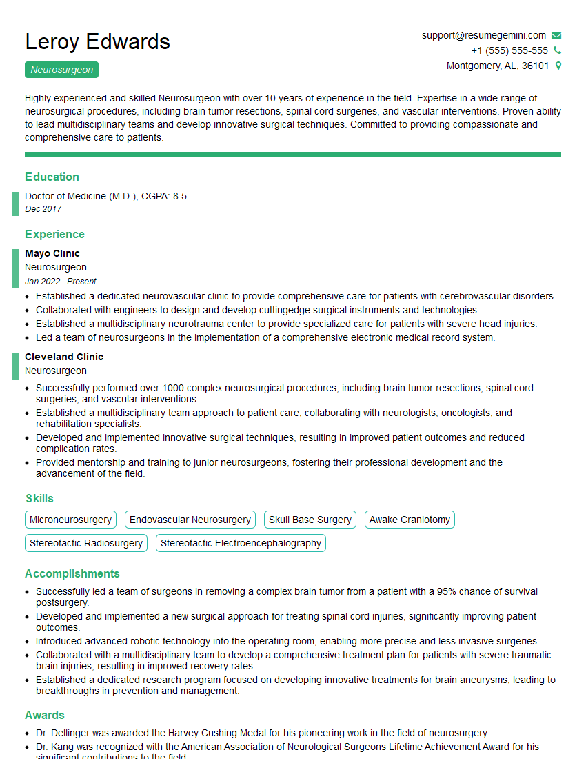
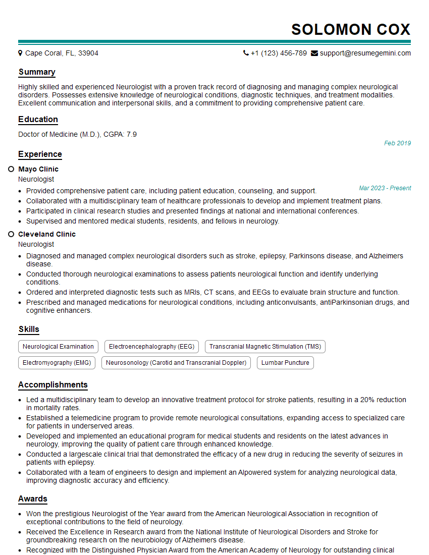
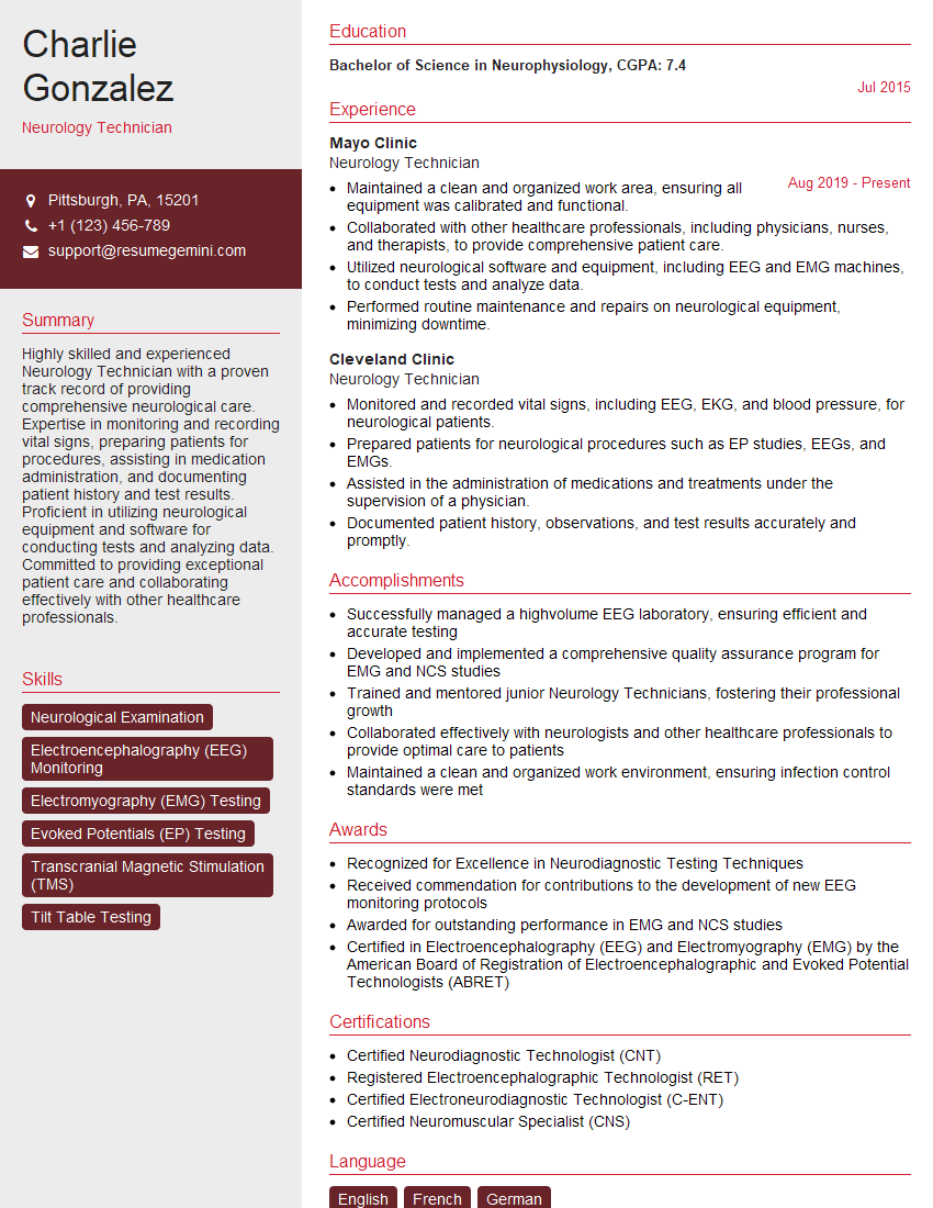
Mastering Electromyography and Nerve Conduction Studies is crucial for career advancement in neurology, physiatry, and other related fields. A strong understanding of these techniques demonstrates expertise and opens doors to exciting opportunities. To maximize your job prospects, focus on building a compelling and ATS-friendly resume that highlights your skills and experience. ResumeGemini is a trusted resource to help you create a professional and effective resume. They offer examples of resumes tailored to Electromyography and Nerve Conduction Studies to help you showcase your qualifications effectively.
Explore more articles
Users Rating of Our Blogs
Share Your Experience
We value your feedback! Please rate our content and share your thoughts (optional).
What Readers Say About Our Blog
To the interviewgemini.com Webmaster.
Very helpful and content specific questions to help prepare me for my interview!
Thank you
To the interviewgemini.com Webmaster.
This was kind of a unique content I found around the specialized skills. Very helpful questions and good detailed answers.
Very Helpful blog, thank you Interviewgemini team.