Preparation is the key to success in any interview. In this post, we’ll explore crucial Diep Flap Surgery interview questions and equip you with strategies to craft impactful answers. Whether you’re a beginner or a pro, these tips will elevate your preparation.
Questions Asked in Diep Flap Surgery Interview
Q 1. Describe the surgical anatomy relevant to a DIEP flap harvest.
Understanding the surgical anatomy for a DIEP (Deep Inferior Epigastric Perforator) flap harvest is crucial for a successful procedure. It involves a thorough knowledge of the abdominal wall’s layers, the location and course of the deep inferior epigastric vessels (DIEAs), and the perforator vessels supplying the abdominal skin and subcutaneous fat. This includes the rectus abdominis muscle, its fascial sheaths, the underlying peritoneum, and the location and variability of the DIEA and its perforating branches.
The DIEA arises from the external iliac artery and travels medially along the inferior border of the rectus abdominis muscle. The perforating branches are key; they are the vessels that nourish the abdominal skin and fat we will use to create the flap. These perforators vary significantly in number, caliber, and location between individuals. This anatomical variability necessitates meticulous preoperative planning, often using advanced imaging techniques like CT angiography or Doppler ultrasound.
Understanding the interconnections with adjacent vasculature, such as the superior epigastric artery, is also vital to avoid inadvertent injury and ensure adequate flap perfusion. The surgical dissection must delicately isolate these vessels and their accompanying veins to minimize damage and maximize the chances of successful flap transfer.
Q 2. Explain the steps involved in planning a DIEP flap procedure.
Planning a DIEP flap procedure is a multi-stage process requiring meticulous attention to detail and patient-specific considerations. It begins with a thorough patient assessment, including evaluation of the recipient site and assessment of the abdominal wall’s suitability for flap harvest. This includes checking the patient’s body mass index (BMI), assessing the quality and thickness of the abdominal skin and fat, and evaluating the presence of prior abdominal surgeries or scars.
- Preoperative Imaging: CT angiography or Doppler ultrasound is essential to map the DIEA and its perforators. This allows for precise planning of the incision and dissection. We carefully analyze the location and caliber of the perforators to select those most suitable for the flap’s vascular pedicle.
- Recipient Site Assessment: The reconstructive need is thoroughly assessed. The size, shape, and vascularity of the recipient site are crucial factors in determining the dimensions and design of the DIEP flap. This might be a breast reconstruction after mastectomy, or reconstruction of other defects.
- Surgical Team Preparation: The surgical team must be proficient in microsurgery. Extensive experience is necessary for anastomosis of the small vessels. Specialized instruments and equipment are necessary, along with a surgical plan detailing the steps of flap elevation, vascular dissection, and microsurgical anastomosis.
- Surgical Simulation: In some cases, surgical simulation tools are used to plan the exact locations of incisions and pedicle dissection.
Careful preoperative planning is paramount to minimize complications and maximize the chances of successful reconstruction. The detailed planning minimizes intraoperative surprises and facilitates efficient surgery.
Q 3. What are the key vascular landmarks for DIEP flap identification?
The key vascular landmarks for DIEP flap identification are primarily the deep inferior epigastric vessels (DIEA) and its perforating branches. Identifying these vessels accurately is critical for successful flap harvest.
- Deep Inferior Epigastric Artery (DIEA): This is the primary landmark. It is located along the inferior border of the rectus abdominis muscle, usually near the arcuate line. Palpating the rectus muscle and tracing it inferiorly helps identify the vessel.
- Deep Inferior Epigastric Vein (DIEV): The DIEV usually runs alongside the DIEA and has a similar course. Identifying this vein helps confirm the location of the artery.
- Perforating Vessels: These are the branches of the DIEA that penetrate the rectus muscle and supply the overlying abdominal skin and fat. These are the vessels we harvest to form the flap’s pedicle. Identification is aided by using a loupe or microscope to observe the characteristic vessels piercing through the rectus muscle.
Careful dissection and preservation of these vessels are crucial to ensure adequate blood supply to the flap. We use Doppler ultrasound intraoperatively to confirm vessel patency and identify suitable perforators.
Q 4. Detail the microsurgical technique used for DIEP flap anastomosis.
The microsurgical technique used for DIEP flap anastomosis involves precise dissection and meticulous vessel handling under magnification using an operating microscope. This is a highly specialized surgical skill.
- Vessel Preparation: Both the donor (DIEP flap) and recipient vessels are carefully dissected and prepared. Any adventitia (outer layer of the vessel) is meticulously removed to allow for optimal apposition of the vessel walls. We use fine micro-instruments like vascular clamps, scissors, and needles.
- Anastomosis: The anastomosis is performed using an end-to-end or end-to-side technique, depending on the recipient vessel’s configuration. We usually employ an 8-0 or 9-0 nylon or polypropylene suture for precise closure. The sutures are placed meticulously to ensure a watertight seal.
- Magnification: The use of an operating microscope provides the necessary magnification to visualize the tiny vessels and perform the delicate anastomosis accurately. It allows for precise suture placement and minimizes the risk of vessel injury.
- Intraoperative Monitoring: We use an intraoperative Doppler to confirm the patency of the anastomosis immediately after the procedure. This ensures adequate blood flow to the flap.
Microsurgical expertise and precision are crucial during this phase. Failure to create a watertight anastomosis can lead to flap necrosis (death of tissue).
Q 5. How do you manage potential complications during a DIEP flap procedure?
Managing potential complications during a DIEP flap procedure requires preparedness and prompt action. Potential complications range from minor bleeding to significant vascular compromise.
- Bleeding Control: Meticulous hemostasis (stopping bleeding) is crucial throughout the procedure. Fine-tipped electrocautery and meticulous suture ligation of bleeders are used.
- Vascular Injury: Should a vessel be inadvertently injured, immediate repair is essential. This may involve direct suture repair or the use of interpositional grafts. Microsurgical expertise is paramount here.
- Flap Ischemia: Monitoring flap perfusion is critical. Any signs of ischemia (lack of blood flow) such as pallor, coolness, or delayed capillary refill necessitate immediate assessment. This might involve revision of the anastomosis or other measures.
- Seromas/Hematoma: These fluid collections are relatively common and are often managed conservatively with close monitoring. Aspiration may be necessary if they are large or symptomatic.
- Infection Prevention: Strict sterile technique is used to minimize infection risk. Prophylactic antibiotics are usually prescribed.
A proactive approach, meticulous technique, and prompt response to any complications are vital for successful outcomes. Experience with various intraoperative challenges is essential.
Q 6. What are the common postoperative complications associated with DIEP flap surgery?
Common postoperative complications associated with DIEP flap surgery can be broadly categorized as vascular, infectious, or related to the donor or recipient site. These complications highlight the complexity of the procedure and require close postoperative monitoring.
- Partial or Complete Flap Necrosis: This is a major complication caused by insufficient blood supply. It can range from minor areas of necrosis to complete loss of the flap, requiring revision surgery or even complete flap failure. Early detection is key.
- Infection: Wound infection is a concern, especially at the donor and recipient sites. Careful wound care, prophylactic antibiotics, and prompt treatment of any signs of infection are essential.
- Seromas/Hematoma: Fluid collections at the surgical sites are relatively frequent and often resolve spontaneously. Aspiration or surgical drainage may be required if symptomatic or large.
- Donor Site Complications: These include hernia formation, abdominal wall weakness, abdominal pain, or unsightly scarring. These are sometimes managed conservatively, and others may require surgical repair.
- Recipient Site Complications: Complications may include poor flap integration, skin flap necrosis, or asymmetry issues requiring revision surgery.
Postoperative care meticulously addresses these potential complications. Patients are monitored closely for any signs of complications, and timely intervention can significantly improve outcomes.
Q 7. Discuss your experience with intraoperative monitoring during DIEP flap surgery.
Intraoperative monitoring during a DIEP flap surgery is crucial for ensuring the success of the procedure. We employ several techniques to assess flap viability and detect potential issues in real-time.
- Doppler Ultrasound: This is used throughout the procedure to assess the patency of the DIEA and its perforators, both before and after anastomosis. It allows for immediate confirmation of blood flow to the flap. It helps us to identify the optimal perforators for the flap pedicle.
- Near-Infrared Spectroscopy (NIRS): This technique allows for assessment of tissue oxygenation. It provides real-time information about flap viability by measuring oxygen saturation in the flap tissue. This allows us to assess the flap’s perfusion in real-time and adjust the procedure accordingly if necessary.
- Visual Inspection: Careful observation of the flap’s color, temperature, and capillary refill is done throughout the procedure. Any signs of ischemia are immediately addressed. Changes in colour and temperature indicate immediate issues with blood supply.
- Fluorescein Dye: The use of fluorescein dye can be helpful in assessing flap perfusion, particularly after anastomosis. We inject the dye intravenously and assess the fluorescence of the flap under ultraviolet light to confirm blood supply to all areas of the flap.
By combining these monitoring techniques, we can detect potential issues early, ensuring timely intervention and maximizing the chances of a successful DIEP flap reconstruction. Continuous monitoring is essential for a safe and successful surgery.
Q 8. Describe your approach to patient selection for DIEP flap breast reconstruction.
Patient selection for DIEP flap breast reconstruction is crucial for optimal outcomes. It’s a multi-faceted process involving a thorough evaluation of the patient’s overall health, body habitus, and specific anatomical features. We begin with a detailed medical history and physical examination, assessing factors such as smoking status (a significant risk factor for complications), comorbidities like diabetes or hypertension, and the patient’s BMI.
Then, we perform a detailed assessment of the abdominal wall. We examine the patient’s abdominal skin and subcutaneous tissue for sufficient quality and quantity. We need enough tissue to create a breast mound of adequate size and projection while maintaining the integrity of the abdominal wall. We utilize imaging techniques, specifically CT scans with 3D reconstruction software, to precisely evaluate the vascular anatomy of the deep inferior epigastric perforators (DIEP) to determine the presence and caliber of suitable perforators. This allows us to predict the likelihood of flap survival. Patients with significant prior abdominal surgeries, previous radiation therapy to the abdomen, or insufficient vascular anatomy are generally not ideal candidates for DIEP flap reconstruction.
Finally, psychological evaluation is critical; the patient needs realistic expectations about the surgery, recovery, and potential complications. A strong support system is beneficial. Ultimately, patient selection is a collaborative decision, balancing the benefits of DIEP flap reconstruction with the individual patient’s risks and suitability.
Q 9. Explain the advantages and disadvantages of DIEP flap compared to other reconstruction options.
DIEP flap breast reconstruction offers several advantages over other techniques, such as implants or latissimus dorsi flaps. A significant advantage is that it utilizes the patient’s own tissue, eliminating the need for implants and reducing the risk of capsular contracture, implant rupture, or other implant-related complications. The resulting breast is more natural in feel and appearance, often better matching the contralateral breast. Furthermore, the DIEP flap provides a better aesthetic result in terms of projection and shape, particularly for larger reconstructions.
However, DIEP flap reconstruction is a major surgical procedure with potential disadvantages. It’s a more complex and technically demanding operation, requiring significant surgical expertise. It carries a longer operative time, increased risk of complications like seroma formation (fluid collection), hematoma (blood collection), and fat necrosis (fat cell death), as well as a small risk of flap failure. The recovery period is also more extensive compared to other methods. There’s also the potential for donor site morbidity, including abdominal wall weakness, numbness, and scarring, although these are generally well-tolerated by most patients.
The decision between DIEP flap and other options depends on individual patient factors like body habitus, medical history, aesthetic goals, and surgical experience. A thorough discussion of the pros and cons is essential for informed consent.
Q 10. How do you counsel patients regarding the risks and benefits of a DIEP flap procedure?
Counseling patients about DIEP flap reconstruction involves a comprehensive and empathetic approach. We begin by clearly explaining the procedure’s purpose, methodology, and potential benefits, emphasizing the creation of a natural-looking breast using the patient’s own tissue. Then, we thoroughly discuss the potential risks and complications, including flap failure (although rare with experienced surgeons), infection, seroma, hematoma, abdominal wall complications, and the need for potential revision surgeries. We use visual aids like photographs and illustrations to help patients visualize the procedure and expected outcomes.
We also discuss the recovery process, including pain management strategies, expected hospital stay, and the need for post-operative follow-up appointments. We encourage patients to ask questions and address any concerns or anxieties they may have. We discuss realistic expectations, emphasizing that individual results may vary. We often provide patient testimonials or connect them with support groups to provide reassurance and shared experiences. The goal is for the patient to feel empowered to make an informed decision that aligns with their individual needs and desires.
Q 11. What is your experience with flap failure and how do you manage it?
Flap failure in DIEP reconstruction, thankfully, is relatively uncommon when performed by experienced surgeons with adequate pre-operative planning and meticulous surgical technique. However, it’s a serious complication that necessitates immediate intervention. The causes of flap failure can include vascular compromise due to thrombosis (blood clot), kinking, or injury to the perforators during surgery, as well as inadequate perfusion (blood supply) pre-operatively.
Our approach to managing flap failure involves immediate assessment of the flap’s viability, often using infrared thermography to evaluate perfusion. If necrosis (tissue death) is evident, surgical exploration is necessary. This might involve thrombectomy (removal of blood clots), revision of the anastomosis (surgical connection of blood vessels), or, in worst-case scenarios, removal of the compromised tissue and consideration of alternative reconstruction techniques. Close monitoring and prompt intervention significantly improve the chances of salvage or minimizing further complications. The experience with flap failure reinforces the importance of meticulous surgical technique and pre-operative assessment.
Q 12. Explain your approach to pain management in DIEP flap patients.
Pain management in DIEP flap patients is a crucial aspect of their postoperative care. We employ a multimodal approach, combining various techniques to effectively control pain while minimizing side effects. This typically involves a combination of regional anesthesia (nerve blocks) during surgery, followed by a regimen of oral analgesics (pain relievers) such as opioids and NSAIDs (nonsteroidal anti-inflammatory drugs) in the post-operative period.
We individualize the pain management plan to each patient’s needs and tolerance, regularly assessing their pain levels using standardized pain scales. We carefully monitor for signs of opioid-induced side effects such as nausea, constipation, and respiratory depression. We also incorporate non-pharmacological pain management techniques, such as heat packs, positioning, and patient education on pain coping strategies. We often use patient-controlled analgesia (PCA) pumps to allow patients to manage their pain effectively. Close monitoring and regular assessment are key to ensuring optimal pain control and patient comfort.
Q 13. How do you assess the viability of the DIEP flap during surgery?
Intraoperative assessment of DIEP flap viability is critical for successful reconstruction. We use a combination of methods to evaluate the flap’s perfusion (blood supply). Visual inspection is important; a healthy flap typically displays a vibrant pink color.
We also assess capillary refill, observing how quickly blood returns to the flap after gentle pressure. Further, we use near-infrared spectroscopy (NIRS) to measure tissue oxygenation, providing a quantitative measure of perfusion. This technology is becoming increasingly important in monitoring flap viability in real-time. We carefully monitor the surgical field for any signs of bleeding or inadequate perfusion, and promptly address any concerns that arise. Intraoperative assessment is crucial in identifying potential problems early, allowing for timely intervention and maximizing the chances of successful flap survival.
Q 14. Describe your experience with the use of imaging modalities in DIEP flap surgery.
Imaging modalities play a crucial role in DIEP flap surgery, both pre-operatively and intra-operatively. Pre-operatively, CT angiography with 3D reconstruction is invaluable in mapping the abdominal vasculature, specifically the DIEP perforators. This detailed visualization allows us to assess the number, caliber, and location of the perforators, facilitating precise surgical planning and optimizing flap design. We can determine the feasibility of the procedure and anticipate potential challenges. This significantly reduces intraoperative complications.
Intraoperatively, we utilize Doppler ultrasound to confirm perfusion within the flap. This provides real-time assessment of blood flow to the flap, helping to identify any potential vascular compromise. While near-infrared spectroscopy (NIRS) is growing in its utilization during surgery, it’s the pre-operative CT angiography with 3D reconstruction that is truly transformative in planning and improving the success rate of DIEP flap breast reconstruction. The use of imaging ensures accurate and comprehensive evaluation, contributing to improved surgical outcomes and patient safety.
Q 15. Discuss your understanding of perfusion pressure and its relevance to DIEP flap success.
Perfusion pressure is the difference between the mean arterial pressure and the venous pressure in the flap. It’s essentially the driving force that pushes blood through the vessels of the DIEP flap, ensuring adequate oxygen and nutrient delivery to the transferred tissue. A successful DIEP flap relies on maintaining sufficient perfusion pressure throughout the procedure and post-operatively. Insufficient perfusion pressure leads to flap ischemia and potentially necrosis.
We meticulously monitor perfusion pressure during the procedure, often by visual assessment of the flap color and capillary refill time. Factors influencing perfusion pressure include the patient’s overall hemodynamic stability, the patency of the anastomosis (the surgical connection of vessels), and the presence of any venous congestion. For instance, if a patient has low blood pressure, we might use fluid resuscitation or vasoactive medications to optimize perfusion. If venous congestion is observed, we might revise the venous anastomosis to improve outflow.
Post-operatively, close monitoring of flap perfusion continues. Signs of compromised perfusion, such as pallor, coolness, or numbness, prompt prompt intervention.
Career Expert Tips:
- Ace those interviews! Prepare effectively by reviewing the Top 50 Most Common Interview Questions on ResumeGemini.
- Navigate your job search with confidence! Explore a wide range of Career Tips on ResumeGemini. Learn about common challenges and recommendations to overcome them.
- Craft the perfect resume! Master the Art of Resume Writing with ResumeGemini’s guide. Showcase your unique qualifications and achievements effectively.
- Don’t miss out on holiday savings! Build your dream resume with ResumeGemini’s ATS optimized templates.
Q 16. What is your approach to managing seroma formation after DIEP flap surgery?
Seroma formation, the accumulation of serous fluid in the surgical site, is a common complication following DIEP flap surgery. Our approach is multifaceted, emphasizing prevention and timely management. Prophylactic measures include meticulous hemostasis (control of bleeding) during the procedure, careful tissue handling to minimize trauma, and the use of closed suction drains. These drains are placed strategically near the flap and are connected to a collection device, which helps remove fluid buildup and prevent seroma formation.
Despite preventative measures, seromas can still occur. If a seroma is small and asymptomatic, we might choose to monitor it closely. Larger or symptomatic seromas often require intervention. This might involve aspiration with a needle, using ultrasound guidance for precision. In cases where aspiration is ineffective or repeated aspirations are necessary, a surgical drainage procedure may be required.
In managing seromas, we emphasize minimally invasive techniques to minimize patient discomfort and reduce the risk of infection.
Q 17. How do you address skin necrosis in a DIEP flap?
Skin necrosis, or death of skin tissue, in a DIEP flap is a serious complication. Early recognition is critical. Clinical signs include discoloration (pallor, cyanosis, or dark-red discoloration), change in temperature (coldness), and changes in the skin texture. We utilize infrared thermography and near-infrared spectroscopy to help identify areas of compromised perfusion. These are non-invasive, providing early warning signs.
Management depends on the extent and severity of necrosis. Minor necrosis may resolve with conservative management, including local wound care, systemic antibiotics, and hyperbaric oxygen therapy. For more extensive necrosis, surgical debridement might be necessary to remove the non-viable tissue. In severe cases, the entire flap might be compromised, necessitating additional reconstructive surgery.
A common scenario: a patient presents with partial necrosis near the flap’s periphery. We would closely monitor for further progression. If it doesn’t improve, we might perform a limited surgical debridement, followed by wound healing measures.
Q 18. Describe your experience with revision surgeries after DIEP flap reconstruction.
Revision surgeries after DIEP flap reconstruction are sometimes necessary. The most common reasons include partial or total flap loss, seroma formation requiring surgical intervention, and venous congestion leading to partial flap necrosis. I have experience with a range of revision procedures.
For example, in a case of partial flap necrosis, I would debride the necrotic tissue, potentially performing a secondary anastomosis to improve perfusion. If a significant portion of the flap is lost, then other reconstructive options might be considered including additional flaps or skin grafts. In cases of persistent seroma that doesn’t respond to conservative measures, surgical revision, such as creating a wider drainage pathway, might be required.
Each case is unique, and the approach is tailored based on the specific complications encountered. The goal is always to achieve optimal aesthetic and functional outcomes while minimizing further complications. We utilize 3D imaging to better plan the revision procedure and improve the chances of success.
Q 19. Explain your knowledge of different types of microsurgical instruments used in DIEP flap surgery.
Microsurgical instruments used in DIEP flap surgery are highly specialized. They are designed for precision and delicate tissue handling. The selection depends on the specific surgical steps. We commonly use:
- Microvascular clamps: These delicate clamps allow for secure occlusion of blood vessels with minimal trauma.
- Microscissors: These are extremely fine scissors designed for precise cutting of tissues during dissection and anastomosis.
- Microsurgical needles and sutures: These needles are incredibly small and sharp, allowing for precise suture placement in tiny vessels.
- Microforceps: These forceps, with fine tips, are utilized for delicate tissue manipulation and handling.
- Operating microscope: This provides magnification, essential for visualizing and working on small vessels.
The choice of specific instruments depends on factors like vessel diameter and tissue fragility. Experience helps in selecting the optimal instruments for each step of the procedure.
Q 20. What is your experience with the use of magnification during DIEP flap anastomosis?
Magnification during DIEP flap anastomosis is absolutely crucial. It significantly improves visualization of the small vessels (often less than 1 mm in diameter), which are essential for successful flap perfusion. Without the magnification provided by an operating microscope, precise anastomosis would be nearly impossible. We typically use a surgical microscope with magnification ranging from 6x to 25x, depending on the vessel size and surgeon preference.
Magnification facilitates accurate suture placement and allows for precise control, minimizing the risk of vessel injury and ensuring a watertight anastomosis. It greatly enhances the success rate and reduces complications. The quality of the images also assists in training of less experienced microsurgeons.
Q 21. How do you manage bleeding during DIEP flap dissection?
Managing bleeding during DIEP flap dissection is paramount. We utilize a combination of techniques. First, meticulous dissection is employed to minimize vessel injury. Gentle handling of tissues and sharp dissection techniques are essential to limit bleeding. We use bipolar electrocautery for hemostasis during dissection. This allows for precise control of bleeding, with minimal thermal injury to the surrounding tissues.
For larger bleeding vessels, we use fine-tipped clamps to temporarily control blood flow while securing the vessel with sutures. In cases of diffuse bleeding, we may employ topical hemostatic agents such as fibrin sealant, which aids in clot formation. Throughout the dissection, the surgical field is kept clean and dry, improving visibility and making it easier to identify and manage bleeding sites. It is important to remember that control of bleeding during dissection directly impacts perfusion and overall flap survival.
Q 22. Describe your approach to donor site closure after DIEP flap harvest.
Donor site closure after DIEP flap harvest is crucial for minimizing complications and achieving optimal aesthetic results. My approach prioritizes meticulous tissue handling and tension-free closure. I begin by assessing the abdominal wall defect, considering the size, shape, and location. The goal is to achieve a flat, stable abdomen with minimal visible scarring.
I typically utilize a layered closure technique, starting with the deep fascia. This involves carefully approximating the rectus abdominis muscles and the transversalis fascia, ensuring proper alignment and minimal tension. Next, the subcutaneous fat layer is closed with absorbable sutures, aiming for even distribution to minimize contour irregularities. Finally, the skin is closed meticulously with meticulous attention to detail. If there’s significant tension, I may consider using mesh reinforcement to reduce the risk of hernia formation. The choice of suture material is dictated by the tissue layer and the individual patient’s characteristics. For example, in a patient with a history of hypertrophic scarring, I might prefer to use finer, absorbable sutures.
Post-operatively, we employ appropriate compression garments to further minimize seroma formation and provide support to the abdominal wall. Patients are carefully monitored for signs of complications such as seroma, hematoma, or wound infection. Regular follow-up appointments are scheduled to assess the healing process and address any concerns.
Q 23. What are the aesthetic considerations during DIEP flap breast reconstruction?
Aesthetic considerations in DIEP flap breast reconstruction are paramount. We strive to create a natural-looking breast that is symmetric with the contralateral breast, has appropriate projection and volume, and integrates seamlessly with the patient’s body. This requires a detailed preoperative planning process, involving careful assessment of the patient’s anatomy, body habitus, and desired aesthetic outcome. This often involves using imaging techniques like MRI or CT scans to better visualize the vascular anatomy and the amount of available tissue.
During surgery, meticulous attention is paid to the flap’s contouring and placement. We aim to create a smooth, natural-looking breast mound with a well-defined inframammary fold. The nipple-areola complex (NAC) reconstruction is also carefully planned, considering the patient’s skin tone and the overall aesthetic harmony. The choice of NAC reconstruction technique (e.g., tattooing, grafting) is individualized based on the patient’s preferences and available tissue. Furthermore, post-operative care includes meticulous wound management, and potentially the use of specialized bras or garments, to support healing and minimize scarring.
Ultimately, achieving optimal aesthetic results often involves a collaborative approach with the patient, ensuring they actively participate in the decision-making process and feel comfortable with the surgical plan and anticipated outcome.
Q 24. How do you address patient concerns regarding sensory changes after DIEP flap surgery?
Sensory changes are a common concern after DIEP flap surgery. The DIEP flap, while providing excellent aesthetic results, is not innervated to the same extent as native tissue. Therefore, patients often experience altered sensation in the reconstructed breast, ranging from numbness to reduced sensitivity. Addressing these concerns requires open communication and realistic expectations from the outset.
I thoroughly explain the potential for sensory changes preoperatively, using simple language and analogies to help patients understand what to expect. I assure patients that, while complete sensory return is not always guaranteed, significant improvements often occur over time. For example, while the initial period might involve numbness, over months, some patients regain a degree of sensation, though it might differ from the feeling in their natural breast. We often explain this by comparing it to a limb that has been temporarily numbed with a local anesthetic – sensation returns gradually.
The importance of monitoring for any unusual or worsening sensory changes and the need for prompt follow-up in case of concerns are emphasized. In some cases, nerve grafting or other reconstructive procedures might be considered to address significant sensory deficits, but this is done only when absolutely necessary and after careful assessment.
Q 25. Describe your understanding of the psychological aspects of breast reconstruction.
The psychological aspects of breast reconstruction are significant and should never be underestimated. Breast cancer surgery and subsequent reconstruction have a profound impact on a patient’s body image, self-esteem, and overall well-being. Many patients experience a range of emotions, including anxiety, depression, and grief, before, during, and after the procedure. Understanding these psychological aspects is vital for providing holistic and patient-centered care.
I ensure a supportive and empathetic environment where patients feel comfortable discussing their feelings. I encourage them to involve their family and support system in the decision-making process. Collaboration with psychologists, therapists or support groups specialized in breast cancer and reconstructive surgery, is often beneficial. Preoperative counseling is essential to manage expectations and address any anxieties. Postoperative support is equally important, with regular follow-up appointments and opportunities for emotional support, which can facilitate better coping with the physical and psychological changes.
Recognizing that the emotional journey is unique for every patient, I tailor my approach to meet their individual needs. A sensitive and supportive approach can greatly enhance patient satisfaction and their overall quality of life after reconstruction.
Q 26. What is your experience with managing patients with comorbidities affecting DIEP flap surgery?
Managing patients with comorbidities that might affect DIEP flap surgery requires a multidisciplinary approach. Careful preoperative assessment is essential to identify and address any potential risks. Conditions like diabetes, hypertension, smoking, obesity, and cardiovascular disease can significantly impact flap viability and surgical outcomes.
For example, patients with diabetes have a higher risk of wound complications and impaired healing. Therefore, we closely monitor their blood glucose levels and optimize their diabetic management before and after surgery. Patients who smoke have a significantly increased risk of flap failure, and we strongly encourage smoking cessation well before the procedure. Obesity can increase surgical challenges due to increased fat tissue, which can obscure anatomical landmarks. Careful pre-operative planning, including imaging, and possibly a multidisciplinary team approach, is required.
In such cases, a tailored surgical plan is developed in collaboration with other specialists, including cardiologists, endocrinologists, and anesthesiologists, to minimize surgical risks and maximize the likelihood of a successful outcome. The patient’s overall health status is carefully assessed to ensure that they are fit enough to undergo surgery. For patients with significant comorbidities, the risks and benefits of DIEP flap reconstruction are discussed thoroughly, and alternative reconstruction options may be considered.
Q 27. How do you stay updated with the latest advancements in DIEP flap surgery?
Staying updated on advancements in DIEP flap surgery is crucial for providing optimal patient care. My approach involves a multi-pronged strategy. I actively participate in national and international conferences, focusing on plastic and reconstructive surgery and microsurgery, presenting my work and learning from other experts.
I regularly read peer-reviewed journals, focusing on publications in prestigious plastic surgery journals and relevant microsurgery publications. This allows me to stay informed about new techniques, technologies, and research findings. Furthermore, I am a member of several professional societies, including the American Society of Plastic Surgeons and the American Society of Reconstructive Microsurgery, whose educational programs allow me to continue learning. These professional associations provide access to educational resources, webinars and workshops. I maintain a network of colleagues who specialize in DIEP flap surgery, which allows me to consult them and learn from their experiences. Finally, I participate in continuing medical education courses to update my knowledge and skills in the field.
This commitment to continuous learning ensures that I can offer my patients the latest, safest, and most effective surgical techniques, resulting in the best possible surgical outcomes.
Key Topics to Learn for Diep Flap Surgery Interview
- Anatomy and Physiology: Thorough understanding of the relevant anatomy, including the superior gluteal artery, perforators, and recipient site vascularity. Consider the variations in anatomy and their implications for surgical planning.
- Surgical Technique: Mastery of the various steps involved in Diep flap surgery, from patient selection and preoperative planning to flap dissection, inset, and closure. Practice explaining your approach to each stage.
- Microsurgery: Demonstrate a strong grasp of microsurgical principles, including vessel anastomosis techniques, and the importance of meticulous surgical technique to minimize complications.
- Patient Selection and Risk Assessment: Explain how you would assess a patient’s suitability for a Diep flap procedure, considering factors such as body habitus, vascularity, and overall health.
- Complications and Management: Be prepared to discuss potential complications (e.g., flap necrosis, hematoma, infection) and your approach to their prevention and management.
- Postoperative Care: Outline the key aspects of postoperative care, including pain management, wound care, and monitoring for complications.
- Aesthetic Considerations: Discuss the importance of achieving optimal aesthetic outcomes, considering factors such as scar placement and tissue contouring.
- Current Research and Advancements: Stay updated on the latest research and advancements in Diep flap surgery techniques and technologies. Be ready to discuss relevant publications or studies.
- Problem-Solving and Decision-Making: Practice explaining how you would approach and solve common intraoperative challenges or unexpected situations during a Diep flap procedure.
Next Steps
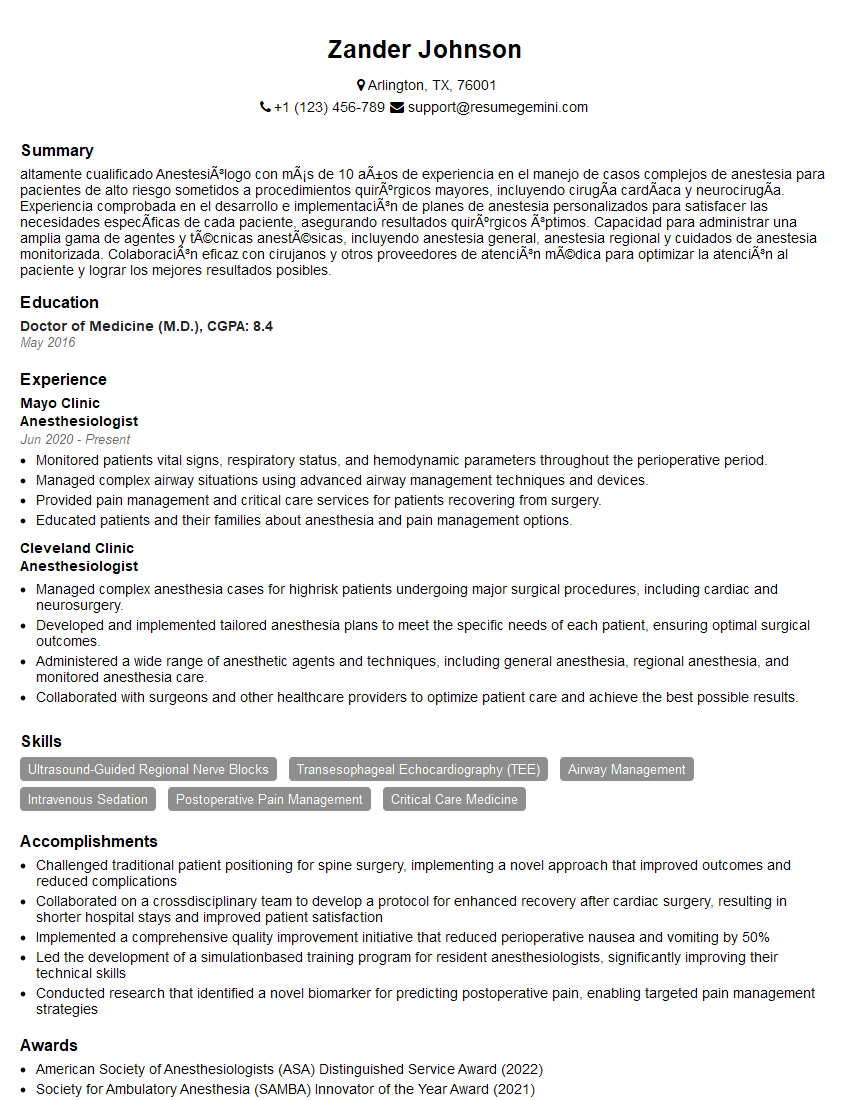
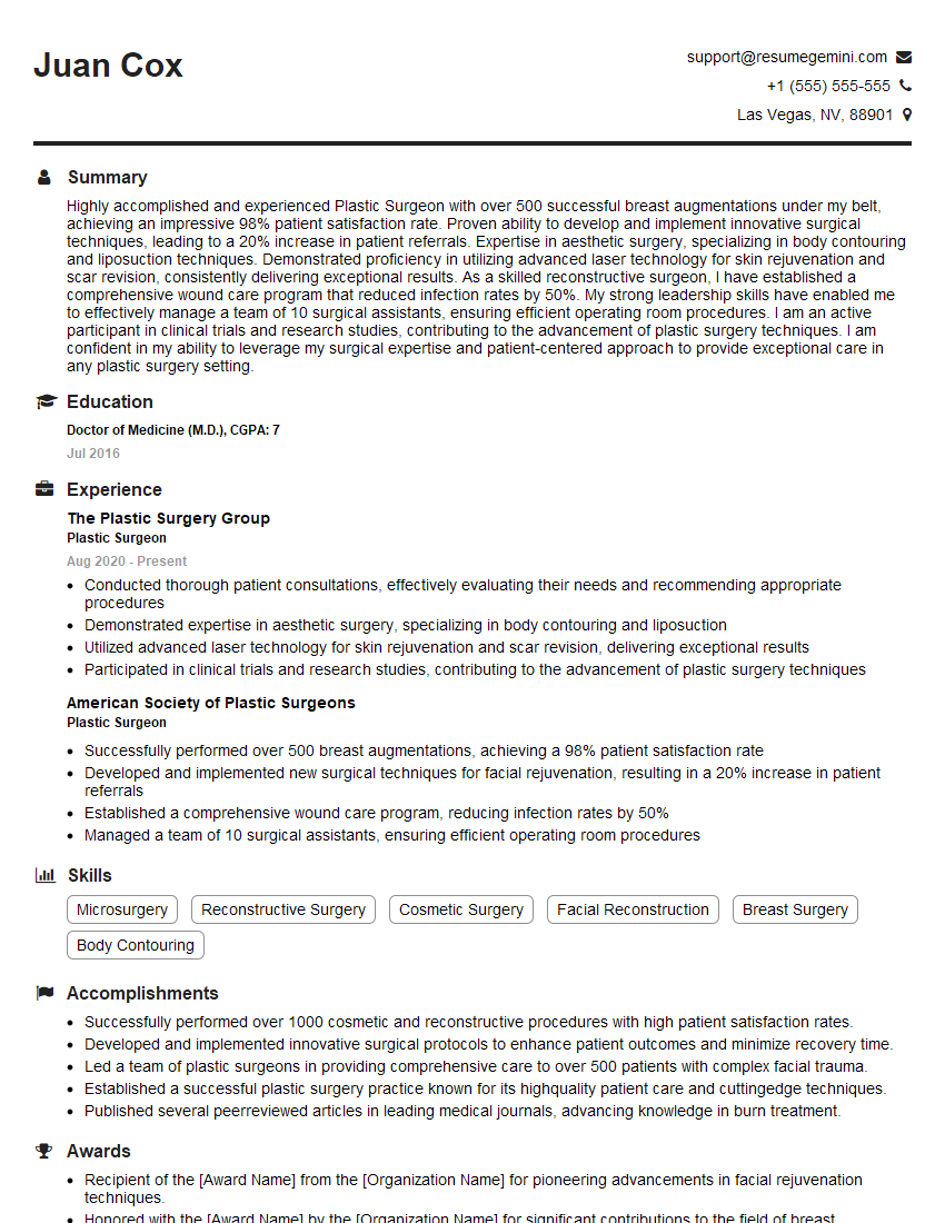
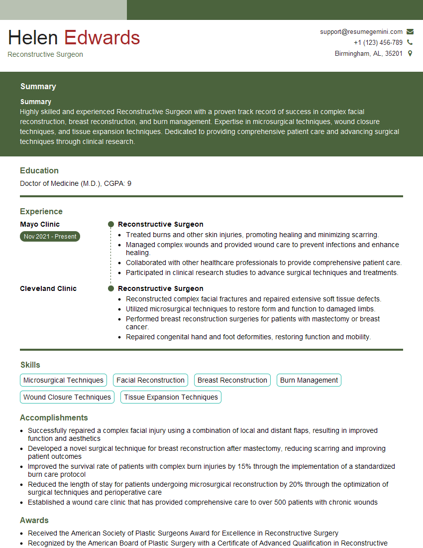
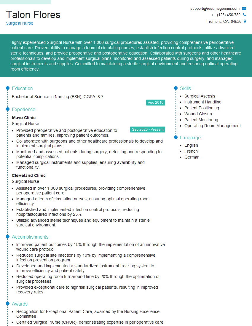
Mastering Diep flap surgery positions you for significant career advancement in reconstructive surgery. It demonstrates a high level of surgical skill and expertise. To maximize your job prospects, focus on creating an ATS-friendly resume that highlights your key skills and experience. ResumeGemini is a trusted resource to help you build a professional and impactful resume. Examples of resumes tailored to Diep Flap Surgery are available to help you showcase your qualifications effectively.
Explore more articles
Users Rating of Our Blogs
Share Your Experience
We value your feedback! Please rate our content and share your thoughts (optional).
What Readers Say About Our Blog
To the interviewgemini.com Webmaster.
Very helpful and content specific questions to help prepare me for my interview!
Thank you
To the interviewgemini.com Webmaster.
This was kind of a unique content I found around the specialized skills. Very helpful questions and good detailed answers.
Very Helpful blog, thank you Interviewgemini team.