Every successful interview starts with knowing what to expect. In this blog, we’ll take you through the top Fertility Analysis interview questions, breaking them down with expert tips to help you deliver impactful answers. Step into your next interview fully prepared and ready to succeed.
Questions Asked in Fertility Analysis Interview
Q 1. Explain the process of oocyte retrieval.
Oocyte retrieval, also known as egg retrieval, is a crucial step in assisted reproductive technologies (ART) like in-vitro fertilization (IVF). It’s a minimally invasive procedure where mature eggs are collected from a woman’s ovaries.
The process typically begins with ovarian stimulation using fertility medications to encourage the development of multiple follicles (fluid-filled sacs containing eggs). Regular monitoring through ultrasound scans tracks follicle growth and hormone levels. Once the follicles reach the optimal size, usually around 18-20mm, human chorionic gonadotropin (hCG) is administered to trigger final egg maturation.
34-36 hours after hCG injection, the oocyte retrieval is performed under ultrasound guidance. A thin needle is inserted transvaginally into each follicle, and the fluid containing the eggs is aspirated. The retrieved oocytes are then carefully examined under a microscope to assess their maturity and quality before being prepared for fertilization.
Think of it like harvesting a ripe crop – the medication helps the ‘crop’ (eggs) grow, ultrasound scans monitor its progress, and the retrieval is the careful harvesting process.
Q 2. Describe the different types of semen analysis and their interpretations.
Semen analysis, or seminal fluid analysis, is a laboratory test that evaluates various aspects of a semen sample to assess male fertility. Several types of analyses exist, each focusing on different parameters.
Routine Semen Analysis: This is the most common type and assesses:
- Volume: The amount of semen produced.
- pH: The acidity or alkalinity of the semen.
- Sperm Concentration: The number of sperm per milliliter of semen.
- Total Sperm Count: The total number of sperm in the entire sample.
- Motility: The percentage of sperm that are moving progressively.
- Morphology: The percentage of sperm with a normal shape.
MAR-test (Mixed Antiglobulin Reaction Test): This test detects antibodies on the surface of sperm that can impair their ability to fertilize an egg.
Sperm DNA Fragmentation Test: This assesses the integrity of the sperm’s DNA, which is crucial for successful fertilization and embryo development.
Computer-Assisted Semen Analysis (CASA): CASA uses computer software to analyze sperm motility and morphology more objectively than manual methods.
Interpreting the results requires comparing the values to established reference ranges. Abnormal results may suggest various underlying issues impacting fertility, requiring further investigation.
For example, a low sperm concentration (oligospermia) or poor motility (asthenospermia) might indicate problems with sperm production or transport, while abnormal morphology (teratospermia) can be due to genetic factors or environmental exposures.
Q 3. What are the key indicators of good embryo quality?
Assessing embryo quality is crucial for successful IVF outcomes. Key indicators include:
Morphology: The embryo’s physical appearance, including the number and symmetry of cells, the presence of fragmentation (cell debris), and the size and regularity of blastomeres (individual cells).
Development Stage: The stage of development the embryo has reached at a given time point (e.g., 2-cell, 4-cell, morula, blastocyst). Reaching the blastocyst stage (a hollow ball of cells) is generally considered a positive sign.
Cell Number: Higher cell numbers are generally associated with better quality, but this should be considered in conjunction with other factors like fragmentation.
Fragmentation: The presence of apoptotic bodies (cell death) or other cellular debris indicates potential developmental problems and is generally undesirable.
Expansion of Blastocyst: In blastocysts, a higher degree of expansion indicates a higher probability of successful implantation.
Trophectoderm and Inner Cell Mass (ICM): In blastocysts, these two parts should be well developed; the ICM forms the fetus and the trophectoderm forms the placenta.
Embryologists use a scoring system to rate embryos based on these factors. A high-quality embryo will exhibit characteristics that suggest a greater likelihood of successful implantation and development into a healthy pregnancy.
Q 4. How do you assess the morphology of sperm?
Sperm morphology assessment involves evaluating the shape and structure of individual sperm cells. It’s a crucial component of semen analysis that helps determine male fertility potential.
Traditionally, this is done using microscopy. A trained embryologist examines stained sperm samples and classifies them based on predefined criteria, such as the size and shape of the head, midpiece, and tail. WHO guidelines provide standardized criteria for assessing sperm morphology. Deviations from these criteria, such as a head that is too large or small, an abnormally shaped tail, or presence of vacuoles, are considered abnormal.
Techniques like strict criteria morphology assessment (Kruger strict criteria) are utilized for more accurate assessment and have become more popular in recent years. These criteria are more stringent than older guidelines, leading to a more accurate reflection of fertilization potential.
Think of it as a quality control check of the sperm’s ‘manufacturing’ process. A well-formed sperm, like a well-designed machine, has a higher chance of functioning correctly and achieving fertilization.
Q 5. What are the common causes of male infertility?
Male infertility can stem from various factors affecting sperm production, function, or delivery. Some common causes include:
Varicocele: Enlarged veins in the scrotum, leading to increased temperature and impaired sperm production.
Infections: Sexually transmitted infections (STIs) or other infections can damage the reproductive system.
Genetic Factors: Chromosomal abnormalities or genetic mutations can affect sperm production or function.
Hormonal Imbalances: Low testosterone or other hormone imbalances can impair sperm production.
Obstructions: Blockages in the reproductive tract can prevent sperm from reaching the ejaculate.
Environmental Factors: Exposure to certain toxins, chemicals, or radiation can damage sperm.
Lifestyle Factors: Smoking, excessive alcohol consumption, and drug use can negatively impact sperm production and quality.
Undescended Testes (Cryptorchidism): Testes that fail to descend into the scrotum can lead to infertility.
Often, multiple factors contribute to male infertility. A thorough evaluation is crucial to identify the underlying causes and develop an appropriate treatment plan.
Q 6. What are the common causes of female infertility?
Female infertility encompasses a wide range of issues that can prevent conception. Some common causes include:
Ovulatory Disorders: Problems with ovulation, such as infrequent or absent ovulation, can prevent the release of eggs for fertilization.
Fallopian Tube Damage or Blockage: Damage or blockage of the fallopian tubes prevents the egg from reaching the sperm or the fertilized egg from reaching the uterus.
Endometriosis: The presence of endometrial tissue outside the uterus can interfere with ovulation and fertilization.
Uterine Fibroids or Polyps: These growths in the uterus can impair implantation of the fertilized egg.
Cervical Factors: Problems with the cervix, such as cervical stenosis (narrowing) or hostile cervical mucus, can prevent sperm from entering the uterus.
Genetic Factors: Genetic abnormalities can affect egg production or development.
Age: Advanced maternal age is associated with reduced egg quality and quantity.
Unexplained Infertility: In some cases, despite thorough investigation, no specific cause for infertility can be identified.
Similar to male infertility, various factors may be at play and proper diagnosis is essential.
Q 7. Explain the different stages of embryo development in vitro.
Embryo development in vitro (in a laboratory dish) mimics the natural process but under controlled conditions. The stages are as follows:
Zygote (Day 0): Fertilization occurs, forming a single-celled zygote.
2-cell stage (Day 1): The zygote divides into two cells.
4-cell stage (Day 2): Further cell division results in four cells.
8-cell stage (Day 3): Eight cells are present, and compaction (cells become tightly bound) begins.
Morula (Day 3-4): A solid ball of cells is formed, comprising 16-32 cells.
Blastocyst (Day 5-6): A hollow sphere develops, with an inner cell mass (ICM) that will form the embryo and a trophectoderm that will form the placenta.
Embryologists closely monitor the embryo’s development at each stage, assessing its morphology (appearance) and rate of progression. Only embryos reaching a certain developmental stage with acceptable morphology are generally transferred to the uterus.
Think of it as a timelapse video of life’s very beginning, observed under carefully controlled conditions to maximize the chance of success.
Q 8. Describe the process of cryopreservation for embryos and gametes.
Cryopreservation, or freezing, is a crucial technique in fertility treatments, allowing the preservation of embryos and gametes (sperm and eggs) for later use. This process involves a controlled reduction of temperature to extremely low levels, halting biological activity and preventing cell damage. For embryos, the process typically involves a slow freezing method using cryoprotective agents (CPAs) – special solutions that prevent ice crystal formation, which can be lethal to cells. These CPAs are gradually added to the embryo, then slowly cooled to sub-zero temperatures, often using liquid nitrogen (-196°C). Once frozen, embryos are stored in liquid nitrogen tanks. The thawing process is the reverse, carefully removing CPAs and gradually warming the embryos. Gamete cryopreservation follows a similar principle, though specific CPA concentrations and freezing protocols are adjusted for sperm and oocytes (eggs) due to their differing sensitivities.
Think of it like putting a delicate plant into a state of suspended animation. The CPAs act as a protective shield, preventing damage from ice crystals forming and destroying the plant’s structure during the freezing process. When we thaw, we carefully bring the plant back to life.
Q 9. What are the ethical considerations in fertility analysis and assisted reproductive technologies?
Ethical considerations in fertility analysis and assisted reproductive technologies (ART) are complex and multifaceted. Key concerns include:
- Embryo selection and disposal: Decisions regarding which embryos to implant or discard raise moral questions about the status of embryos and the potential for discarding viable life.
- Genetic screening and selection: Prenatal diagnosis and preimplantation genetic testing (PGT) raise concerns about eugenics and the potential for selecting embryos based on desirable traits, potentially overlooking considerations of disability.
- Reproductive autonomy and access: Access to ART is often influenced by socioeconomic factors, raising concerns about equitable access and reproductive justice.
- Third-party involvement (donors): The use of donor gametes or gestational carriers introduces ethical challenges regarding anonymity, compensation, and the psychological impact on all parties involved.
- Safety and efficacy of procedures: The potential risks and side effects of ART procedures must be carefully considered and balanced against potential benefits.
These ethical issues highlight the necessity of informed consent, rigorous ethical review boards, and ongoing dialogue to ensure responsible and ethical practices in fertility treatments.
Q 10. How do you perform a sperm concentration and motility assessment?
Assessing sperm concentration and motility is a fundamental part of semen analysis. Concentration refers to the number of sperm per milliliter (mL) of semen, while motility describes the percentage of sperm that move progressively. We typically use a computer-assisted semen analysis (CASA) system for accurate and objective measurements. The process involves:
- Sample Preparation: A semen sample is allowed to liquefy at room temperature. A small aliquot is then diluted with a special solution to facilitate counting under a microscope.
- Concentration Measurement: Using a hemocytometer or CASA system, we count sperm in a defined volume to determine the concentration (e.g., millions of sperm/mL). CASA systems automate this process, offering higher precision and reducing human error.
- Motility Assessment: We observe the sperm under a microscope, classifying their movement into categories: progressive motility (forward movement), non-progressive motility (movement in place), and immotility (no movement). CASA systems analyze the speed, direction, and linearity of sperm movement.
For example, a report might show a concentration of 50 million sperm/mL with 40% progressive motility. This would indicate a good sperm concentration, but the motility is somewhat below average, potentially impacting fertility.
Q 11. What are the different methods used for fertilization?
Fertilization, the fusion of sperm and egg, can be achieved through several methods, primarily:
- In-vivo fertilization (natural fertilization): This is the natural process of fertilization that occurs within the woman’s fallopian tubes.
- In-vitro fertilization (IVF): Eggs are retrieved from the woman’s ovaries, fertilized with sperm in a laboratory dish, and then transferred to the uterus.
- Gamete intrafallopian transfer (GIFT): Eggs and sperm are placed together in the fallopian tube, allowing fertilization to occur in the body.
- Zygote intrafallopian transfer (ZIFT): Eggs are fertilized in the lab, and the resulting zygotes (fertilized eggs) are then placed in the fallopian tube.
The choice of method depends on the couple’s specific infertility diagnosis and individual circumstances.
Q 12. Explain the process of intracytoplasmic sperm injection (ICSI).
Intracytoplasmic sperm injection (ICSI) is a specialized IVF technique used when there are issues with sperm quality or quantity. Instead of allowing natural fertilization to occur in a dish, a single sperm is directly injected into an egg using a microscopic needle. The process involves:
- Egg retrieval: Eggs are retrieved from the ovaries using ultrasound-guided aspiration.
- Sperm preparation: A semen sample is prepared, and individual sperm are selected under a microscope based on their morphology (shape) and motility.
- Injection: A single, presumably healthy sperm is injected into the cytoplasm (the egg’s interior) using a microneedle.
- Incubation: The injected eggs are incubated in a specialized laboratory environment.
- Embryo transfer: After several days, embryos that have successfully fertilized and developed are transferred to the uterus.
ICSI is particularly helpful for men with severe male factor infertility, such as azoospermia (absence of sperm in semen) where sperm may be surgically retrieved from the testes or epididymis (testicular appendage). ICSI provides a more direct method of fertilization compared to standard IVF where many sperm are allowed to interact with the egg.
Q 13. What are the challenges associated with using frozen embryos?
Using frozen embryos presents several challenges:
- Reduced implantation rates: Frozen embryos may have a lower chance of implanting and resulting in a successful pregnancy compared to fresh embryos. This can be attributed to potential damage during the freezing and thawing process.
- Increased risk of chromosomal abnormalities: The freezing and thawing process can potentially increase the risk of chromosomal abnormalities in the embryos. This is an area of ongoing research to optimize cryopreservation techniques further.
- Difficulty in assessing embryo quality post-thaw: It’s sometimes more challenging to accurately assess embryo quality after thawing compared to fresh embryos, which may influence the selection for transfer.
- Cryopreservation cost: The cost of freezing and storing embryos adds to the overall expense of IVF treatment.
Despite these challenges, advancements in cryopreservation techniques continue to improve the success rates of using frozen embryos. Careful selection of suitable embryos for freezing and optimized thawing protocols contribute to maximizing the chances of a successful outcome.
Q 14. How do you identify and manage common complications during IVF procedures?
Common complications during IVF procedures require prompt identification and management. These can include:
- Ovarian hyperstimulation syndrome (OHSS): OHSS is a condition characterized by ovarian enlargement and fluid buildup in the abdomen. Management includes fluid monitoring, medication to reduce fluid retention, and in severe cases, hospitalization.
- Multiple gestation: Implantation of multiple embryos can result in a twin, triplet, or higher-order multiple pregnancy, increasing the risks for mother and babies. Strategies to mitigate this involve selective single-embryo transfer (SET).
- Ectopic pregnancy: Implantation of the embryo outside the uterus, typically in the fallopian tubes, requires prompt diagnosis and treatment, often involving surgery.
- Infection: Infections at the site of egg retrieval or embryo transfer are relatively uncommon but can be serious. Prophylactic antibiotics are often used to minimize this risk.
- Failed fertilization or implantation: If fertilization or implantation does not occur, further investigation may be required to identify underlying issues and adjust treatment strategies.
Careful monitoring throughout the IVF process, along with experienced medical professionals, is essential to promptly identify and manage these potential complications to ensure the patient’s safety and the best possible outcome.
Q 15. What are the different types of assisted hatching techniques?
Assisted hatching techniques aim to improve the chances of successful implantation by making it easier for the embryo to hatch from its protective shell, the zona pellucida. Different methods exist, each with its own nuances and success rates.
Mechanical Assisted Hatching: This involves creating a small opening in the zona pellucida using a laser or a sharp instrument (e.g., microneedle). Laser assisted hatching is the most common; it offers precise control and minimizes damage. Think of it like carefully making a small hole in an eggshell to help the chick emerge.
Chemical Assisted Hatching: This method uses chemicals, like Tyrode’s solution, to weaken the zona pellucida. The solution partially dissolves the zona, facilitating hatching. This approach is less precise than laser hatching.
Acid Tyrode’s Solution: A variation of chemical assisted hatching utilizing a slightly acidic solution to weaken the zona pellucida. The acidity helps create a small opening, but control is less precise than laser methods.
The choice of technique depends on several factors, including the embryo’s quality, the patient’s history, and the clinic’s expertise. It’s important to remember that assisted hatching isn’t always necessary and its efficacy remains a subject of ongoing research.
Career Expert Tips:
- Ace those interviews! Prepare effectively by reviewing the Top 50 Most Common Interview Questions on ResumeGemini.
- Navigate your job search with confidence! Explore a wide range of Career Tips on ResumeGemini. Learn about common challenges and recommendations to overcome them.
- Craft the perfect resume! Master the Art of Resume Writing with ResumeGemini’s guide. Showcase your unique qualifications and achievements effectively.
- Don’t miss out on holiday savings! Build your dream resume with ResumeGemini’s ATS optimized templates.
Q 16. What are the risks associated with assisted reproductive technologies?
Assisted reproductive technologies (ART), while offering hope for many couples facing infertility, do carry certain risks. These risks can vary depending on the specific procedure and the patient’s individual health.
Multiple pregnancies: The transfer of multiple embryos increases the risk of twins, triplets, or higher-order multiple pregnancies, which can pose significant health risks to both the mother and the babies.
Ovarian hyperstimulation syndrome (OHSS): This is a complication of ovarian stimulation medications used in IVF and other ART procedures. OHSS can range in severity from mild discomfort to life-threatening complications.
Ectopic pregnancy: Implantation of the embryo outside the uterus, often in a fallopian tube, is a serious risk. Ectopic pregnancies require immediate medical intervention.
Miscarriage: The risk of miscarriage is generally higher in ART cycles compared to natural conception.
Birth defects: While the overall risk remains low, some studies suggest a slightly increased risk of certain birth defects in babies conceived through ART. However, it’s important to note that many of these studies lack conclusive evidence on a causal relationship.
Emotional stress: The ART process can be emotionally and psychologically taxing for couples. The uncertainty and potential for disappointment can significantly impact mental well-being.
It’s crucial for couples considering ART to have a thorough discussion with their fertility specialist about the potential risks and benefits of each procedure. A well-informed decision is essential for minimizing potential complications.
Q 17. Explain the role of preimplantation genetic testing (PGT).
Preimplantation genetic testing (PGT) is a powerful tool used to analyze embryos created through in vitro fertilization (IVF) before implantation. It helps identify genetic abnormalities that could lead to miscarriage, birth defects, or inherited diseases. Different types of PGT exist, each targeting specific genetic issues.
PGT-A (Aneuploidy): Screens for the correct number of chromosomes in the embryo. A balanced number is crucial for healthy development. Think of it as a chromosome count to ensure the right number of each type is present.
PGT-M (Monogenic): Tests for specific gene mutations known to cause inherited diseases, such as cystic fibrosis or Huntington’s disease. This is particularly helpful for couples carrying genes for these conditions.
PGT-SR (Structural Rearrangements): Identifies balanced or unbalanced chromosome translocations or inversions in the parents that could lead to abnormal chromosome numbers in the offspring.
PGT allows for the selection of genetically healthy embryos for transfer, thereby increasing the chances of a successful pregnancy and reducing the risk of having a child with a genetic disorder. However, it’s a complex procedure with limitations and potential for errors. It’s a significant decision that couples should carefully discuss with their genetic counselor and fertility doctor.
Q 18. How do you interpret the results of a karyotype analysis?
Karyotype analysis is a laboratory test that examines the number and structure of chromosomes in a cell. The results are presented as a karyotype, a visual representation of the chromosomes arranged in pairs according to their size and shape. Interpreting a karyotype involves several steps.
Chromosome Number: The normal human karyotype contains 46 chromosomes, arranged in 23 pairs (22 pairs of autosomes and one pair of sex chromosomes). Any deviation from this number (e.g., 47, 45) indicates aneuploidy.
Chromosome Structure: The karyotype reveals structural abnormalities such as deletions, duplications, inversions, and translocations. These changes can impact gene function and lead to various genetic disorders.
Sex Chromosomes: The sex chromosomes (XX in females, XY in males) are carefully examined for abnormalities. This helps in diagnosing conditions such as Turner syndrome (XO), Klinefelter syndrome (XXY), and others.
For example, a karyotype of 47,XX,+21 indicates trisomy 21, also known as Down syndrome. A detailed report from a qualified geneticist is essential for accurate interpretation and proper clinical management.
Q 19. Describe the different types of infertility treatments.
Infertility treatments vary widely depending on the cause of infertility. They range from simple lifestyle changes to complex medical interventions. The approach is always personalized based on a thorough evaluation of the couple’s situation.
Lifestyle Modifications: Addressing factors such as weight, diet, smoking, and alcohol consumption can significantly improve fertility in some cases.
Ovulation Induction Medications: Drugs like Clomiphene Citrate or Letrozole stimulate ovulation in women with irregular cycles or anovulation.
Intrauterine Insemination (IUI): This procedure involves placing sperm directly into the uterus to increase the chances of fertilization.
In Vitro Fertilization (IVF): Eggs are retrieved from the ovaries, fertilized with sperm in a laboratory, and then transferred back into the uterus.
Intracytoplasmic Sperm Injection (ICSI): A single sperm is injected directly into an egg, often used when male factor infertility is present.
Gamete Intrafallopian Transfer (GIFT) and Zygote Intrafallopian Transfer (ZIFT): Older techniques, less commonly used now, involving the transfer of gametes or zygotes into the fallopian tubes.
Surgical Procedures: Procedures such as laparoscopy or hysteroscopy can address physical problems like endometriosis or blocked fallopian tubes.
The choice of treatment requires a careful evaluation of factors such as age, medical history, type and severity of infertility, and cost. A collaborative approach between the couple and their fertility specialist is essential for making informed decisions.
Q 20. What is the role of hormonal assays in fertility analysis?
Hormonal assays play a crucial role in fertility analysis. By measuring hormone levels in blood or urine, doctors can assess various aspects of reproductive function in both men and women. These assays provide valuable information for diagnosis and treatment planning.
Women: Hormones like Follicle-Stimulating Hormone (FSH), Luteinizing Hormone (LH), Estrogen, and Progesterone are monitored to evaluate ovarian function, assess ovulation, and detect hormonal imbalances.
Men: Testosterone, Follicle-Stimulating Hormone (FSH), and Luteinizing Hormone (LH) levels are evaluated to assess testicular function and sperm production. Prolactin levels are also checked, as elevated prolactin can lead to decreased libido and infertility.
For example, elevated FSH levels in women may indicate diminished ovarian reserve, while low progesterone levels can suggest luteal phase deficiency. In men, low testosterone levels can point to hypogonadism. The interpretation of hormone levels requires careful consideration of factors such as age and menstrual cycle phase.
Q 21. Explain the use of time-lapse imaging in embryo assessment.
Time-lapse imaging is a sophisticated technology used in IVF laboratories to monitor embryo development in real time. Instead of assessing embryos at specific time points, it captures images continuously, providing a comprehensive view of the embryo’s growth and developmental events.
This technology offers several advantages:
Improved Embryo Selection: By tracking subtle morphological changes and developmental kinetics, embryologists can identify embryos with higher implantation potential. This is analogous to watching a plant grow and assessing its health over time, versus only looking at it at specific time points.
Reduced Handling: Because time-lapse imaging minimizes the need for frequent handling and assessment, it reduces stress and potential damage to the developing embryos.
More Detailed Information: The continuous monitoring allows for the identification of subtle developmental events and patterns that may not be apparent during standard microscopic evaluation.
Data-Driven Decisions: Time-lapse data allows for more objective embryo selection based on established algorithms and developmental parameters.
However, the cost of time-lapse imaging systems and the need for specialized training to analyze the data are potential drawbacks. While promising, further research is needed to fully understand the long-term impact of this technology on IVF success rates.
Q 22. How do you maintain quality control in a fertility laboratory?
Maintaining quality control in a fertility lab is paramount, as even minor errors can have significant consequences for patients. It’s a multi-layered approach involving stringent protocols, meticulous record-keeping, and regular audits.
- Personnel Training and Competency: We implement rigorous training programs covering all aspects of laboratory procedures, including aseptic techniques, equipment operation, and data management. Regular competency assessments ensure that everyone maintains proficiency. For example, we conduct blind proficiency testing where technicians analyze samples without knowing the expected result, ensuring accuracy and reducing bias.
- Equipment Calibration and Maintenance: All equipment – incubators, microscopes, centrifuges, etc. – undergoes regular calibration and preventative maintenance according to manufacturer’s specifications and internal schedules. We maintain detailed logs tracking calibrations and any maintenance performed. This ensures equipment is functioning optimally and delivering accurate results. For instance, we might use standardized temperature probes to verify the accuracy of our incubators multiple times a day.
- Reagent Quality Control: We meticulously source reagents from reputable suppliers and implement a system of incoming quality control checks. This includes verifying expiration dates, assessing physical appearance for signs of degradation, and sometimes running control experiments. Documentation is crucial, allowing us to trace any issues back to the source.
- Internal Audits and External Accreditation: We conduct regular internal audits to review our processes and identify areas for improvement. Furthermore, we actively seek accreditation from reputable organizations like CAP (College of American Pathologists) or equivalent, demonstrating our commitment to internationally recognized quality standards. This includes external audits that scrutinize our entire workflow.
- Data Management and Tracking: A robust electronic data management system is essential. We use a LIMS (Laboratory Information Management System) that tracks samples, tests, and results, ensuring data integrity and providing an audit trail of all activities. This facilitates error detection and enables comprehensive data analysis for quality improvement initiatives.
By combining these elements, we create a system that minimizes errors and ensures the reliability of our lab results, maximizing the chances of successful IVF cycles for our patients.
Q 23. Describe your experience with different types of culture media.
My experience with culture media spans various formulations, each with specific advantages and disadvantages for different stages of gamete and embryo development. The choice of media depends on several factors, including the specific needs of the patient and the protocol being followed.
- Basic Culture Media: I’ve worked extensively with standard culture media such as Ham’s F-10 and HTF (human tubal fluid) media. These are widely used for supporting gamete and early embryo development. They provide essential nutrients and maintain a suitable pH and osmolality.
- Sequential Media: I have substantial experience with sequential media systems that mimic the changes in the uterine environment during embryo development. These media formulations often change throughout the culture period, providing optimized conditions at each stage. This helps improve embryo quality and developmental competence.
- Specialty Media: I’ve utilized specialized media formulations, such as media supplemented with growth factors or antioxidants, tailored for specific clinical situations like poor-quality embryos or patients with a history of recurrent implantation failure. The results are often carefully analyzed for effectiveness.
- Commercial vs. In-house: I’ve used both commercially available and in-house prepared media. While commercial media offer convenience and standardization, in-house preparations sometimes offer greater control over the precise composition, though this necessitates strict quality control measures. The choice depends on the lab resources and preferences.
In my experience, the key is not just using a specific type of media but understanding its properties and how to optimize its use for individual patients’ needs, always following rigorous quality control procedures to ensure consistency and efficacy.
Q 24. What is your experience with troubleshooting IVF laboratory equipment?
Troubleshooting IVF laboratory equipment requires a systematic approach combining technical expertise, methodical problem-solving, and good record-keeping. It’s not just about fixing the immediate problem but also preventing future occurrences.
- Documentation is Key: I always start by thoroughly reviewing maintenance logs, calibration records, and any previous error reports. This historical context is crucial for identifying patterns and potential root causes.
- Systematic Approach: My approach follows a logical sequence: 1. Identify the problem precisely, 2. Check the obvious – power supply, connections, reagents, etc., 3. Consult manuals and technical support, 4. If needed, isolate components for testing. For example, if an incubator’s temperature is unstable, I’d check the sensor, the heating element, and the control system one by one.
- Preventative Maintenance: A proactive approach is vital. Regular preventative maintenance significantly reduces the frequency and severity of equipment malfunctions. This includes cleaning, lubrication, and periodic replacement of consumable parts.
- Calibration and Validation: Calibration and validation procedures are critical in ensuring the accuracy of results. Regularly scheduled calibration prevents inaccurate readings and ensures the equipment is functioning correctly within accepted tolerances.
- Understanding the Equipment: A deep understanding of the equipment’s operational principles is essential. This enables me to diagnose problems more effectively and make informed decisions about repairs or replacements. For instance, knowing the functionality of a micromanipulator helps troubleshoot issues with its precision and stability.
Ultimately, efficient equipment troubleshooting is not just about fixing a broken machine; it’s about ensuring the lab’s operations remain consistent and reliable, protecting the quality of patient care.
Q 25. How do you handle and prevent laboratory contamination?
Preventing and handling contamination in a fertility lab is absolutely critical; any contamination can compromise embryo viability and lead to treatment failure. Our strategy focuses on a combination of preventative measures and rigorous contamination control procedures.
- Aseptic Techniques: All personnel receive extensive training in aseptic techniques, including proper handwashing, gowning procedures, and the use of laminar flow hoods. We strictly enforce these protocols to minimize the introduction of contaminants.
- Environmental Control: We maintain a clean and controlled environment using HEPA filters, regular cleaning and disinfection protocols, and monitoring of environmental parameters such as air quality and temperature. Regular surface disinfection with appropriate agents is vital.
- Reagent Handling: Reagents are handled with care, using sterile techniques to prevent contamination. Aliquoting reagents into smaller sterile containers minimizes exposure to contamination and reduces waste.
- Equipment Sterilization: Equipment is regularly sterilized using appropriate methods, such as autoclaving, UV sterilization, or chemical sterilization. The choice of sterilization method depends on the equipment’s material and sensitivity.
- Monitoring and Reporting: We actively monitor for contamination through regular environmental testing and quality control checks. Any contamination incident is meticulously documented and thoroughly investigated to identify the source and prevent recurrence. For example, we track the types and frequency of contaminants found to help identify possible sources such as air vents or faulty equipment.
Our contamination control procedures are not just reactive but highly proactive. By prioritizing prevention and consistently adhering to stringent protocols, we ensure a clean and safe environment for the sensitive work involved in fertility treatments.
Q 26. How do you interpret the results of a sperm chromatin dispersion (SCD) test?
The Sperm Chromatin Dispersion (SCD) test assesses the integrity of DNA within sperm cells. It’s a valuable tool in evaluating male fertility, as DNA fragmentation can significantly impair fertilization and embryo development.
The test involves exposing sperm to a denaturing solution which causes the DNA to unwind. The degree of DNA dispersion is then assessed microscopically, typically scored on a scale (e.g., 0-100%, or categorized into groups reflecting different levels of DNA fragmentation).
- Interpretation: A high percentage of sperm with highly dispersed chromatin (i.e., low DNA fragmentation) indicates good DNA integrity, suggesting a higher likelihood of successful fertilization and pregnancy. Conversely, a high percentage of sperm with poorly dispersed chromatin (i.e., high DNA fragmentation) implies compromised DNA integrity, which can lead to reduced fertilization rates, poor embryo quality, and increased rates of miscarriage.
- Clinical Significance: SCD test results are interpreted in conjunction with other semen parameters like sperm concentration, motility, and morphology. It’s crucial to consider the overall semen analysis profile when counseling patients. A low SCD score may necessitate further investigation into the causes of DNA damage and exploration of assisted reproduction techniques, such as intracytoplasmic sperm injection (ICSI) which bypasses some of the natural selection processes.
- Limitations: It’s essential to be aware of the limitations of SCD and not rely solely on it. It provides a snapshot of DNA damage at the time of testing, and the clinical significance might vary across laboratories due to differing protocols.
In practice, the SCD test helps us identify patients who may benefit from ICSI or other assisted reproductive techniques, potentially increasing their chances of a successful outcome. We always counsel patients thoroughly on the test’s implications and incorporate its results into a broader fertility assessment.
Q 27. Explain the significance of the zona pellucida in fertilization.
The zona pellucida (ZP) is a glycoprotein layer surrounding the mammalian oocyte (egg). It plays a crucial role in several key steps of fertilization.
- Sperm Binding and Recognition: The ZP contains specific receptors that bind to complementary molecules on the sperm surface. This species-specific binding ensures that only sperm of the same species can fertilize the egg. It’s the initial step in the fertilization process.
- Acrosome Reaction: Following binding, the sperm undergoes the acrosome reaction, releasing enzymes that digest a pathway through the ZP, allowing the sperm to penetrate it. This is a critical step, and failure here can prevent fertilization.
- Prevention of Polyspermy: The ZP plays a vital role in preventing polyspermy – the fertilization of an egg by multiple sperm. After a sperm successfully penetrates the ZP, changes occur in the ZP, creating a ‘block’ that prevents further sperm entry. This ensures the correct number of chromosomes are inherited.
- Protection of Oocyte: The ZP acts as a protective barrier, shielding the oocyte from mechanical damage and harmful substances in the reproductive tract.
In assisted reproduction technologies (ART), the ZP’s properties are carefully considered. For example, techniques like ICSI bypass the need for sperm to penetrate the ZP, which is helpful in cases of severe male factor infertility. Furthermore, techniques like assisted hatching may involve partially removing the ZP to improve the chances of embryo implantation.
Understanding the zona pellucida’s role in fertilization is essential for both natural conception and successful ART procedures. Its multifaceted functions are crucial for ensuring successful fertilization and the development of a healthy embryo.
Key Topics to Learn for Fertility Analysis Interview
- Ovarian Reserve Assessment: Understanding various methods like antral follicle count (AFC), anti-Müllerian hormone (AMH) levels, and their clinical implications in predicting ovarian response to stimulation.
- Semen Analysis: Mastering the interpretation of semen parameters (volume, concentration, motility, morphology) and their correlation with male fertility potential. Practical application includes understanding the limitations of basic semen analysis and when to recommend further investigations.
- Endometrial Receptivity: Learning about the factors influencing endometrial receptivity, including hormonal profiles, endometrial thickness, and the use of endometrial biopsy or other assessments for evaluating implantation potential.
- Ovulation Detection: Familiarize yourself with different methods for ovulation detection, including basal body temperature charting, ovulation predictor kits (OPKs), and ultrasound monitoring. Understand the strengths and weaknesses of each method.
- Infertility Diagnostics: Gain a comprehensive understanding of various diagnostic tests used in infertility investigations, such as hormonal assays, hysterosalpingography (HSG), and laparoscopy. Be prepared to discuss their indications, interpretation, and limitations.
- Assisted Reproductive Technologies (ART): Understand the principles and procedures of common ART techniques, including in-vitro fertilization (IVF), intracytoplasmic sperm injection (ICSI), and gamete intrafallopian transfer (GIFT). Focus on the practical aspects and potential complications.
- Data Analysis and Interpretation: Develop your skills in interpreting complex fertility data, identifying trends, and drawing meaningful conclusions. This includes understanding statistical concepts relevant to fertility analysis.
- Ethical Considerations: Be prepared to discuss ethical considerations related to fertility treatments, including patient counseling, informed consent, and the responsible use of technology.
Next Steps
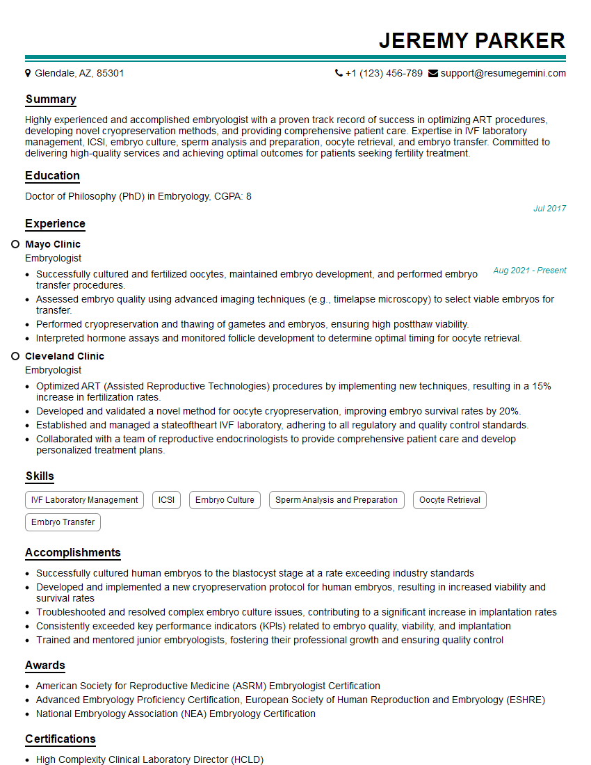
Mastering Fertility Analysis is crucial for a successful and rewarding career in reproductive medicine. A strong understanding of these concepts opens doors to exciting opportunities and positions you as a valuable asset in this growing field. To maximize your job prospects, creating an ATS-friendly resume is essential. ResumeGemini is a trusted resource that can help you build a professional and effective resume that highlights your skills and experience. We provide examples of resumes tailored to Fertility Analysis to guide you in crafting a compelling application. Take the next step towards your dream career today!
Explore more articles
Users Rating of Our Blogs
Share Your Experience
We value your feedback! Please rate our content and share your thoughts (optional).
What Readers Say About Our Blog
To the interviewgemini.com Webmaster.
Very helpful and content specific questions to help prepare me for my interview!
Thank you
To the interviewgemini.com Webmaster.
This was kind of a unique content I found around the specialized skills. Very helpful questions and good detailed answers.
Very Helpful blog, thank you Interviewgemini team.