Feeling uncertain about what to expect in your upcoming interview? We’ve got you covered! This blog highlights the most important Fetal Autopsy interview questions and provides actionable advice to help you stand out as the ideal candidate. Let’s pave the way for your success.
Questions Asked in Fetal Autopsy Interview
Q 1. Describe the process of a standard fetal autopsy.
A standard fetal autopsy is a meticulous examination of a deceased fetus to determine the cause of death and identify any underlying conditions. It’s a systematic process involving external and internal examinations.
The external examination begins with noting the sex, size, weight, and overall appearance of the fetus. We carefully assess the skin for any anomalies, such as birthmarks or lesions. Then, we measure the crown-rump length (CRL) – a crucial indicator of gestational age.
The internal examination involves a systematic dissection to examine major organs. This includes opening the skull to inspect the brain, examining the heart and great vessels, inspecting the lungs for evidence of breathing, and examining the abdominal organs like the liver, spleen, kidneys, and gastrointestinal tract. We carefully look for any malformations or abnormalities. Finally, detailed photographs and measurements are recorded throughout the process. This meticulous approach helps build a comprehensive picture of the fetal development and the cause of death.
Q 2. What are the key differences between a fetal autopsy and a neonatal autopsy?
The key difference between a fetal and a neonatal autopsy lies primarily in the gestational age at the time of death and the level of organ development. A fetal autopsy involves fetuses that die in utero before birth, while a neonatal autopsy focuses on newborns who die within the first 28 days of life.
Fetal autopsies often concentrate on identifying developmental issues, congenital anomalies, and placental abnormalities that contributed to the demise. For example, we meticulously examine the umbilical cord for abnormalities. In contrast, neonatal autopsies might delve deeper into infectious causes, birth complications (like hypoxia), and issues related to the transition to extrauterine life, and might involve more sophisticated techniques like microbiological cultures.
The extent of organ development significantly differs. For instance, a full-term fetus will have more mature organs compared to a first-trimester fetus, influencing the focus and depth of the autopsy examination.
Q 3. Explain the significance of placental examination in a fetal autopsy.
Placental examination is absolutely crucial in a fetal autopsy as the placenta is the lifeline of the fetus. It plays a vital role in providing the fetus with oxygen and nutrients, and removing waste products. Examining the placenta allows us to identify potential problems that may have contributed to fetal demise.
We assess the placenta’s size, weight, shape, and appearance. We carefully examine the umbilical cord for abnormalities like knots, true knots, or abnormalities in the number of vessels. We check for signs of infection (chorioamnionitis), placental abruption (premature separation of the placenta), or placenta previa (placenta covering the cervix). Microscopic examination of placental tissue sections may be necessary to confirm certain conditions like infarction or infection.
For example, a finding of significant placental abruption can explain intrauterine growth restriction (IUGR) and fetal distress leading to death. Similarly, evidence of infection can suggest the cause of fetal demise. In essence, a thorough placental examination is essential to build a comprehensive understanding of the events leading to the fetal death.
Q 4. How do you determine gestational age in a fetal autopsy?
Determining gestational age accurately is paramount in a fetal autopsy. Several methods are used, often in combination, to arrive at the most accurate estimate.
Crown-rump length (CRL): This is the most accurate method in early pregnancy (up to 20 weeks). We measure the distance from the crown of the head to the bottom of the buttocks. Biparietal diameter (BPD): The distance between the two sides of the fetal skull is measured using ultrasound imaging (if available pre-mortem). Femur length: The length of the fetal thigh bone is another valuable measurement. Ossification centers: We assess the development of ossification centers in various bones – these centers appear at specific gestational ages. Finally, external characteristics such as the presence of lanugo hair, skin texture, and the development of external genitalia can also be used to estimate gestational age. All these measurements are then compared with established growth charts to estimate the gestational age at the time of death.
It’s important to remember that these methods provide estimates; the level of accuracy varies depending on the gestational age and fetal development.
Q 5. What are the common causes of fetal demise, and how are they identified during autopsy?
Common causes of fetal demise are diverse and can be broadly categorized as infections, chromosomal abnormalities, genetic disorders, and various other complications.
Infections: Viral or bacterial infections (like cytomegalovirus or toxoplasmosis) can severely affect fetal development, leading to death. In autopsy, we might find evidence of infection through histological examination of tissues or microbiological cultures. Chromosomal abnormalities: Trisomy 21 (Down syndrome), trisomy 18 (Edwards syndrome), and trisomy 13 (Patau syndrome) are some common examples. These are often identified through karyotyping of fetal tissues post-mortem. Genetic disorders: These can manifest in various ways, leading to fetal death. Specific genetic testing might be required to identify the exact genetic defect. Other complications: These include placental abruption, placental insufficiency, umbilical cord problems (prolapse or compression), severe congenital anomalies incompatible with life, multiple gestation complications, maternal conditions (like uncontrolled diabetes or hypertension), and unknown causes. We assess these factors through a combination of external and internal examination, including microscopic analysis and special studies, to determine their role in fetal death.
Determining the exact cause of death often requires a multidisciplinary approach, combining autopsy findings with maternal history and prenatal care records.
Q 6. Describe your experience with fetal autopsy techniques, including tissue sampling and preservation.
My experience encompasses a wide range of fetal autopsy techniques, emphasizing precision and adherence to established protocols. We routinely perform external and internal examinations, including detailed dissection of major organ systems. We utilize various imaging techniques, including radiography and ultrasound, when appropriate. Tissue sampling is crucial. We meticulously collect samples for histological examination, including sections from various organs, the placenta, and umbilical cord. These samples are carefully processed and stained for microscopic evaluation. We also collect samples for karyotyping (chromosomal analysis), genetic testing, and microbiological cultures when indicated. Proper tissue preservation is paramount. Samples are fixed in formalin, embedded in paraffin, sectioned, and stained according to established protocols to maintain tissue integrity and facilitate accurate microscopic examination. The meticulous collection, processing, and interpretation of these samples allow for a thorough understanding of the cause of fetal demise.
Q 7. How do you handle cases involving suspected congenital anomalies?
Suspected congenital anomalies require a particularly detailed and comprehensive approach. The autopsy protocol is tailored to thoroughly investigate the specific anomalies. For example, if a cardiac anomaly is suspected, we perform a detailed examination of the heart, including dissection of the chambers, valves, and great vessels. Similarly, if a neural tube defect is suspected, we meticulously examine the brain, spinal cord, and overlying tissues.
In such cases, we may employ specialized techniques such as detailed imaging studies (radiography, ultrasound), genetic testing (karyotyping, chromosomal microarray analysis), and advanced microscopic techniques (immunohistochemistry) to gain more insight into the development and severity of the anomaly. We might also involve specialists like geneticists and pediatric cardiologists for consultation to understand the complexities of these cases. The goal is to provide the family with as much information as possible regarding the anomaly, its severity, and its potential impact on fetal development. Furthermore, we work to identify any underlying causes or contributing factors that may have led to the anomaly’s development. This comprehensive approach helps not only in clarifying the cause of fetal death but also informs future pregnancies and genetic counseling for the family.
Q 8. Explain the importance of accurate documentation in fetal autopsy.
Accurate documentation in fetal autopsy is paramount. It forms the bedrock of the entire process, ensuring the findings are reliable, legally defensible, and contribute meaningfully to medical knowledge and future preventative care. Without meticulous documentation, the autopsy loses its value – both diagnostically and legally.
- Complete and Detailed Examination Notes: Every step, from initial external examination (measuring crown-rump length, noting anomalies) to internal organ dissection and microscopic analysis, must be meticulously recorded. Any deviations from normal anatomy or developmental milestones should be described precisely.
- High-Quality Imaging: Photographs and radiographic images (X-rays, ultrasounds) are crucial to visualize macroscopic findings. These images must be labeled clearly and correlated with the written description.
- Clear Specimen Labeling: Tissues collected for further testing (histology, microbiology, cytogenetics) require unambiguous labels to maintain traceability and avoid confusion. This includes details like the specimen type, gestational age, and patient identifiers.
- Standardized Terminology: Employing standardized medical terminology ensures consistency and prevents ambiguity in interpreting the findings. This improves communication between pathologists and clinicians.
- Legal and Ethical Considerations: The documentation should also record consent obtained from the parents (or legal guardians) for the autopsy procedure. This adheres to ethical and legal requirements.
For example, in a case of suspected anencephaly, meticulous documentation of the absent cranial vault, malformed brain tissue, and associated anomalies would be critical for diagnosis and counseling future pregnancies.
Q 9. How do you interpret fetal autopsy findings in the context of maternal history?
Interpreting fetal autopsy findings requires a holistic approach, integrating the autopsy results with the maternal history. The maternal history provides crucial context, helping to explain the findings and establish a potential etiology. Ignoring the maternal history significantly limits the diagnostic accuracy and completeness of the autopsy report.
- Maternal Infections: A history of maternal infections (e.g., rubella, cytomegalovirus) can explain congenital anomalies or inflammation found during the autopsy.
- Maternal Medications: Exposure to teratogenic drugs during pregnancy may cause specific fetal abnormalities. The maternal medication history allows us to assess potential drug-related effects.
- Maternal Medical Conditions: Pre-existing maternal conditions like diabetes, hypertension, autoimmune disorders, and thyroid diseases can affect fetal development and health. Understanding these conditions is essential.
- Environmental Exposures: Maternal exposure to toxins or environmental hazards can affect the developing fetus. This information should be obtained to explore potential links.
- Obstetrical History: Information about previous pregnancies, including any miscarriages or stillbirths, provides a valuable context for interpreting the current case.
For instance, finding evidence of hydrops fetalis in a fetal autopsy might be better understood if the maternal history revealed a pre-existing cardiac condition or severe anemia.
Q 10. Discuss your familiarity with different fetal autopsy reporting formats.
My familiarity with fetal autopsy reporting formats spans various styles, including those from academic institutions, government agencies, and private hospitals. While the core elements remain consistent (identifying information, external examination, internal examination, ancillary test results, diagnosis, and summary), the specific formatting and level of detail might vary.
- Standardized Forms: Many institutions utilize standardized forms to ensure consistency and completeness in reporting. These forms often include sections for demographics, clinical history, gross findings, microscopic findings, and diagnostic impressions.
- Narrative Reports: Some settings prefer narrative reports, allowing for more detailed and descriptive accounts of the findings. These reports demand excellent writing skills and meticulous organization.
- Electronic Reporting Systems: Many modern pathology departments employ electronic reporting systems that streamline data entry, improve efficiency, and enable easier data analysis.
- International Classification of Diseases (ICD) Codes: Appropriate ICD codes are crucial for proper statistical reporting and epidemiological studies.
Regardless of the format, I prioritize clarity, conciseness, and accuracy to ensure the report is easily understood by clinicians, researchers, and bereaved parents.
Q 11. How do you ensure the chain of custody is maintained during a fetal autopsy?
Maintaining the chain of custody is crucial for the integrity and legal admissibility of the fetal autopsy findings. Any break in the chain can compromise the evidence and potentially affect legal proceedings or future investigations.
- Secure Transportation: The fetus is transported securely from the delivery location to the autopsy suite, documented with time stamps and signatures at each transfer.
- Unique Identification Numbers: Each specimen, including the fetus itself, receives a unique identification number, consistently tracked throughout the autopsy.
- Detailed Logs and Records: A comprehensive log records who handled the fetus, at what time, and for what purpose, ensuring complete transparency.
- Secure Storage: Tissues and other specimens are stored securely, with appropriate access restrictions to prevent loss, damage, or unauthorized tampering.
- Witnessing Procedures: When appropriate, specific procedures may involve witnesses to ensure the integrity of the process.
For example, the designated individual transferring the fetus from the hospital to the morgue would sign a transfer log, noting the time, date, and condition of the fetus. The same meticulous approach is followed for each step of the process.
Q 12. Describe your experience with fetal autopsy in cases of suspected infection.
My experience includes numerous cases of suspected fetal infection. In these cases, the autopsy plays a vital role in identifying the causative agent and understanding the extent of the infection’s impact on fetal development. The approach involves a combination of macroscopic and microscopic examinations, along with specialized laboratory tests.
- Macroscopic Examination: We look for signs of inflammation, organomegaly (enlarged organs), abscesses, or other abnormalities that might suggest infection.
- Microscopic Examination: Tissue samples from various organs (placenta, liver, lungs, brain) are examined microscopically to identify inflammatory cells or microorganisms.
- Culture and Sensitivity Testing: Cultures are performed to identify the specific infectious agent. Sensitivity testing determines the effectiveness of various antimicrobial agents.
- Immunohistochemistry (IHC): IHC is used to detect specific antigens associated with certain infectious agents.
- Molecular Diagnostics: Polymerase chain reaction (PCR) tests detect specific viral or bacterial DNA/RNA, even if the microorganisms are not readily visible.
A classic example is a case of suspected congenital cytomegalovirus (CMV) infection, where microscopic examination of the fetal tissues for characteristic CMV inclusion bodies, coupled with PCR confirmation, would provide a definitive diagnosis. The placental examination would also be crucial.
Q 13. How do you address ethical considerations in fetal autopsy cases?
Ethical considerations are central to fetal autopsy. The process involves a deceased fetus, adding a layer of sensitivity and respect. These ethical considerations require careful handling and communication.
- Informed Consent: Obtaining informed consent from the parents (or legal guardians) is paramount. This involves explaining the purpose, procedures, and potential benefits and risks of the autopsy in a compassionate and understandable manner.
- Respect for the Fetus and Family: Treating the fetus with dignity and respect throughout the entire process is essential. This includes careful handling of the body and sensitive communication with the family.
- Confidentiality: Maintaining strict confidentiality concerning the autopsy findings and the family’s personal information is crucial. All information remains protected and shared only with those directly involved in the care and investigation.
- Transparency and Communication: Open and honest communication with the family regarding the autopsy process and findings is paramount. The findings should be explained clearly and with empathy, addressing their questions and concerns.
- Emotional Support: It is vital to recognize the emotional distress experienced by the parents. Providing them with emotional support, grief counseling, and appropriate referrals are significant elements of ethical care.
For example, ensuring that the parents understand that the autopsy is not intended to place blame but rather to understand the cause of the fetal demise and provide them with information for future pregnancies is a key ethical aspect.
Q 14. Explain your approach to communicating autopsy findings to bereaved parents.
Communicating autopsy findings to bereaved parents is one of the most challenging yet crucial aspects of my role. Empathy, sensitivity, and clear communication are essential.
- Timing and Setting: The meeting should take place in a private and comfortable setting. The timing should be appropriate, considering the parents’ emotional state. Allowing time for the family to process the information is crucial.
- Clear and Simple Language: Avoid medical jargon and use plain language the parents can easily understand. If complex information is necessary, explain it clearly and patiently, using analogies or visual aids if helpful.
- Empathetic Approach: Acknowledge the parents’ grief and pain. Listen attentively to their concerns and validate their feelings. Creating a supportive and non-judgmental atmosphere is paramount.
- Addressing Questions: Allow ample time for the parents to ask questions and answer them honestly and compassionately. If unsure about a specific finding, acknowledge this and offer to obtain additional information.
- Written Summary: Provide a written summary of the autopsy findings. This will allow the parents to review the information at their own pace and provide a lasting record of the events.
For example, instead of saying “there was evidence of placental abruption,” I might say, “The placenta separated from the uterine wall prematurely, which is likely what caused the baby’s death.” This clear and simple language allows for better understanding.
Q 15. What are the limitations of fetal autopsy in determining cause of death?
Fetal autopsy, while invaluable, has limitations in definitively determining the cause of death, particularly in cases of early gestation or extensive fetal decomposition. Several factors contribute to this:
- Resolution of Findings: Sometimes, the subtle changes associated with certain conditions might be difficult to detect, especially in very small fetuses. For example, a subtle cardiac anomaly might not be readily apparent without specialized imaging or microscopic examination.
- Postmortem Changes: Autolysis (self-digestion) and putrefaction can significantly alter the appearance of tissues, obscuring underlying pathology. This is especially problematic in cases of prolonged gestation outside of the uterus.
- Multiple Contributing Factors: Fetal death is often multifactorial, involving a combination of genetic, environmental, and placental factors. Isolating the primary cause from secondary effects can be challenging.
- Limitations of Technology: While advanced techniques like chromosomal analysis and molecular testing are helpful, they don’t always provide a complete picture. There are still some conditions for which we lack definitive diagnostic markers.
- Incomplete Information: The accuracy of the autopsy findings depends heavily on the information available about the pregnancy and the mother’s health. A lack of complete maternal history can hinder the interpretation of autopsy results.
For example, a fetus found with hydrops fetalis (fluid accumulation) might have a range of potential causes, including cardiac defects, chromosomal abnormalities, or infections. The autopsy might reveal the hydrops but require further testing to identify the underlying cause.
Career Expert Tips:
- Ace those interviews! Prepare effectively by reviewing the Top 50 Most Common Interview Questions on ResumeGemini.
- Navigate your job search with confidence! Explore a wide range of Career Tips on ResumeGemini. Learn about common challenges and recommendations to overcome them.
- Craft the perfect resume! Master the Art of Resume Writing with ResumeGemini’s guide. Showcase your unique qualifications and achievements effectively.
- Don’t miss out on holiday savings! Build your dream resume with ResumeGemini’s ATS optimized templates.
Q 16. Describe your experience with specialized fetal autopsy techniques, such as chromosomal analysis or molecular testing.
My experience encompasses a broad range of specialized fetal autopsy techniques. Chromosomal analysis, using karyotyping or fluorescent in situ hybridization (FISH), is routinely employed to detect chromosomal abnormalities like trisomy 21 (Down syndrome), trisomy 18 (Edwards syndrome), and trisomy 13 (Patau syndrome), which are major causes of fetal demise.
Molecular testing, including polymerase chain reaction (PCR) and next-generation sequencing (NGS), is crucial for detecting infectious agents such as cytomegalovirus (CMV), toxoplasmosis, and other pathogens. We also utilize molecular autopsy techniques to identify specific gene mutations linked to fetal disorders, where clinical suspicion exists. For example, a fetus presenting with multiple anomalies might undergo targeted gene sequencing for known syndromic causes.
I’ve personally been involved in numerous cases where these advanced techniques provided critical insights that would have been impossible to obtain through traditional autopsy methods alone. In one case, a fetus with multiple congenital anomalies was found to have a previously undescribed gene mutation after NGS testing, significantly impacting genetic counseling for the family. This highlights the power of molecular diagnostics in fetal autopsy.
Q 17. How do you manage unexpected findings during a fetal autopsy?
Unexpected findings during a fetal autopsy require a systematic and methodical approach. The first step involves careful documentation of the unexpected finding, including detailed photographic and macroscopic descriptions. The next step is to consider all possible explanations for the finding, which often involves extensive review of the available clinical information.
If the finding suggests a potentially life-threatening condition for future pregnancies, I communicate this to the family’s physician and recommend appropriate genetic counseling. We also utilize our multidisciplinary team; discussing cases with geneticists, pathologists specializing in other areas (cardiologists or neonatologists for example), and other specialists.
For example, finding an unexpected brain malformation might necessitate further investigation through microscopic analysis, genetic testing, or even consultation with a neuropathologist. The goal is to provide the family with as complete and accurate an explanation as possible, even if a definitive answer cannot be reached immediately.
Transparency and empathetic communication with the family are paramount. We explain the findings clearly, address any concerns, and outline the potential next steps, including further investigations if necessary.
Q 18. What quality control measures do you implement in your fetal autopsy practice?
Quality control in fetal autopsy is paramount to ensure accurate and reliable results. We employ a multifaceted approach that includes:
- Standard Operating Procedures (SOPs): Strict adherence to standardized protocols for specimen handling, dissection, and tissue processing is crucial for consistency. We regularly review and update our SOPs.
- Internal Quality Control Audits: Regular internal audits are conducted to ensure adherence to protocols, review technique, and assess the accuracy of our diagnoses. This internal review provides a system of checks and balances.
- External Quality Assurance Programs: Participation in external quality assurance programs allows us to compare our results with those of other laboratories and identify areas for improvement. External benchmarking ensures our standards are aligned with best practices.
- Regular Calibration and Maintenance of Equipment: Our equipment, including microscopes and analytical instruments, undergoes regular calibration and maintenance to ensure accuracy and reliability of testing.
- Continuing Medical Education (CME): Our team actively participates in CME activities to stay abreast of the latest advancements in fetal pathology and autopsy techniques.
These measures are vital not only for the accuracy of individual cases but also for contributing to the broader advancement of our understanding of fetal pathology.
Q 19. Describe your experience with using imaging techniques (e.g., ultrasound) in conjunction with fetal autopsy.
Ultrasound imaging plays a crucial role in conjunction with fetal autopsy. Prenatal ultrasound findings provide valuable context for the autopsy, guiding the examination and helping to interpret autopsy results. For example, if the prenatal ultrasound showed a suspected cardiac anomaly, the autopsy will focus on a detailed examination of the fetal heart.
Postmortem imaging techniques, such as fetal MRI or CT scans, can also be useful, particularly in cases where there is significant fetal decomposition or when subtle anomalies are difficult to identify macroscopically. They can provide high-resolution images to complement the findings of the traditional autopsy. In many cases, the information gained from prenatal and postmortem imaging can be integrated to create a more comprehensive understanding of the cause of death.
For instance, prenatal ultrasound might reveal hydrocephalus, and the post-mortem MRI will provide more detailed images of the brain to assess the extent of the ventricular enlargement and help determine if any other structural anomalies were present that might contribute to the issue. This correlation improves diagnostic accuracy and completeness.
Q 20. How do you differentiate between antepartum, intrapartum, and postpartum death in a fetal autopsy?
Differentiating between antepartum (before labor), intrapartum (during labor and delivery), and postpartum (after delivery) fetal death requires a thorough examination and integration of multiple findings. Several factors are crucial:
- Macroscopic Findings: Signs of antepartum death might include maceration (softening and breakdown of tissues), skin desquamation (peeling), and evidence of autolysis. Intrapartum death might show evidence of meconium aspiration, or signs of trauma related to the delivery process. Postpartum death is rare in viable fetuses and typically suggests issues related to postnatal care.
- Microscopic Findings: Histological examination can reveal evidence of ischemic changes (lack of blood supply) indicative of antepartum death or inflammation, indicative of an infection.
- Placental Examination: The placenta plays a vital role; placental abruption (separation from the uterine wall) is associated with intrapartum death, while chronic placental insufficiency might suggest antepartum death. Infections can also be found in the placenta.
- Clinical History: A detailed maternal history is essential to interpret autopsy results. Information about the pregnancy timeline, maternal illnesses, and labor complications are extremely important.
For instance, a fetus showing significant maceration and placental abruption would strongly suggest antepartum death, whereas one with meconium aspiration and evidence of shoulder dystocia (difficulty during delivery) would point towards intrapartum death.
Q 21. What is your experience with different types of placental pathology?
My experience encompasses a wide spectrum of placental pathology, including:
- Placental Abruption: Premature separation of the placenta from the uterine wall, often associated with maternal hypertension, trauma, or cocaine use.
- Placenta Accreta/Increta/Percreta: Abnormal adherence of the placenta to the uterine wall, ranging from mild (accreta) to severe (percreta), potentially causing significant postpartum hemorrhage.
- Placental Insufficiency: Reduced blood flow to the placenta, leading to fetal growth restriction and hypoxia. Causes can range from maternal vascular disease to infections.
- Placental Infections: Infections like chorioamnionitis (infection of the fetal membranes) can lead to severe complications, including fetal death.
- Vasa Previa: Fetal blood vessels crossing the cervical os (opening of the cervix), putting the vessels at risk of rupture during labor.
- Placental Tumors: Rare tumors can occur in the placenta, with varying implications for fetal development.
Accurate diagnosis of placental pathology is crucial, as it often provides important clues about the cause of fetal death. For example, finding evidence of chronic placental insufficiency on histological examination of the placenta can point toward a long-standing problem that contributed to fetal compromise.
Q 22. Discuss the role of fetal autopsy in improving obstetric care.
Fetal autopsy plays a crucial role in improving obstetric care by providing answers to unexplained fetal deaths, identifying underlying causes of pregnancy complications, and ultimately contributing to preventing similar tragedies in the future. It helps us understand the reasons behind stillbirths, neonatal deaths, and pregnancy losses, allowing healthcare providers to refine prenatal care strategies and improve maternal and fetal outcomes.
For example, identifying a genetic anomaly through fetal autopsy might lead to genetic counseling for future pregnancies within the family. Similarly, discovering an infection during autopsy can prompt changes in infection control protocols to protect other pregnancies. The information gained directly informs clinical practice, potentially saving lives.
- Improved Diagnosis: Pinpointing the cause of fetal demise allows for more accurate diagnosis and management of future pregnancies.
- Refined Prenatal Care: Findings from autopsies contribute to the development of better screening and diagnostic tools.
- Prevention Strategies: Identifying risk factors helps in developing preventative measures.
Q 23. How familiar are you with relevant legal and regulatory requirements related to fetal autopsy?
I am very familiar with the legal and regulatory requirements surrounding fetal autopsy. These regulations vary slightly depending on jurisdiction but generally involve obtaining informed consent from the parents or legal guardians before proceeding. Strict protocols are in place regarding the handling, storage, and disposal of fetal remains, adhering to ethical and legal standards related to human tissue. Documentation is paramount, requiring meticulous record-keeping of the entire process, from initial consent to final report. I am well-versed in HIPAA regulations concerning patient confidentiality and data security, ensuring the protection of sensitive information throughout the process.
Furthermore, I am aware of the potential for legal ramifications if proper procedures are not followed, including potential lawsuits related to wrongful death or negligence. Understanding and adhering to these guidelines is critical to maintaining ethical and professional standards in my work.
Q 24. Describe a challenging fetal autopsy case you have handled and how you overcame the challenges.
One particularly challenging case involved a stillborn fetus with multiple congenital anomalies. The initial external examination revealed several inconsistencies, making it difficult to determine a single cause of death. The challenges included the severity and complexity of the anomalies, requiring extensive microscopic examination and specialized staining techniques to understand the interrelationship between various defects. We also faced difficulties in definitively establishing the chronological order of events leading to the stillbirth.
To overcome these challenges, we employed a multidisciplinary approach. We consulted with geneticists, cardiologists, and other specialists to fully analyze the findings. Advanced imaging techniques, such as fetal MRI scans (if available prenatally) were reviewed to ascertain the developmental timeline of the anomalies. Ultimately, we were able to piece together a comprehensive picture, concluding that the combined effect of multiple, severe anomalies led to fetal demise. This case highlighted the importance of collaboration and the use of advanced technologies in complex fetal autopsy cases.
Q 25. What are the latest advancements in fetal autopsy techniques and technologies?
Recent advancements in fetal autopsy techniques and technologies have significantly improved our ability to diagnose and understand the causes of fetal loss. These advancements include:
- Advanced Imaging Techniques: High-resolution ultrasound, fetal MRI, and 3D reconstruction techniques provide detailed anatomical information before and during the autopsy process.
- Molecular Pathology: Genetic testing, including microarray analysis and next-generation sequencing, helps identify chromosomal abnormalities and genetic mutations.
- Microbiome Analysis: Investigation of the fetal microbiome is emerging as a potentially valuable tool for understanding infection-related causes of fetal death.
- Improved Histochemical and Immunohistochemical techniques: Enhanced staining methods allow for more precise detection of subtle pathological changes at the cellular level.
These advancements lead to a more comprehensive understanding of complex cases, improving diagnostic accuracy and our ability to advise families and improve future obstetric care.
Q 26. How do you stay updated with the latest developments in fetal pathology?
Staying updated in fetal pathology is crucial. I actively participate in professional organizations such as the Society for Pediatric Pathology and the College of American Pathologists, attending conferences and workshops to learn about the latest research and techniques. I regularly review peer-reviewed journals, such as the American Journal of Obstetrics and Gynecology and the Journal of Pathology, to keep abreast of the newest findings. I also participate in continuing medical education courses specifically focused on fetal pathology and related fields like genetics and teratology.
Furthermore, I maintain professional networks with colleagues specializing in fetal pathology and related disciplines, engaging in discussions and exchanging information on challenging cases and cutting-edge research.
Q 27. How do you ensure accurate and reliable results in fetal autopsy?
Ensuring accurate and reliable results in fetal autopsy requires a meticulous approach to every step of the process. This begins with careful documentation of the case history, including maternal medical history, pregnancy details, and any prenatal findings. The autopsy itself is performed using standardized protocols, involving a thorough external examination, followed by a systematic internal examination of all organs and systems. Multiple samples are taken for histological examination, and specialized stains are employed to highlight specific features. Genetic testing is often integrated to provide a comprehensive diagnosis.
Quality control measures are implemented throughout the process, including double-checking results and utilizing appropriate reference materials. Cases are reviewed by experienced colleagues to ensure accuracy and consistency. A comprehensive report is generated summarizing all findings and interpretations, allowing for clear communication with the referring physician and family.
Q 28. Describe your experience collaborating with other healthcare professionals in managing fetal autopsy cases.
Collaboration is essential in managing fetal autopsy cases. I routinely work closely with obstetricians, geneticists, neonatologists, and other specialists to obtain a complete clinical picture and to ensure accurate interpretation of findings. For instance, discussions with geneticists are crucial when genetic abnormalities are suspected, and collaboration with obstetricians helps establish the context of the pregnancy and delivery. Sharing information and insights ensures that families receive comprehensive and accurate explanations of their loss.
Effective communication is key, ensuring all members of the team are on the same page and that the family receives clear, compassionate, and timely updates.
Key Topics to Learn for Fetal Autopsy Interview
- Fetal Anatomy & Development: Thorough understanding of normal fetal development at various gestational ages, including organogenesis and anatomical variations.
- Autopsy Techniques & Procedures: Mastering the proper techniques for external examination, internal examination, organ weighing, and tissue sampling in fetal autopsies. Practical experience is invaluable.
- Causes of Fetal Death: Comprehensive knowledge of common causes of fetal death, including genetic abnormalities, infections, placental complications, and environmental factors. Understanding the differential diagnosis is crucial.
- Pathological Findings & Interpretation: Ability to identify and interpret significant pathological findings during autopsy, correlating them with clinical history and maternal factors. This includes understanding macroscopic and microscopic pathology.
- Reporting & Documentation: Precise and detailed report writing skills are essential. Knowing how to effectively communicate findings to clinicians and other healthcare professionals is vital.
- Ethical Considerations: Familiarity with ethical considerations related to fetal autopsy, including informed consent, handling of fetal remains, and communication with bereaved families.
- Case Studies & Problem-Solving: Ability to analyze case studies, interpret autopsy findings, and develop differential diagnoses to determine the cause of death. Practice approaching complex scenarios systematically.
- Instrumentation & Technology: Familiarity with the instruments and technologies used in fetal autopsies, including imaging techniques that might be used pre-autopsy.
Next Steps
Mastering fetal autopsy techniques and knowledge significantly enhances your career prospects in pathology and perinatal care. It demonstrates a high level of expertise and dedication to understanding the complexities of fetal development and mortality. To further advance your career, a strong, ATS-friendly resume is essential for getting noticed by recruiters. ResumeGemini is a trusted resource to help you craft a compelling and effective resume that showcases your skills and experience. Examples of resumes tailored specifically for Fetal Autopsy positions are available to guide you through the process.
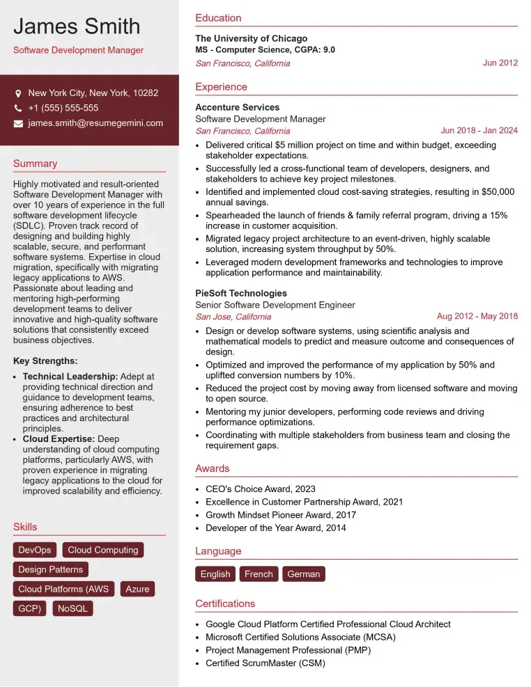
Check Out Resume Samples at ResumeGemini

Check Out Resume Samples at ResumeGemini

Check Out Resume Samples at ResumeGemini

Check Out Resume Samples at ResumeGemini

Check Out Resume Samples at ResumeGemini
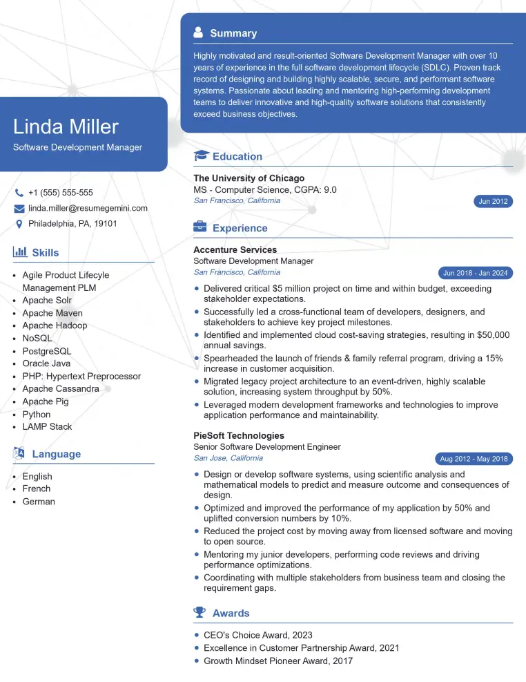
Check Out Resume Samples at ResumeGemini
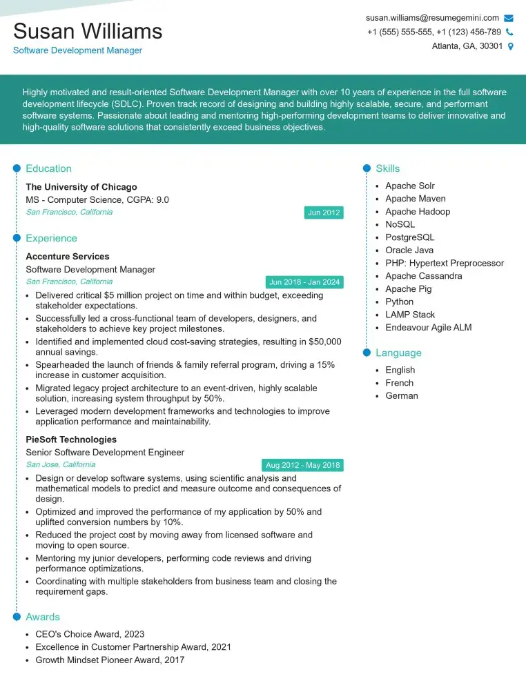
Check Out Resume Samples at ResumeGemini
Explore more articles
Users Rating of Our Blogs
Share Your Experience
We value your feedback! Please rate our content and share your thoughts (optional).
What Readers Say About Our Blog
To the interviewgemini.com Webmaster.
Very helpful and content specific questions to help prepare me for my interview!
Thank you
To the interviewgemini.com Webmaster.
This was kind of a unique content I found around the specialized skills. Very helpful questions and good detailed answers.
Very Helpful blog, thank you Interviewgemini team.