The right preparation can turn an interview into an opportunity to showcase your expertise. This guide to Forensic Physical Medicine interview questions is your ultimate resource, providing key insights and tips to help you ace your responses and stand out as a top candidate.
Questions Asked in Forensic Physical Medicine Interview
Q 1. Describe the process of determining time of death.
Determining the time of death, or post-mortem interval (PMI), is a complex process that relies on a combination of factors and is rarely an exact science. There’s no single test; instead, we utilize a multidisciplinary approach.
- Lividity (Livor Mortis): The settling of blood due to gravity. It helps estimate the position of the body after death and can give an approximate time since death if the livor is fixed (after roughly 8-12 hours).
- Rigor Mortis: The stiffening of muscles. It starts in smaller muscles, progresses to larger ones, and then eventually resolves. The timing is temperature-dependent and can help narrow down the PMI, but it’s also affected by factors like the individual’s activity level before death.
- Algor Mortis: The cooling of the body after death. This is affected by environmental temperature, body size, clothing, and other variables, making it a less precise indicator.
- Decomposition: The breakdown of tissues by bacterial action. The stages of decomposition (autolysis, putrefaction, etc.) provide clues about the PMI, but the rate is highly variable and impacted by factors such as temperature, humidity, and insect activity. We often use entomological evidence (insect life cycles) to help refine PMI estimates.
- Stomach contents analysis: The type and amount of food can provide some indication of the time elapsed since the last meal. It is not reliable but may provide a context.
- Witness statements and circumstantial evidence: This is crucial, and in many cases is the most reliable information.
In practice, we consider all these factors simultaneously. For example, if we find fixed lividity, early rigor, and minimal decomposition in a body found in a temperate climate, we might conclude a PMI of roughly 12-24 hours. However, this is an approximation, and a detailed report outlines all the considered elements along with their limitations.
Q 2. Explain the difference between manner and cause of death.
The cause of death is the specific disease, injury, or other event that directly caused the death. It is a medical determination. For example, the cause of death might be a gunshot wound to the chest, or acute myocardial infarction (heart attack).
The manner of death, on the other hand, describes the circumstances surrounding the cause of death. It’s a legal classification. The manners are typically categorized as: natural, accidental, suicidal, homicidal, and undetermined. A natural death might have a cause of heart disease. A homicide might have a cause of multiple stab wounds. The manner and cause work together to paint a complete picture of what happened.
Q 3. How do you identify and document injuries consistent with blunt force trauma?
Blunt force trauma refers to injuries caused by a blow from a non-penetrating object. Documentation is critical and involves a systematic approach.
- Visual Examination: We meticulously document the location, size, shape, and color of injuries. Photographs are taken at various angles and with scale markers. The presence of abrasions (scrapes), contusions (bruises), lacerations (tears), or fractures is noted. The distribution of injuries can provide clues about the type of object used and the direction of the force.
- Palpation: We carefully feel the area of injury to assess the depth and consistency of the tissue damage. This helps differentiate between superficial and deep injuries. We note any tenderness or crepitation (a crackling sound indicating fractured bone).
- Detailed Description: A thorough written report details each injury, its characteristics, and its location relative to anatomical landmarks (e.g., 2cm superior to the left eyebrow). This information is essential for reconstructing the events leading up to death.
- Photography and Videography: High quality photos and videos from multiple angles are essential. These serve as objective documentation that can be reviewed later by the team and the legal system.
- Radiological Examination: X-rays or CT scans may be needed to visualize underlying fractures or internal bleeding not visible on the surface. This is particularly helpful with blunt force injuries to the head or trunk.
For example, multiple contusions in a linear pattern on the back might suggest a beating with a rod or pipe, while a depressed skull fracture might suggest a forceful blow from a heavy object.
Q 4. What are the key features of a strangulation injury?
Strangulation injuries, whether by ligature (e.g., rope, wire) or manual (e.g., hands), result from compression of the neck’s blood vessels and/or airway. Key features include:
- Petechiae: Tiny, pinpoint hemorrhages (bleeding) in the conjunctiva (white of the eye), and in the skin of the face and neck. These are caused by increased pressure in the blood vessels. Their presence suggests strangulation, but their absence doesn’t rule it out.
- Ligature Marks: In ligature strangulation, a distinct groove may be present around the neck, reflecting the pressure from the constricting object. Its depth, width, and location provide valuable insights.
- Fractures: Fractures of the hyoid bone (a U-shaped bone in the neck) or thyroid cartilage are more common in manual strangulation, but they are not always present.
- Soft tissue injury: Abrasions and contusions can be present on the neck, depending on the method and force of strangulation.
- Internal Injuries: Internal injuries like hemorrhage in the neck muscles or larynx are often present but not always visibly apparent and require a careful autopsy.
Differentiating between manual and ligature strangulation often relies on the presence or absence of a ligature mark and the pattern of injuries. The absence of a ligature mark does not exclude strangulation as it can happen using soft materials. Careful and detailed examination are critical for accurate assessment and interpretation.
Q 5. Describe the process of collecting and preserving forensic evidence from a body.
Collecting and preserving forensic evidence from a body is a meticulous process that requires adherence to strict protocols to maintain the integrity of the evidence and prevent contamination.
- Scene Security: The scene must be secured to prevent unauthorized access. A detailed photographic and video record is made before any evidence is collected.
- Personal Protective Equipment (PPE): The forensic team must wear appropriate PPE, including gloves, masks, gowns, and eye protection, to avoid contamination of both the scene and themselves.
- Evidence Collection: Evidence is collected systematically, documented with photographs and written notes, and labeled appropriately. This includes clothing, hair, fibers, and any other items that may be relevant. Blood and other biological samples are collected using sterile techniques.
- Body Examination: A complete external examination of the body is conducted, documenting all injuries, wounds, and other significant findings. The body is photographed and video recorded.
- Autopsy: An autopsy is performed to determine the cause and manner of death. During the autopsy, further evidence is collected, including internal organ samples, tissue samples, and toxicology samples.
- Chain of Custody: A detailed chain of custody must be maintained for every piece of evidence to ensure its integrity. This means tracking who handled the evidence, when it was handled, and where it was stored.
- Packaging and Transportation: Evidence is packaged carefully to prevent damage or contamination during transportation to the laboratory.
The process is crucial because any lapse in procedure can compromise the entire investigation. Each step must be carried out with precision and attention to detail. For instance, improper collection of trace evidence at the scene or inappropriate handling during the autopsy can lead to the destruction of DNA profiles or crucial evidence.
Q 6. Explain your understanding of post-mortem changes.
Post-mortem changes are the alterations that occur in the body after death. These changes are influenced by factors such as environmental temperature, humidity, and the presence of insects. Understanding these changes is crucial for estimating the PMI and determining the cause and manner of death.
- Early Post-Mortem Changes: These include algor mortis (cooling of the body), rigor mortis (stiffening of the muscles), and livor mortis (settling of the blood).
- Decomposition: This is a complex process involving autolysis (self-digestion of tissues by enzymes) and putrefaction (breakdown of tissues by bacteria). The stages of decomposition are characterized by changes in skin color, bloating, and the release of gases.
- Skeletalization: This is the final stage of decomposition where only the skeleton remains. The rate of skeletalization depends on several factors including environmental conditions, soil composition, and animal scavenging.
- Mummification: In certain conditions (e.g., dry and hot climates), the body can undergo mummification, slowing down decomposition.
- Adipocere (Grave Wax): In moist environments, the body fat can convert into a waxy substance called adipocere, which can preserve the body for extended periods.
It’s important to note that the rate and progression of these changes vary significantly based on factors like environmental temperature, humidity, and insect activity. Therefore, understanding these influencing factors is essential when interpreting post-mortem changes.
Q 7. How do you differentiate between antemortem and postmortem injuries?
Differentiating between antemortem (before death) and postmortem (after death) injuries is essential for accurate determination of cause and manner of death. Several factors aid in this distinction:
- Evidence of Healing: Antemortem injuries will show signs of healing, such as the formation of a scab or scar tissue. Postmortem injuries lack these features.
- Presence of Blood Clots: Blood clots are typically present in antemortem injuries but absent in postmortem injuries. Postmortem injuries may have blood that remains liquid, rather than clotting normally.
- Inflammation: Inflammation is characteristic of antemortem injuries, but not typically seen in postmortem injuries.
- Microscopic Examination: Microscopic analysis of tissue surrounding the injury can help differentiate between antemortem and postmortem injuries. For example, the presence of inflammatory cells is indicative of antemortem injuries.
- Consideration of the circumstances and circumstances of injuries: The location and nature of the injuries, together with other evidence from the scene, help determine their timing.
For instance, a stab wound with a well-formed scab and surrounding inflammation would be classified as antemortem. In contrast, a clean-cut injury on the skin without evidence of inflammation or healing would be suspicious of a post-mortem injury – perhaps inflicted after death.
Q 8. Describe your experience with documenting wound characteristics.
Documenting wound characteristics is crucial in forensic physical medicine. It involves a systematic approach to record the precise details of any injury, forming a critical piece of evidence. This goes beyond simply stating ‘a cut on the arm’; it requires meticulous observation and recording.
- Location: Precise anatomical location using standardized terminology (e.g., ‘2cm proximal to the left ulnar styloid process’).
- Size and Shape: Measurements in centimeters (length, width, depth), shape (linear, stellate, circular), and any unusual features.
- Appearance: Color (red, purple, black), texture (smooth, ragged), presence of bruising (contusions), abrasions, or lacerations. The presence of foreign material embedded in the wound must also be meticulously documented.
- Edges: Sharp, irregular, or abraded edges can indicate the type of weapon used.
- Photography and Diagrams: High-quality photographs and detailed diagrams are essential. Scale markers must be included. Photos should be taken from multiple angles and distances, capturing the entire wound and its surrounding area.
- Additional details: Evidence of bleeding, healing, or infection should be noted. The presence of any foreign bodies within the wound should be described and documented.
For example, a description might read: ‘A 5cm long, linear laceration on the anterior aspect of the left forearm, 3cm proximal to the wrist crease. The wound edges are clean and sharply defined, consistent with a sharp object. Minimal bleeding is present. A photograph and diagram are attached.’ This level of detail is vital for accurate interpretation and for presenting the information effectively in court.
Q 9. How do you interpret toxicological findings in the context of a death investigation?
Toxicological findings are pivotal in death investigations. They reveal the presence and concentration of drugs, poisons, and other substances in the body, providing essential context for understanding the cause and manner of death. The interpretation isn’t simply about identifying substances; it’s about understanding their effects within the individual’s specific circumstances.
- Identifying Substances: The lab report will list detected substances and their concentrations. This requires knowledge of the various substances, their known effects, and possible interactions.
- Therapeutic vs. Toxic Levels: The concentration of a substance is compared to therapeutic (safe) levels. Concentrations exceeding therapeutic levels suggest potential toxicity. However, therapeutic drug monitoring is not always possible postmortem.
- Contextual Factors: Individual factors (age, health conditions, tolerance) and circumstances (time of ingestion, route of administration) significantly influence the interpretation. A lethal dose of a drug for one person might be subtherapeutic for another.
- Correlating with other evidence: Toxicological results are crucial when integrated with other findings (autopsy, scene investigation) to construct a holistic picture. For instance, a high blood alcohol concentration might explain the behavior described by witnesses at a scene of a motor vehicle accident.
- Postmortem redistribution: Drugs and chemicals can redistribute in the body after death, affecting concentration levels found in various samples. Expert knowledge is needed to account for such changes and reach accurate conclusions.
In a case where an individual is found deceased with a high concentration of a specific opioid, the toxicologist’s report is not just a list of chemicals, but a crucial piece of evidence indicating a possible overdose. This might require further investigation, such as interviewing potential witnesses or examining the scene to verify the timeline and possible route of administration.
Q 10. Explain the significance of Livor Mortis, Rigor Mortis, and Algor Mortis.
Livor mortis, rigor mortis, and algor mortis are post-mortem changes that help estimate the time of death. Understanding these changes is crucial for establishing a timeline of events in an investigation.
- Livor Mortis (Lividity): This is the settling of blood due to gravity after the heart stops. It begins within 30 minutes to 2 hours after death and becomes fixed after about 8-12 hours. The color of the lividity can provide clues, for example, bright red lividity might indicate carbon monoxide poisoning.
- Rigor Mortis (Stiffening): This is the stiffening of muscles due to chemical changes after death. It begins within 2-4 hours, becomes complete in 12-24 hours, and then gradually dissipates over the next 24-36 hours. The speed of onset and dissipation can be influenced by factors like temperature and physical activity before death.
- Algor Mortis (Cooling): This is the cooling of the body to ambient temperature after death. The rate of cooling depends on factors such as ambient temperature, body size, clothing, and whether the body was exposed to the elements. It generally follows a predictable pattern, which can be used to estimate the time since death.
By observing the degree of livor mortis, the presence and extent of rigor mortis, and the body temperature, a forensic pathologist can estimate the post-mortem interval (PMI) – the time elapsed since death. This estimate is rarely precise but provides crucial information for the investigation.
For example, a body showing fixed lividity, complete rigor mortis, and a near-ambient body temperature suggests a longer post-mortem interval than a body exhibiting early lividity and minimal rigor.
Q 11. How do you handle a case involving suspected child abuse?
Suspected child abuse cases require a highly sensitive and thorough approach. The priority is always the child’s safety and well-being. My approach involves:
- Detailed Medical Examination: A comprehensive physical examination meticulously documents all injuries, including their location, size, shape, age, and any underlying conditions. Imaging studies (X-rays, CT scans) might be necessary to identify internal injuries.
- Photography and Documentation: Detailed photographs and medical diagrams are essential for recording the injuries. This needs to be handled carefully to ensure the privacy and dignity of the child.
- Interviewing: While interviewing the child is crucial, it must be conducted by a trained professional, using age-appropriate techniques to avoid leading questions or traumatizing the child further. Interviews with parents and other caregivers are also critical to gather their perspectives, though caution is needed to avoid contamination of evidence.
- Multidisciplinary Approach: Child abuse investigations are almost always multidisciplinary. I work closely with law enforcement, child protective services, and social workers to ensure the child’s safety and to gather all relevant information.
- Child’s History: This includes the child’s medical history, developmental history and any previous encounters with the healthcare system.
- Reporting: My findings are thoroughly documented and reported to the appropriate authorities to ensure the child is protected and legal action is taken where necessary.
In these cases, my role goes beyond simply diagnosing injuries; it’s about providing evidence to support child protection efforts and the legal process. The welfare of the child is always paramount.
Q 12. Describe your familiarity with various types of weapons and their associated injuries.
Familiarity with various weapons and their associated injuries is crucial. Different weapons leave distinct patterns of injury, providing valuable clues in investigations.
- Sharp Weapons (Knives, Blades): Often produce incised wounds (cuts) with sharp, clean edges. Depth and length vary depending on the blade and force used. The wound trajectory can help determine the angle and direction of the attack.
- Blunt Weapons (Bats, Hammers): These create contusions (bruises), lacerations (tears), and sometimes fractures. The pattern of injury might reflect the shape of the weapon.
- Firearms: Produce gunshot wounds with characteristic features like entrance and exit wounds, powder burns, and stippling. The type of firearm and the distance of the shot leave distinctive markings.
- Projectile Weapons (Arrows, Bolts): These cause penetrating wounds, often with a distinct trajectory. The presence of the projectile itself provides strong evidence.
- Other Weapons: This encompasses tools, broken glass, and other instruments which create injuries that may help determine the causative object.
For example, a wound with a deep, clean-edged incision is suggestive of a sharp weapon, whereas a large, irregular laceration with surrounding bruising could indicate a blunt weapon. Careful examination and correlation with the scene investigation are key to accurately interpreting these injuries.
Q 13. How would you handle a situation where conflicting evidence is presented?
Conflicting evidence is not uncommon in forensic investigations. My approach involves a systematic and unbiased evaluation of all available information.
- Comprehensive Review: I meticulously review all evidence—medical records, witness statements, scene investigation findings, and laboratory results—paying close attention to potential biases or limitations.
- Critical Evaluation: Each piece of evidence is critically evaluated for its validity, reliability, and relevance. This involves considering the potential for contamination, error, or misinterpretation.
- Alternative Explanations: I consider all possible explanations for the conflicting evidence, actively seeking to identify potential sources of error or discrepancies. This may include statistical analysis of alternative hypotheses.
- Weight of Evidence: Based on my analysis, I assess the weight of evidence—determining which findings are most reliable and best support a specific conclusion. This often involves evaluating the relative strength of different pieces of evidence.
- Collaboration and Consultation: When faced with particularly complex or challenging cases, I may consult with other experts in relevant fields to gain additional perspectives and insights.
In the end, my report will reflect a comprehensive, reasoned analysis of the conflicting evidence, presenting all possibilities with their relative weight and supporting rationale. The goal is to provide a clear, objective assessment of the facts, even if there isn’t a definitive conclusion.
Q 14. Describe your approach to testifying in court.
Testifying in court requires clear, concise, and objective communication of my findings. My approach centers on:
- Preparation: Thorough review of the case file, including all medical records, reports, and photographs. I anticipate potential questions and prepare clear, concise answers.
- Clarity and Objectivity: I communicate my findings using simple, non-technical language where possible, avoiding jargon that a jury might not understand. I remain neutral and objective, presenting facts without personal opinions or speculation.
- Adherence to Ethical Guidelines: I adhere strictly to ethical guidelines for expert witnesses, ensuring impartiality and honesty. I refrain from offering opinions outside my expertise.
- Responding to Questions: I respond to questions directly and honestly, avoiding ambiguity. If I don’t know the answer to a specific question, I state it clearly.
- Visual Aids: I often use visual aids, such as photographs, diagrams, and charts, to illustrate my findings and make the information easier to understand.
My goal is not to ‘win’ the case, but to provide the court with a clear and accurate account of my medical findings, assisting in achieving justice. This ensures the scientific integrity of my work remains at the forefront, even when subjected to intense scrutiny.
Q 15. Explain your understanding of chain of custody protocols.
Chain of custody refers to the meticulous documentation of the handling, storage, and transfer of evidence from the crime scene to the courtroom. Think of it like a carefully maintained relay race; every person who touches the evidence must be accounted for, ensuring its integrity and admissibility in court. This process prevents contamination, tampering, or loss of evidence, protecting the legal process.
- Initial Securement: The first responder documents the location and condition of the evidence, sealing it in tamper-evident packaging.
- Transfer and Receipt: Each transfer of evidence is documented with a detailed record, including the date, time, names of individuals involved, and a description of the evidence. Signatures are usually required at each stage.
- Storage and Security: Secured storage facilities maintain the integrity of the evidence, preventing unauthorized access or environmental damage.
- Chain of Custody Form: A dedicated form meticulously tracks each step in the chain, creating an unbroken trail from crime scene to court. Any break in the chain can compromise the admissibility of the evidence.
For example, imagine a blood sample collected at a crime scene. The officer collecting it must document the location, time, and relevant case information, then sign the chain of custody form and transfer it to a lab technician who repeats the process. This continues until the evidence is presented in court.
Career Expert Tips:
- Ace those interviews! Prepare effectively by reviewing the Top 50 Most Common Interview Questions on ResumeGemini.
- Navigate your job search with confidence! Explore a wide range of Career Tips on ResumeGemini. Learn about common challenges and recommendations to overcome them.
- Craft the perfect resume! Master the Art of Resume Writing with ResumeGemini’s guide. Showcase your unique qualifications and achievements effectively.
- Don’t miss out on holiday savings! Build your dream resume with ResumeGemini’s ATS optimized templates.
Q 16. How do you document and interpret findings from a forensic autopsy?
Documenting a forensic autopsy involves a detailed, systematic approach that combines visual observations, measurements, and photography to build a comprehensive record. This record provides the foundation for interpreting the findings and determining the cause and manner of death.
- External Examination: This includes a thorough description of the body’s appearance, noting any injuries, identifying marks, or unusual features. Detailed photographs are crucial at this stage.
- Internal Examination: This involves a systematic dissection of the body, examining each organ for abnormalities, taking samples for toxicology and histology, and documenting any injuries or diseases found.
- Photography and Videography: Images document the body’s condition at each stage, providing visual evidence for the autopsy report.
- Autopsy Report: The culmination of the process, the report synthesizes all observations and findings into a concise and comprehensive summary, providing the cause and manner of death and other relevant information.
Interpreting the findings requires analyzing the correlation between the internal and external findings, the toxicology results, and any relevant case history. For example, the presence of specific injuries alongside toxicology findings can help determine if a death was accidental, homicidal, or suicidal.
Q 17. What are the ethical considerations in forensic physical medicine?
Ethical considerations in forensic physical medicine are paramount, ensuring objectivity, impartiality, and respect for the deceased and their families. The core principles involve maintaining the integrity of the scientific process, adhering to professional standards, and ensuring justice is served without compromising ethical responsibilities.
- Impartiality and Objectivity: Forensic pathologists must remain impartial and base their conclusions solely on evidence, avoiding bias or personal opinions.
- Confidentiality: Information obtained during the autopsy and investigation must remain confidential, protecting the privacy of the deceased and their families.
- Respect for the Deceased: The body must be treated with dignity and respect throughout the entire process.
- Transparency and Accountability: Forensic pathologists must be transparent in their methods and conclusions, accountable for their actions and findings.
For instance, a pathologist should not allow personal feelings about a case to influence their findings or report. They must also be transparent with the investigators and legal teams, ensuring they have access to all relevant information.
Q 18. Explain your experience with different types of forensic imaging techniques.
Forensic imaging plays a critical role in providing non-invasive and detailed visual information about injuries and underlying conditions. My experience encompasses a range of techniques, including:
- X-rays: Essential for detecting fractures, foreign objects, and certain types of internal injuries.
- Computed Tomography (CT) scans: Provide detailed cross-sectional images of the body, revealing internal injuries and organ damage in great detail.
- Magnetic Resonance Imaging (MRI): Excellent for visualizing soft tissues, such as brain injuries, spinal cord damage, and internal organ pathologies.
- Ultrasound: Useful for visualizing internal organs and identifying fluid accumulation, particularly in situations where radiation exposure is undesirable.
In practice, the choice of imaging modality depends on the specific circumstances of the case and the questions we are trying to answer. For example, a CT scan might be used to visualize a gunshot wound, while an MRI might be employed to assess brain injuries following a motor vehicle accident.
Q 19. Describe your knowledge of forensic entomology and its applications.
Forensic entomology utilizes the study of insects and other arthropods to aid in criminal investigations, primarily in determining the post-mortem interval (PMI) – the time elapsed since death. The stages of insect development on a decomposing body are predictable and provide valuable clues.
- PMI Estimation: By identifying the insect species present and their developmental stages, entomologists can estimate the time of death. Different insects colonize a body at different stages of decomposition.
- Location of Death: Insect species are specific to certain environments. The insects found on a body may indicate where the death occurred.
- Movement of a Body: The presence of certain insect species might suggest that a body was moved after death.
- Drug Detection: Insects can ingest and retain drugs present in the body, providing additional toxicological information.
For example, the presence of blowfly larvae of a specific age range on a body can provide a narrow window for the PMI. Combining this with other evidence from the autopsy and crime scene helps to construct a timeline of events.
Q 20. How would you determine the presence of alcohol or drugs in a deceased individual?
Determining the presence of alcohol or drugs in a deceased individual is a critical aspect of forensic toxicology. Samples are collected from various bodily fluids and tissues during the autopsy.
- Sample Collection: Blood, urine, vitreous humor (fluid in the eye), and other tissues are collected and stored in appropriate containers to prevent degradation.
- Analytical Techniques: Gas chromatography-mass spectrometry (GC-MS) and liquid chromatography-mass spectrometry (LC-MS) are commonly used to identify and quantify drugs and alcohol in these samples.
- Interpretation of Results: The concentrations of substances found are analyzed in conjunction with other autopsy findings to determine their significance in the cause and manner of death.
- Postmortem Redistribution: It is crucial to consider postmortem redistribution of drugs and alcohol, which can alter their concentrations in different tissues after death.
The results are vital in determining whether intoxication played a role in the cause of death, differentiating between accidental overdose and other causes. For example, high blood alcohol content may indicate alcohol poisoning, while the presence of certain prescription medications might help establish whether a death was accidental or intentional.
Q 21. How do you assess the reliability and validity of forensic evidence?
Assessing the reliability and validity of forensic evidence is essential for ensuring the integrity of the justice system. This involves careful consideration of various factors.
- Chain of Custody: A complete and unbroken chain of custody is vital to ensure the evidence has not been tampered with.
- Scientific Method: Forensic evidence must be collected and analyzed using scientifically validated methods.
- Validation of Techniques: The techniques used to analyze evidence must be scientifically valid and reliable.
- Error Rate: The potential for error in both the collection and analysis of evidence should be considered.
- Contextual Interpretation: Evidence must be interpreted in the context of the entire case, considering other evidence and circumstances.
For instance, DNA evidence is considered highly reliable when properly collected and analyzed, but its interpretation should be cautious, taking into account potential contamination or partial matches. Similarly, the interpretation of microscopic trace evidence needs to consider the possibility of transfer and the prevalence of similar materials in the environment.
Q 22. What are the legal requirements for conducting a forensic autopsy?
Conducting a forensic autopsy requires strict adherence to legal protocols, which vary slightly depending on jurisdiction but generally involve several key steps. First, there must be legal authority for the autopsy, typically a coroner’s or medical examiner’s order. This order is issued after a death is deemed suspicious, unexpected, or unexplained. The process always begins with clear identification of the deceased, usually through comparison of personal effects and fingerprints against official records.
Second, comprehensive documentation is paramount. This includes detailed photographic records of the body’s external appearance, including any wounds or injuries. A detailed written report is created alongside the photos that includes everything from the body’s condition to the findings of internal examination. Chain of custody procedures must be rigorously followed to maintain the integrity of any evidence collected during the autopsy. This means meticulous logging of who handled what, when, and under what circumstances. Finally, all findings must be presented clearly and objectively in a final autopsy report which serves as a legal document and is often used in criminal investigations or civil lawsuits.
For example, in a case of suspected homicide, the legal order for the autopsy will specify the need for a thorough examination focused on identifying potential signs of trauma, including microscopic examination of tissues. The report will then detail these findings, which may become crucial evidence in a subsequent criminal trial.
Q 23. Describe your experience with documenting and interpreting skeletal injuries.
My experience with skeletal injuries encompasses a wide range of cases, from ancient remains to recent trauma victims. The process begins with a careful visual inspection for fractures, dislocations, and other signs of damage. Advanced imaging techniques, such as X-rays and CT scans, provide invaluable supplemental data, revealing subtle fractures or internal damage invisible to the naked eye.
Interpreting these injuries requires a thorough understanding of skeletal anatomy and biomechanics. For instance, a spiral fracture of a long bone is highly suggestive of a twisting force, often seen in assaults or accidents. Conversely, a compression fracture in the vertebrae might indicate a fall from height or a significant impact. I also consider the context in which the injuries occurred, considering factors like the age and overall health of the individual.
For example, in one case, I examined skeletal remains where a depressed skull fracture was discovered; the pattern and severity of the fracture indicated a high-velocity impact, such as a gunshot wound. I carefully documented the exact location, size, and shape of the fracture using photographic documentation and 3D scanning to allow precise comparison to other evidence found at the scene.
Q 24. Explain your understanding of different types of asphyxia.
Asphyxia refers to a condition where the body is deprived of oxygen, leading to cellular damage and ultimately death. Several distinct types of asphyxia exist, each with its unique characteristics:
- Suffocation: This involves blocking the airway, preventing air from entering the lungs. Examples include smothering, choking, or being trapped in a confined space.
- Strangulation: This involves compression of the neck, obstructing blood flow to the brain. It can be either manual (with hands or a ligature) or accidental (such as hanging).
- Drowning: Involves the presence of fluid in the airways and lungs, preventing the uptake of oxygen.
- Chemical Asphyxia: This occurs when exposure to certain chemicals or gases, such as carbon monoxide or cyanide, prevents oxygen from binding to the hemoglobin in red blood cells.
The forensic examination of asphyxia often involves a combination of external and internal findings, including evidence of ligature marks, petechiae (small red spots on the skin), lung findings (such as pulmonary edema), and chemical analysis of blood and tissues. Distinguishing between different types of asphyxia is crucial for determining the cause and manner of death.
Q 25. How do you approach the investigation of a sudden unexpected death?
Investigating a sudden unexpected death (SUD) demands a systematic and thorough approach. The first step involves securing the scene and documenting everything – the body’s position, any surrounding objects, and the overall environment. Next, a detailed history is collected from family, witnesses, and healthcare providers, if available. This is followed by a complete external examination of the body, noting any visible injuries or unusual findings.
A forensic autopsy is then performed, including a thorough internal examination of all organs and systems. Toxicological testing is often crucial to detect the presence of drugs, alcohol, or other poisons that may have contributed to death. Microscopic examination of tissues (histopathology) can reveal subtle changes in organs not evident during the gross examination. In many SUD cases, a thorough investigation involving law enforcement, medical professionals, and potentially other specialists may be needed. The goal is to determine the precise cause and manner of death; this could range from a natural cause like a heart attack to something more complex such as an undiagnosed disease or even an unexpected adverse effect of a medication.
For example, a seemingly healthy young adult found dead in their bed might require an extensive investigation involving toxicology screening to rule out drug overdose or a thorough cardiological analysis to determine whether there was an undetected cardiac condition. A meticulous approach, combining clinical knowledge, forensic techniques, and collaboration with other specialists, is key to resolving such cases.
Q 26. Describe your knowledge of various types of traumatic brain injuries.
Traumatic brain injuries (TBIs) can range from mild concussions to severe, life-threatening injuries. They are broadly classified into two categories:
- Closed head injuries: These occur without penetration of the skull. Examples include contusions (bruises), diffuse axonal injury (shearing of nerve fibers), and cerebral edema (swelling of the brain).
- Open head injuries: These involve penetration of the skull, such as skull fractures, lacerations, and penetration by foreign objects (e.g., gunshot wounds).
The severity of a TBI is assessed based on factors such as the extent of the injury, the presence of associated neurological deficits (e.g., loss of consciousness, amnesia), and the duration of post-traumatic symptoms. Forensic examination involves a combination of external observations of the skull and scalp, imaging studies (CT scans and MRI), and a detailed neuropathological examination of the brain itself, which provides information about the location and severity of the brain damage.
For instance, a significant subdural hematoma (bleeding under the dura mater) often results from a severe blow to the head. This might require advanced imaging, like a CT scan, for accurate identification and assessment.
Q 27. Explain your experience with performing forensic examinations on various age groups.
My experience encompasses forensic examinations across various age groups, each presenting unique challenges and considerations. Infants and young children often have more fragile bones and less developed organ systems, requiring a particularly delicate approach during both the external and internal examinations. The interpretation of injuries in these age groups also necessitates a thorough understanding of child development and the types of injuries that might be inflicted through abuse or neglect.
Conversely, older adults may have pre-existing medical conditions that can complicate the determination of cause and manner of death. For example, a seemingly minor fall in an elderly person with osteoporosis could result in a fatal hip fracture.
In each age group, detailed documentation is essential. In cases involving suspected child abuse, the photographs and reports are particularly important, as they may become critical evidence in legal proceedings. In all cases, a careful and comprehensive approach is vital, considering the specific physiological characteristics and vulnerabilities of each age group.
Q 28. How do you handle stressful situations in the field of Forensic Physical Medicine?
Forensic Physical Medicine often involves exposure to traumatic scenes and emotionally challenging situations. Developing coping mechanisms is crucial for maintaining professional well-being. My strategies include:
- Maintaining a structured approach: A systematic approach to investigations helps minimize emotional overwhelm by providing a framework for processing information. The emphasis is on objective observation and data collection.
- Peer support and supervision: Discussing challenging cases with colleagues or supervisors provides a valuable opportunity for emotional processing and professional guidance.
- Self-care: Maintaining a healthy work-life balance, engaging in regular exercise, and seeking mental health support if needed are crucial for long-term well-being. This also includes taking appropriate time away from particularly disturbing cases.
- Professional boundaries: Establishing clear professional boundaries helps to avoid emotional entanglement with the cases being investigated, preventing emotional burnout.
It’s vital to remember that seeking help and acknowledging the emotional toll of this profession is not a sign of weakness; it’s a sign of resilience and self-awareness. Creating a supportive network and prioritizing self-care are crucial for maintaining both physical and mental health in this demanding field.
Key Topics to Learn for Forensic Physical Medicine Interview
- Trauma Analysis: Understanding mechanisms of injury, patterns of injury, and their correlation with the circumstances of death or assault. Practical application includes analyzing injury patterns in autopsies to reconstruct events.
- Death Investigation: Mastering the principles of death certification, including manner, cause, and mechanism of death. Practical application involves interpreting autopsy findings to determine the cause and manner of death in diverse cases.
- Forensic Pathology Techniques: Familiarizing yourself with autopsy procedures, sampling techniques, and the use of various imaging modalities. Practical application includes understanding the limitations and strengths of different techniques.
- Forensic Toxicology: Understanding the principles of drug metabolism, detection, and interpretation of toxicology reports. Practical application includes correlating toxicological findings with clinical presentation and injury patterns.
- Evidence Collection and Preservation: Understanding proper procedures for evidence collection, chain of custody, and preservation techniques to ensure admissibility in court. Practical application includes understanding the legal implications of improper handling of evidence.
- Legal and Ethical Considerations: Thorough understanding of medico-legal aspects, ethical dilemmas, and professional responsibilities in forensic physical medicine. Practical application includes navigating complex ethical situations encountered during investigations.
- Case Presentation and Communication: Developing clear and concise communication skills for presenting findings to legal professionals, investigators, and other stakeholders. Practical application includes presenting complex medical information in a clear and understandable manner.
Next Steps
Mastering Forensic Physical Medicine opens doors to a rewarding career path, offering unique challenges and opportunities to contribute significantly to the justice system. To maximize your job prospects, a strong and ATS-friendly resume is crucial. ResumeGemini is a trusted resource that can help you craft a compelling resume showcasing your skills and experience effectively. We provide examples of resumes tailored specifically to Forensic Physical Medicine to guide you in creating a document that stands out. Take the next step towards your ideal career—build a resume that reflects your expertise and passion.
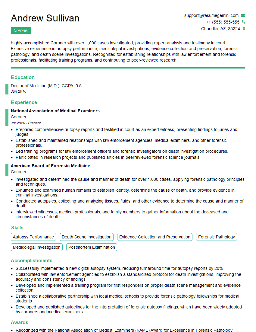
Coroner Resume Sample
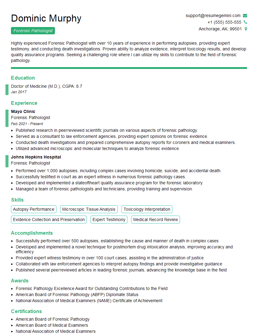
Forensic Pathologist Resume Sample
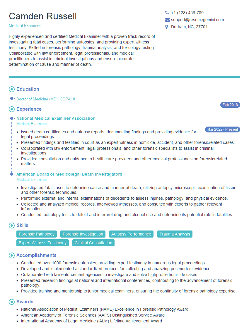
Medical Examiner Resume Sample
Forensic Chemist Resume Sample
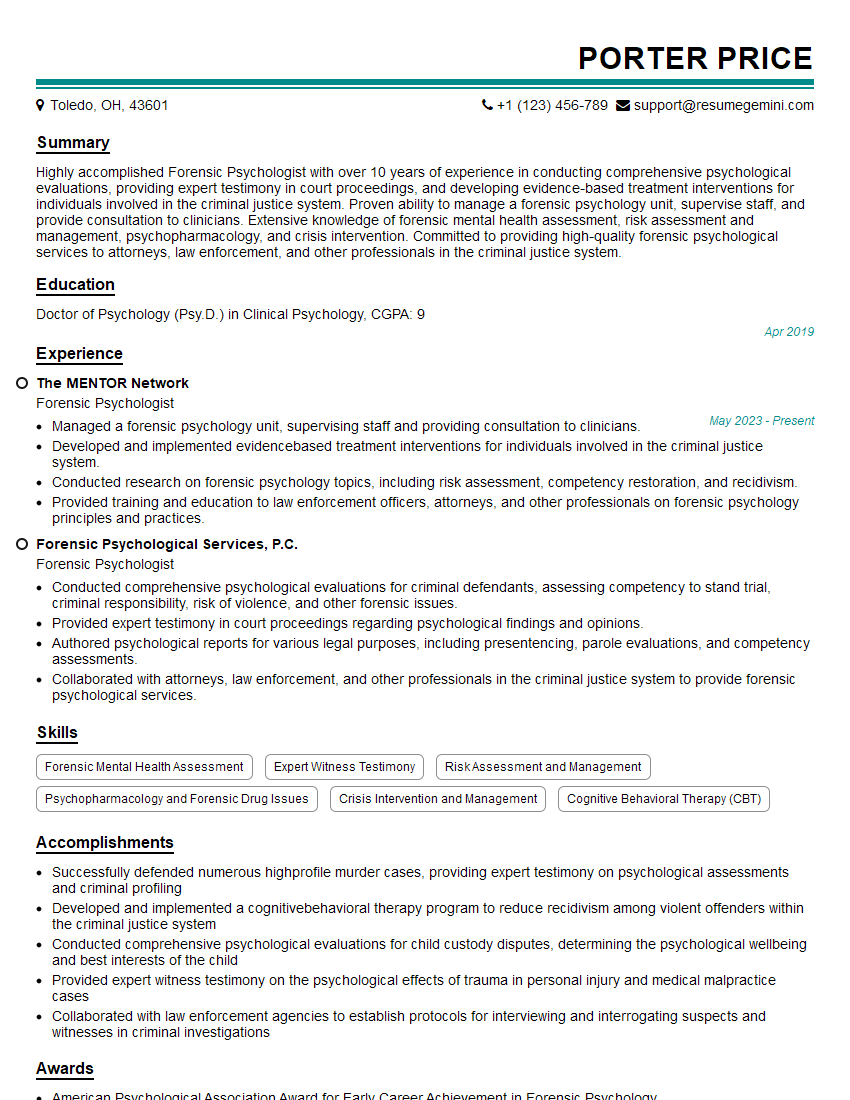
Forensic Psychologist Resume Sample
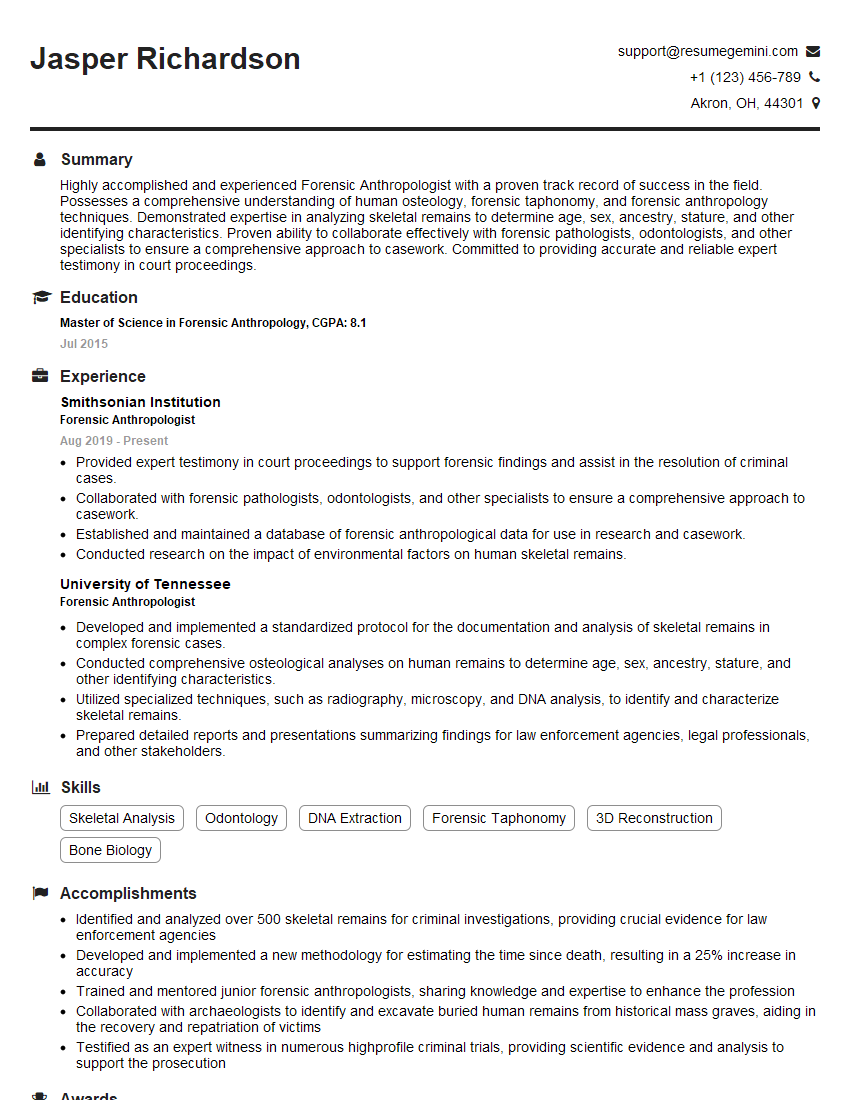
Forensic Anthropologist Resume Sample
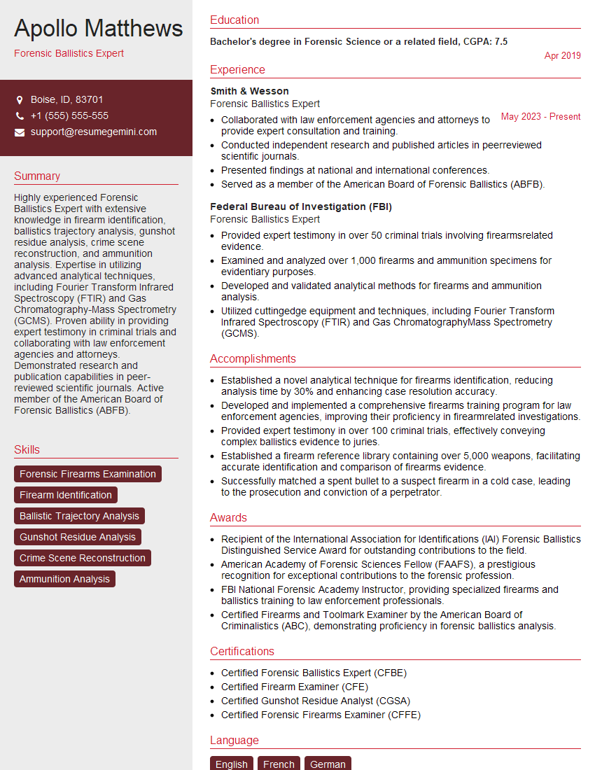
Forensic Ballistics Expert Resume Sample
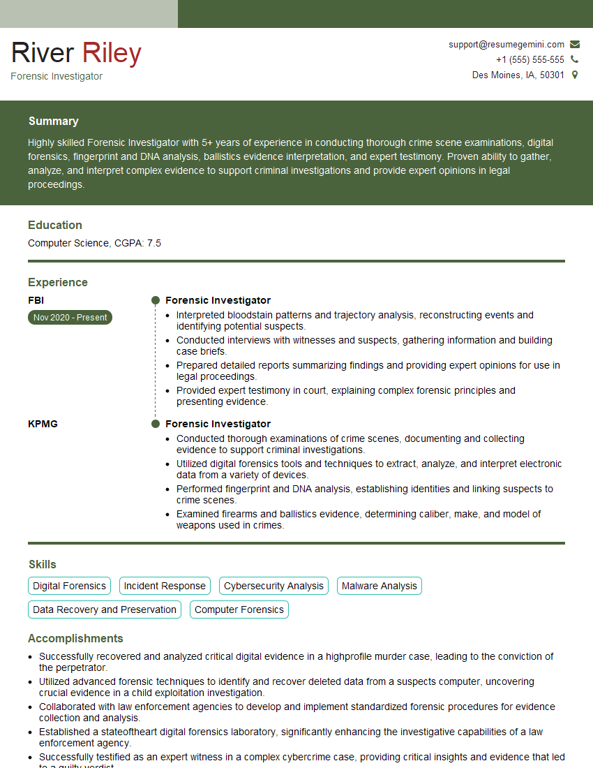
Forensic Investigator Resume Sample
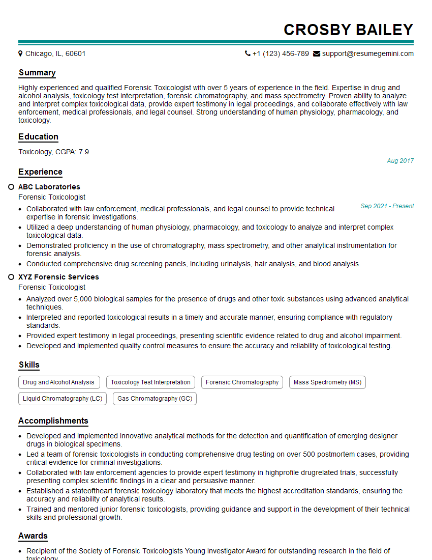
Forensic Toxicologist Resume Sample
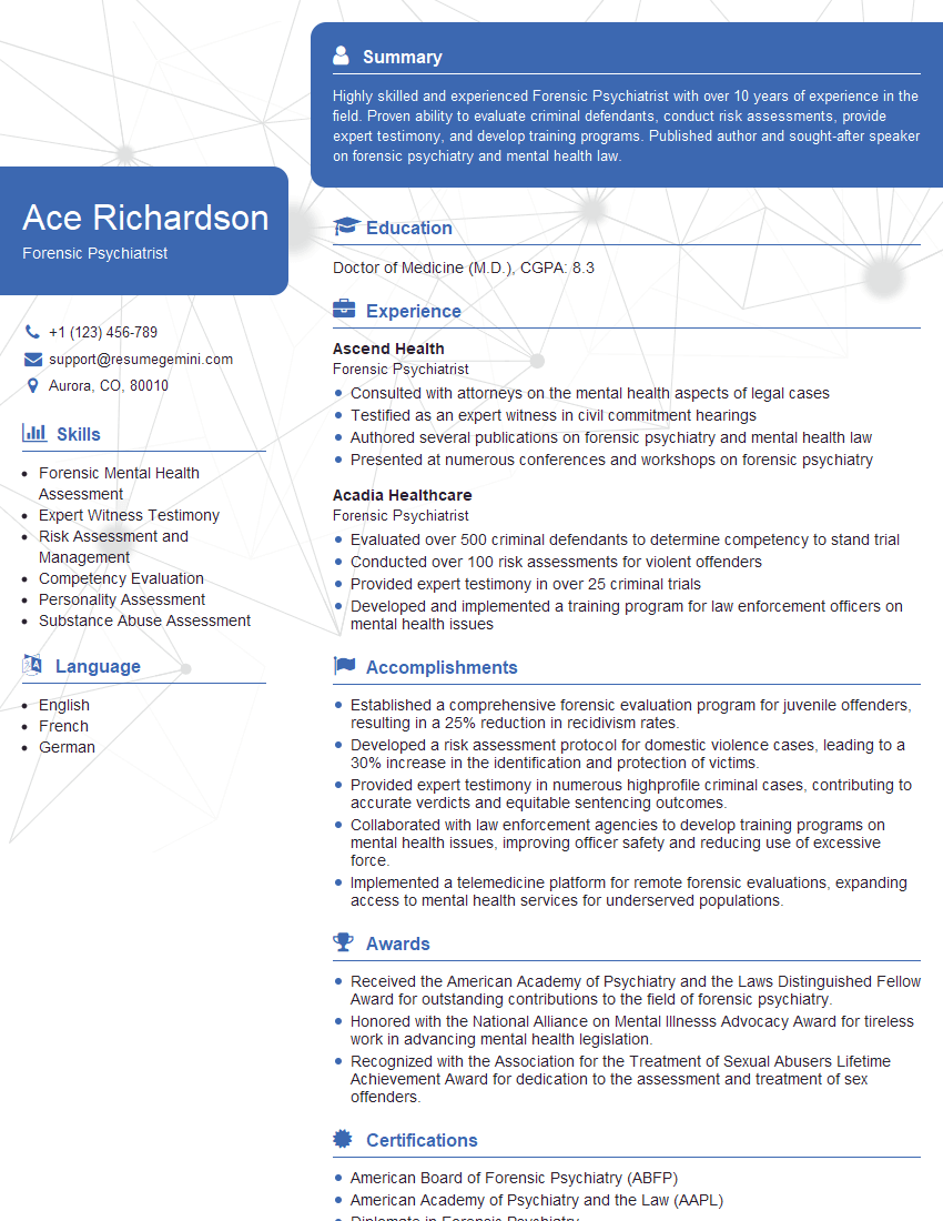
Forensic Psychiatrist Resume Sample
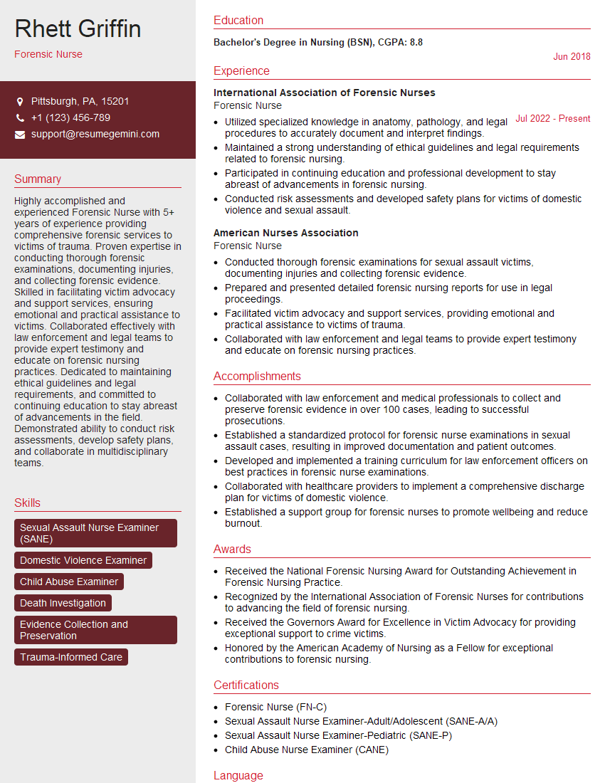
Forensic Nurse Resume Sample
Explore more articles
Users Rating of Our Blogs
Share Your Experience
We value your feedback! Please rate our content and share your thoughts (optional).
What Readers Say About Our Blog
To the interviewgemini.com Webmaster.
Very helpful and content specific questions to help prepare me for my interview!
Thank you
To the interviewgemini.com Webmaster.
This was kind of a unique content I found around the specialized skills. Very helpful questions and good detailed answers.
Very Helpful blog, thank you Interviewgemini team.