Preparation is the key to success in any interview. In this post, we’ll explore crucial Hepatectomy interview questions and equip you with strategies to craft impactful answers. Whether you’re a beginner or a pro, these tips will elevate your preparation.
Questions Asked in Hepatectomy Interview
Q 1. Describe the different surgical approaches for hepatectomy.
Hepatectomy, the surgical removal of a portion or all of the liver, can be approached in several ways, primarily dictated by the location and extent of the disease, as well as patient factors. These approaches can be broadly categorized as open, laparoscopic, or robotic-assisted.
- Open Hepatectomy: This traditional approach involves a large abdominal incision, providing excellent visualization and access to the liver. It’s often preferred for complex cases involving large tumors or extensive liver involvement. Imagine needing to open a large box to carefully extract a fragile object – open hepatectomy gives surgeons the most control.
- Laparoscopic Hepatectomy: This minimally invasive technique uses smaller incisions and specialized instruments to perform the surgery. Laparoscopic hepatectomy is generally suitable for smaller tumors in accessible locations. The benefits include less pain, shorter hospital stays, and faster recovery times, akin to performing a complex operation using only small tools and a camera for guidance.
- Robotic-Assisted Hepatectomy: Combining laparoscopic techniques with robotic technology, this method offers enhanced precision, dexterity, and visualization, particularly beneficial in complex anatomical situations. Think of a surgeon having a highly precise and stable ‘hand’ to work with within a confined space.
The choice of approach is a careful consideration based on factors such as tumor size and location, patient’s overall health, and surgeon’s expertise. A multidisciplinary team, including surgeons, radiologists, and oncologists, usually determines the optimal approach for each patient.
Q 2. Explain the Pringle maneuver and its limitations.
The Pringle maneuver is a temporary occlusion of the hepatic inflow (portal vein and hepatic artery) using a clamp placed across the hepatoduodenal ligament. This technique helps to control bleeding during liver resection by reducing blood flow to the liver. It’s a crucial tool in many hepatectomies, providing a ‘bloodless’ surgical field allowing for better visualization and precision.
However, the Pringle maneuver has limitations. Its duration must be limited to avoid ischemic injury to the liver. Typically, the maneuver is only clamped for periods of 15-20 minutes, followed by release to allow for reperfusion. Prolonged occlusion can lead to hepatocellular damage and potentially liver failure. Furthermore, tumors that have a rich collateral blood supply may not be effectively controlled by this maneuver. Finally, the Pringle maneuver can only be used in selected patients, and meticulous monitoring of the liver function is needed during and after the application of the maneuver.
Q 3. Discuss the role of pre-operative imaging in hepatectomy planning.
Pre-operative imaging plays a pivotal role in hepatectomy planning. It provides crucial information about tumor location, size, vascular supply, and the relationship to surrounding structures such as major blood vessels and bile ducts. The information helps in determining the feasibility and extent of resection.
- CT Scan with Contrast: This is often the first imaging modality used to identify the tumor and assess its size and extent. It helps define vascular anatomy, especially the hepatic veins and portal venous branches.
- MRI with Contrast: MRI offers better soft tissue contrast than CT, allowing for better delineation of tumor margins and assessment of tumor infiltration into surrounding structures, particularly the biliary system.
- Angiography (Hepatic Arteriography): This helps map the hepatic arterial supply to the tumor and surrounding liver parenchyma, crucial in planning the resection to preserve healthy liver tissue and avoid damaging essential arteries.
- 3D-Imaging: Reconstructed 3D images from CT or MRI data allow for more comprehensive planning of the resection, particularly for complex anatomical situations. It assists in evaluating resectability in relation to hepatic veins and major bile ducts.
These imaging modalities are used in conjunction with clinical examination to determine the best surgical approach and assess the patient’s overall fitness for surgery.
Q 4. How do you assess the feasibility of a hepatectomy based on imaging?
Assessing the feasibility of hepatectomy based on imaging relies on evaluating several key factors. The goal is to determine if the tumor can be completely resected while leaving behind sufficient healthy liver tissue to maintain adequate liver function.
- Tumor Size and Location: Large tumors that extend into major blood vessels or biliary structures may not be resectable. The location in relation to major hepatic veins, portal veins, and bile ducts also plays a significant role.
- Extent of Liver Involvement: The percentage of the liver occupied by the tumor is vital. Resection is generally not feasible if the remaining liver volume after resection is deemed insufficient for adequate liver function. This is often assessed using pre-operative liver function tests and imaging analysis.
- Vascular Anatomy: The imaging must show whether major vessels can be preserved after resection. If critical vessels are involved, resection might not be possible without potentially life-threatening complications.
- Biliary Anatomy: Similarly, the imaging assesses the involvement of bile ducts. Resection is challenging if there’s significant involvement, requiring complex biliary reconstruction, which may increase surgical risk.
The assessment is often performed by a multidisciplinary team, which includes surgeons, radiologists, hepatologists, and oncologists. They analyze the imaging data and combine it with the patient’s clinical status to arrive at a joint decision regarding the feasibility of the surgery.
Q 5. What are the key considerations for intraoperative blood management during hepatectomy?
Intraoperative blood management during hepatectomy is critical due to the liver’s high vascularity. Significant blood loss can lead to hypovolemic shock and potentially life-threatening complications. Strategies for effective blood management include:
- Pre-operative Autologous Blood Donation: Patients can donate their own blood before surgery, reducing the need for allogeneic transfusions. This decreases the risk of transfusion-related complications.
- Cell Salvage: During the surgery, a cell salvage device can collect shed blood, process it, and re-infuse it into the patient. This minimizes blood loss and reduces the need for allogeneic transfusions.
- Hypotensive Anesthesia: Controlled hypotension can be used to decrease bleeding during certain phases of the procedure, though it needs careful monitoring to avoid organ ischemia.
- Surgical Techniques: Precise surgical techniques, including the use of harmonic scalpel and meticulous dissection, minimize bleeding.
- Pringle Maneuver (as discussed previously): This maneuver plays a significant role in controlling blood flow.
- Use of Hemostatic Agents: Fibrin sealant and other hemostatic agents can help control bleeding from small vessels.
Careful monitoring of blood pressure, heart rate, and hemoglobin levels throughout the procedure is crucial for successful blood management.
Q 6. Detail the steps involved in performing a right hepatectomy.
A right hepatectomy involves the resection of the right lobe of the liver, encompassing segments V, VI, VII, and VIII. The procedure is complex and involves a step-by-step approach:
- Exposure and Mobilization: A large right subcostal incision is typically made. The right lobe is carefully mobilized from adjacent structures.
- Control of the Hepatic Vascular Supply: The right hepatic artery and portal vein branches are identified and controlled, either by clamping or using ligatures. The Pringle maneuver is often employed.
- Dissection of the Biliary System: The right hepatic duct is dissected and identified, often requiring careful dissection to avoid damage.
- Division of the Hepatic Parenchyma: Using ultrasonic shears, radiofrequency ablation, or electrocautery, the parenchyma is divided along the planned resection line, creating a relatively bloodless surgical field.
- Hepatic Vein Division: The right hepatic vein is carefully divided using sutures, ensuring hemostasis (stopping the bleeding) to prevent injury to the left hepatic vein.
- Resection and Closure: The resected specimen is removed. The raw liver surface is inspected carefully, and any bleeding points are controlled. The abdominal cavity is irrigated and closed in layers.
Postoperative care includes monitoring liver function, managing pain, and preventing complications such as infection and bleeding. The specific steps and details might vary depending on the individual case and the surgeon’s preference.
Q 7. Describe the different types of liver resections (e.g., wedge resection, segmentectomy).
Liver resections vary in extent, ranging from removal of a small wedge to a major hepatectomy. The choice of resection is tailored to the location and size of the lesion, preserving as much healthy liver tissue as possible.
- Wedge Resection: This involves the removal of a small, wedge-shaped piece of liver tissue. It’s suitable for small, superficial lesions that are not close to major vessels or bile ducts. Imagine removing a small, triangular piece of cake.
- Segmentectomy: This procedure involves the removal of one or more liver segments (functional units of the liver). This offers a more precise resection than wedge resection and preserves more liver parenchyma. Think of taking out a defined section of a cake instead of a random wedge.
- Lobe Resection (e.g., Right Hepatectomy, Left Hepatectomy): This involves removing a larger portion of the liver, encompassing several segments. It’s performed for larger tumors or those involving multiple segments. This is like removing a large portion of the cake.
- Extended Hepatectomy: This includes removing portions of the liver that extend beyond the typical anatomical boundaries, potentially involving resection of nearby structures such as parts of the inferior vena cava or adjacent organs. This is a more extensive procedure reserved for rare, complex cases.
The choice of resection strategy is carefully planned using pre-operative imaging and a comprehensive assessment of the patient’s overall health. The goal is to remove all diseased tissue while maximizing the chance of a complete cure and preserving enough healthy liver tissue to sustain liver function post-surgery.
Q 8. What are the common postoperative complications of hepatectomy?
Postoperative complications following hepatectomy are unfortunately common and can range in severity from minor to life-threatening. The most frequent complications can be broadly categorized into:
- Bleeding: This is a major concern, especially in cases involving larger resections or challenging vascular anatomy. Early recognition and prompt intervention are crucial.
- Biliary complications: These include bile leaks, strictures, and cholangitis (infection of the bile ducts). They often necessitate further procedures, such as endoscopic stenting or surgery.
- Infection: Surgical site infections, pneumonia, and other infections are possible, especially in patients with underlying comorbidities or prolonged hospital stays. Prophylactic antibiotics are commonly used.
- Liver failure: Postoperative liver failure is a serious complication, particularly in patients with underlying liver disease or insufficient liver remnant function. This requires intensive supportive care.
- Renal failure: Reduced blood flow to the kidneys post-surgery, especially in patients with pre-existing renal impairment, can lead to kidney failure.
- Abdominal compartment syndrome: Fluid accumulation in the abdomen can lead to increased abdominal pressure, potentially compromising organ function.
- Thromboembolic events: Deep vein thrombosis (DVT) and pulmonary embolism (PE) are risks following major abdominal surgery. Prophylactic anticoagulation is often employed.
The incidence and severity of these complications depend on various factors, including the extent of resection, the patient’s overall health, and the surgical technique employed. Careful preoperative planning and meticulous surgical technique are key to minimizing these risks.
Q 9. How do you manage biliary complications following hepatectomy?
Management of biliary complications post-hepatectomy requires a multidisciplinary approach and depends on the specific complication. The initial assessment involves thorough clinical evaluation, imaging studies (e.g., CT scan, MRCP), and often, endoscopic retrograde cholangiopancreatography (ERCP).
- Bile leaks: Small bile leaks may resolve spontaneously with conservative management (e.g., drainage, nutritional support). Larger leaks typically require endoscopic or surgical intervention, often involving placement of stents or drains.
- Biliary strictures: These narrowings of the bile ducts often require endoscopic dilation or placement of stents. In some cases, surgical reconstruction may be necessary.
- Cholangitis: This infection requires prompt treatment with antibiotics, often broad-spectrum initially, tailored to culture results. Drainage procedures (e.g., percutaneous drainage or ERCP) may be necessary to remove infected bile.
Close monitoring of liver function tests, serum bilirubin levels, and clinical status is crucial in the management of biliary complications. Regular follow-up with imaging studies helps assess the efficacy of treatment and identify any recurrence or new complications.
Q 10. Describe the management of post-hepatectomy liver failure.
Post-hepatectomy liver failure (PHLF) is a life-threatening complication characterized by insufficient liver function to support life. Management is largely supportive and focuses on optimizing organ function while allowing the remaining liver to regenerate. Early recognition and prompt intervention are crucial.
- Intensive Care Unit (ICU) admission: Close monitoring of vital signs, fluid and electrolyte balance, and hepatic function is necessary.
- Nutritional support: Adequate nutrition is essential for liver regeneration. This may involve parenteral nutrition (TPN) in cases of severe liver failure or inability to tolerate oral intake.
- Medical management: Addressing underlying causes and supportive measures like medications to manage encephalopathy, ascites, and bleeding are vital.
- Liver transplantation: In cases of severe and refractory PHLF, liver transplantation may be the only life-saving option. Careful assessment of patient suitability is crucial, considering other medical conditions and the availability of a suitable donor liver.
The prognosis of PHLF is dependent on various factors, including the extent of the initial liver resection, the patient’s pre-operative liver function, and the presence of other comorbidities. Early diagnosis and aggressive management significantly improve survival rates.
Q 11. Explain your approach to assessing liver function pre- and post-hepatectomy.
Pre- and postoperative assessment of liver function is crucial in planning and managing hepatectomy. A thorough assessment involves a combination of:
- Liver function tests (LFTs): These include serum bilirubin, albumin, alanine aminotransferase (ALT), aspartate aminotransferase (AST), and prothrombin time (PT). These tests help assess the liver’s synthetic and metabolic capacity.
- Imaging studies: CT scans, MRI scans, and sometimes PET scans, provide detailed anatomical information about the liver, including the location and extent of the lesion, as well as the assessment of the future liver remnant (FLR).
- Liver volumetry: This technique quantifies the volume of the FLR and helps determine the amount of liver tissue that will remain after resection. This is critical for assessing the risk of PHLF.
- Indocyanine green clearance test (ICG): This test helps assess hepatic blood flow and function, providing an additional measure of liver reserve capacity.
Postoperatively, similar tests are used to monitor the recovery of liver function, detect any complications (such as liver failure), and guide further management. Serial measurements of LFTs are critical for evaluating the healing process and adjusting treatment as needed. Imaging studies can be used to detect complications like biloma or abscess.
Q 12. What are the indications and contraindications for hepatectomy?
Hepatectomy is a major surgical procedure with specific indications and contraindications.
- Indications: Hepatectomy is indicated for the treatment of various benign and malignant liver diseases, including:
- Hepatocellular carcinoma (HCC): A primary liver cancer.
- Cholangiocarcinoma: Cancer of the bile ducts.
- Metastatic liver disease: Cancer that has spread to the liver from another primary site.
- Benign liver tumors: Such as hemangiomas, focal nodular hyperplasia (FNH), and adenomas.
- Liver trauma: Severe liver injuries may require resection of damaged liver tissue.
- Liver abscesses: Large or non-responsive liver abscesses.
- Contraindications: Absolute contraindications include:
- Severe liver disease: Patients with advanced cirrhosis or significant liver dysfunction have an unacceptably high risk of PHLF.
- Extensive vascular involvement: Tumors with significant involvement of major hepatic vessels may be inoperable.
- Inability to achieve adequate future liver remnant (FLR): If the remaining liver tissue isn’t sufficient to maintain life, resection isn’t possible.
Relative contraindications, where the benefits may outweigh the risks depending on the specific situation, include significant cardiovascular or pulmonary comorbidities, disseminated intravascular coagulation (DIC), and uncontrolled infection.
Q 13. How do you determine the extent of resection needed in a patient with liver metastases?
Determining the extent of resection for liver metastases requires a careful assessment balancing the need for complete tumor removal with the preservation of adequate liver function. This is a complex decision that considers several factors.
- Number and size of metastases: Smaller, fewer metastases are more likely to be amenable to complete resection.
- Location of metastases: Metastases located in critical areas (e.g., near major blood vessels or the biliary tree) may be technically challenging or impossible to remove completely.
- Future liver remnant (FLR): The FLR must be sufficient to maintain adequate liver function. Preoperative assessment using imaging and liver function tests is critical.
- Patient’s overall health: The patient’s general health and ability to tolerate a major surgical procedure play a significant role in determining the extent of possible resection.
Advanced imaging techniques, such as CT scans and MRI scans with contrast enhancement, are used to accurately delineate the extent of the disease. Surgical planning often involves multidisciplinary discussions between surgeons, oncologists, radiologists, and other specialists to determine the optimal approach.
In some cases, a staged approach may be employed, with smaller resections performed over time to allow for liver regeneration. This is especially important when maximizing the potential to remove all visible disease without compromising hepatic function is the goal.
Q 14. Discuss the role of minimally invasive techniques in hepatectomy.
Minimally invasive techniques, such as laparoscopic and robotic hepatectomy, are playing an increasingly important role in the management of liver tumors. These techniques offer several potential advantages compared to open surgery.
- Smaller incisions: Leading to less pain, reduced blood loss, and shorter hospital stays.
- Improved cosmesis: Smaller incisions result in smaller and less visible scars.
- Reduced risk of postoperative complications: Some studies suggest lower rates of infection and other complications with minimally invasive techniques, though the evidence isn’t universally conclusive across all scenarios.
However, minimally invasive hepatectomy is not always feasible. The complexity of the procedure, the size and location of the tumor, and the surgeon’s expertise are critical considerations. Laparoscopic and robotic approaches are particularly well-suited for smaller resections and specific tumor locations. For more extensive or complex procedures, open surgery may still be the preferred approach. The choice of technique should be individualized based on a thorough assessment of the patient and the nature of the disease.
Ongoing research and technological advancements continue to expand the applications of minimally invasive techniques in hepatectomy, making them a valuable option for appropriately selected patients.
Q 15. What are the advantages and disadvantages of robotic-assisted hepatectomy?
Robotic-assisted hepatectomy offers several advantages over open surgery, primarily improved precision and minimally invasive access. The smaller incisions lead to less pain, faster recovery times, and reduced blood loss. The magnified 3D vision provided by the robotic system allows for intricate dissection in challenging anatomical areas, particularly beneficial in complex liver resections. However, disadvantages exist. The high initial cost of the robotic system and specialized training required for surgeons are significant barriers. Furthermore, the lack of tactile feedback compared to open surgery can pose a challenge, potentially increasing the risk of inadvertent injury to vital structures. Additionally, the longer operative time compared to some experienced open surgeons, though often offset by faster recovery, needs consideration. For example, in a patient with a complex tumor near the hepatic veins, the robotic approach allows for precise dissection minimizing the risk of bleeding and injury to these vital vessels. Conversely, in a patient with a large, easily accessible tumor, an open approach might be equally effective and less costly.
Career Expert Tips:
- Ace those interviews! Prepare effectively by reviewing the Top 50 Most Common Interview Questions on ResumeGemini.
- Navigate your job search with confidence! Explore a wide range of Career Tips on ResumeGemini. Learn about common challenges and recommendations to overcome them.
- Craft the perfect resume! Master the Art of Resume Writing with ResumeGemini’s guide. Showcase your unique qualifications and achievements effectively.
- Don’t miss out on holiday savings! Build your dream resume with ResumeGemini’s ATS optimized templates.
Q 16. How do you manage a bleeding complication during hepatectomy?
Managing bleeding during hepatectomy is critical, as it can rapidly lead to significant morbidity and mortality. Our approach is multi-pronged and begins with meticulous surgical technique, using careful dissection, optimal visualization, and precise hemostasis at every step. We utilize various methods including electrocautery, ultrasonic shears, and pressure to control bleeding from smaller vessels. For larger bleeding vessels, we employ techniques such as suture ligation, clips, and advanced methods like argon beam coagulation. In cases of uncontrollable bleeding, we may use temporary Pringle maneuver (occluding the hepatic artery and portal vein) to control inflow. In extremely challenging situations, we may resort to packing the bleeding site temporarily to gain better control and then proceed with definitive repair. Blood product transfusion is readily available if necessary. Rapid response and proactive management are crucial to minimizing the impact of bleeding complications. For instance, in a case involving a large liver hemangioma, the initial steps will involve careful dissection away from the lesion to avoid rupture before applying ligation to any feeding vessels.
Q 17. Describe your experience with different types of liver tumors.
My experience encompasses a wide spectrum of liver tumors. Hepatocellular carcinoma (HCC) is the most common primary liver malignancy I encounter, ranging from small, solitary lesions amenable to resection to large, multifocal tumors requiring extensive hepatectomy or transplantation. I have also managed various benign tumors such as focal nodular hyperplasia (FNH) and hepatic adenomas, requiring individualized approaches based on tumor size, location, and patient characteristics. In addition, I have experience with metastatic liver disease from various primary sources, including colorectal cancer, breast cancer, and lung cancer. These cases often require complex resections, involving multiple segments of the liver, and the surgical approach is tailored to achieve maximal tumor removal while preserving sufficient liver function. Each case presents unique challenges and requires careful preoperative planning and intraoperative decision-making to optimize patient outcomes. For example, I recently managed a patient with a large HCC that involved the hepatic vein; this required a complex anatomical resection involving reconstruction of the hepatic vein.
Q 18. What are the criteria for liver transplantation in patients with liver disease requiring hepatectomy?
The criteria for liver transplantation in patients requiring hepatectomy are complex and depend on several factors, including tumor characteristics, liver function, overall health status, and the presence of other comorbidities. For HCC, the Milan criteria (single tumor ≤5 cm or up to three tumors, each ≤3 cm, with no vascular invasion) are often used as a guideline for eligibility, although other factors are considered. If a patient has advanced disease exceeding the Milan criteria or evidence of vascular invasion, liver transplantation is usually not an option. Patients who have significant underlying liver disease compromising their liver function might be considered for a transplant as an alternative to hepatectomy, particularly if hepatectomy would leave insufficient liver reserve. Furthermore, the patient’s overall health status and suitability for major surgery and subsequent immunosuppression are critical considerations. For instance, a patient with a small HCC but severe cirrhosis might be a better candidate for transplantation despite meeting the criteria for hepatectomy. Each case requires individualized assessment and a multidisciplinary team approach.
Q 19. How do you counsel patients about the risks and benefits of hepatectomy?
Counseling patients about hepatectomy involves a careful discussion of the risks and benefits tailored to their individual circumstances. I begin by explaining the diagnosis and the rationale for surgery in a clear and concise manner, using anatomical models and diagrams to aid understanding. I then explain the potential benefits of surgery, including tumor removal, improved survival rates, and symptom relief. However, I also openly discuss potential complications, such as bleeding, infection, bile leaks, liver failure, and the need for blood transfusions. I provide realistic expectations about recovery time, potential limitations in post-operative activity, and long-term outcomes. This shared decision-making process empowers patients to make informed choices about their care. A patient with a large tumor may be informed that while the potential benefits are significant, the risks associated with surgery are higher due to the complexity of the procedure. In contrast, a patient with a small, easily accessible tumor would receive a more favorable prognosis.
Q 20. What are the current guidelines for post-operative pain management after hepatectomy?
Post-operative pain management after hepatectomy is crucial for patient comfort and optimal recovery. We utilize a multimodal approach, combining analgesics such as opioids with non-opioid pain relievers like NSAIDs and acetaminophen. Regional anesthesia techniques, such as paravertebral blocks or transversus abdominis plane (TAP) blocks, are often employed to provide effective pain control while reducing the need for systemic opioids. Patient-controlled analgesia (PCA) pumps allow patients to manage their pain effectively. We carefully monitor patients for pain levels, side effects of analgesics, and potential signs of complications. Regular assessment and adjustment of the analgesic regimen based on the patient’s response are essential. The transition from intravenous to oral analgesics is managed systematically to ensure adequate pain control during the recovery phase. For instance, we often initiate a TAP block in the operating room, which provides significant pain relief in the early post-operative period, minimizing the need for high doses of opioids and preventing opioid-related side effects.
Q 21. Discuss the role of adjuvant therapies (chemotherapy, radiation) in hepatocellular carcinoma.
Adjuvant therapies play a crucial role in the management of hepatocellular carcinoma (HCC), particularly after surgical resection. The decision to use adjuvant chemotherapy or radiation therapy depends on several factors, including the stage of the disease, the presence of microscopic disease, and the patient’s overall health. In advanced-stage HCC, systemic chemotherapy, often using regimens combining drugs like sorafenib or lenvatinib, is commonly used to improve survival rates. Radiation therapy can be used in select cases to target residual disease or unresectable tumors. However, radiation therapy is less common due to the risk of damage to the liver itself. Targeted therapy, which aims to selectively inhibit tumor growth while limiting damage to healthy cells, is increasingly used in the treatment of HCC. These adjuvant therapies are used in conjunction with regular surveillance to monitor for disease recurrence. The selection of adjuvant therapy is tailored to individual patients, taking into account tumor characteristics, patient health, and potential side effects. For example, a patient with a large HCC with microscopic residual disease after resection might receive adjuvant chemotherapy to reduce the chance of recurrence.
Q 22. What are the latest advancements in hepatectomy techniques?
Hepatectomy, the surgical removal of part or all of the liver, has seen significant advancements. These improvements center around minimally invasive techniques, improved surgical planning, and enhanced patient recovery.
- Minimally Invasive Approaches: Laparoscopic and robotic hepatectomies are becoming increasingly prevalent. These methods offer smaller incisions, reduced blood loss, less postoperative pain, and faster recovery times compared to open surgery. For example, robotic surgery allows for greater precision and dexterity, particularly in complex cases involving intricate liver anatomy.
- Advanced Imaging and Planning: Preoperative imaging techniques like 3D CT scans and MRI, coupled with sophisticated software, allow surgeons to create detailed 3D models of the liver. This allows for precise surgical planning, minimizing the risk of damage to surrounding structures like blood vessels and bile ducts. This is particularly crucial in complex cases where tumors are located near critical vascular structures.
- Improved Surgical Techniques: New techniques like radiofrequency ablation (RFA) and microwave ablation are being used more frequently to pre-treat tumors or to perform resections in less invasive ways, particularly for smaller tumors. These techniques cause less tissue damage compared to traditional surgical resection.
- Enhanced Recovery After Surgery (ERAS) Protocols: ERAS protocols involve a multidisciplinary approach to optimize patient care before, during, and after surgery. This includes strategies like enhanced analgesia, early mobilization, and optimized nutrition, contributing to significantly reduced hospital stays and improved patient outcomes.
Q 23. How do you manage a patient with a suspected bile duct injury after hepatectomy?
Managing a suspected bile duct injury after hepatectomy is a surgical emergency requiring immediate attention. Prompt diagnosis and appropriate intervention are crucial to prevent serious complications like cholangitis (infection of the bile ducts), liver abscess, and sepsis.
- Immediate Assessment: A high index of suspicion is needed. Postoperative symptoms like fever, jaundice, abdominal pain, and elevated bilirubin levels should prompt immediate investigation.
- Imaging Studies: MRCP (Magnetic Resonance Cholangiopancreatography) or ERCP (Endoscopic Retrograde Cholangiopancreatography) are used to precisely locate and characterize the injury.
- Surgical Repair: The approach depends on the severity and location of the injury. Options include direct repair of the bile duct, Roux-en-Y hepaticojejunostomy (connecting the bile duct to the jejunum), or placement of a biliary stent.
- Postoperative Management: Postoperative management includes aggressive fluid resuscitation, broad-spectrum antibiotics, and close monitoring for signs of infection or other complications. Regular imaging studies may be needed to assess the effectiveness of the repair.
For instance, in one case I encountered, a patient developed a bile leak post-hepatectomy. ERCP revealed a small tear in the common hepatic duct. We successfully repaired the leak using a stent, and the patient recovered without further complications.
Q 24. Describe your approach to perioperative infection prevention in hepatectomy.
Perioperative infection prevention in hepatectomy is paramount, given the liver’s proximity to the gut and its high vascularity. Our approach is multifaceted and incorporates several key strategies:
- Preoperative Optimization: This involves careful assessment and management of any existing infections, optimization of nutritional status, and smoking cessation counseling.
- Surgical Technique: Maintaining meticulous surgical technique, minimizing tissue trauma, and achieving hemostasis (stopping bleeding) are crucial to prevent infection. This includes using appropriate surgical instruments, carefully handling tissues, and employing efficient bleeding control techniques.
- Prophylactic Antibiotics: Appropriate prophylactic antibiotics are administered before the incision and continued for a limited duration postoperatively. The choice of antibiotic depends on local susceptibility patterns.
- Strict Aseptic Techniques: Adherence to strict sterile techniques in the operating room, including proper gowning and gloving, is essential.
- Postoperative Wound Care: Meticulous wound care, including proper dressing changes and monitoring for signs of infection, is vital.
- Intensive Monitoring: Close monitoring of the patient’s vital signs, inflammatory markers, and clinical status postoperatively is necessary to detect infection early.
Q 25. Explain your experience with using different types of surgical instruments and equipment in hepatectomy.
My experience spans a wide range of surgical instruments and equipment used in hepatectomy. The selection depends on the complexity of the procedure and the individual patient’s anatomy.
- Dissecting Instruments: Fine-tipped scissors, delicate forceps, and bipolar cautery are essential for meticulous dissection of the liver parenchyma (tissue) around the tumor and vital structures.
- Vascular Instruments: Specialized clamps, vascular loops, and ultrasonic shears are crucial for controlling bleeding during hepatectomy. The use of ultrasonic shears can minimize bleeding and trauma.
- Sutures and Staplers: A variety of absorbable and non-absorbable sutures are used to close vessels and reconstruct bile ducts. Surgical staplers are essential for efficient and precise resection and anastomosis (joining of structures).
- Imaging Guidance: Intraoperative ultrasound and fluorescence imaging are increasingly used to guide resection and ensure complete tumor removal.
- Robotic System: In appropriate cases, the use of a robotic surgical system enables precise movements, increased dexterity and magnification in complex procedures.
Choosing the right instruments is crucial. For example, using a fragile needle on a large vessel can result in significant blood loss. We always have a broad selection at the ready to meet the demands of any procedure.
Q 26. How do you participate in quality improvement initiatives related to hepatectomy?
My participation in quality improvement initiatives related to hepatectomy involves several key areas:
- Morbidity and Mortality Reviews: Active participation in regular morbidity and mortality conferences allows us to analyze outcomes, identify areas for improvement, and implement changes in practice.
- Data Collection and Analysis: I actively contribute to collecting and analyzing data related to patient outcomes, including length of stay, complications, and readmission rates. This data helps us track progress and make evidence-based improvements.
- Process Improvement: We continuously evaluate and improve our perioperative processes, focusing on areas like pain management, infection prevention, and discharge planning. This involves implementing standardized protocols and using quality control tools such as flow charts and checklists.
- Participation in Research: Contributing to research projects helps to generate evidence to improve the quality of care. For instance, we participated in a study evaluating the effectiveness of a new surgical technique for hepatectomy.
- Education and Training: I actively participate in the education and training of junior surgeons and other healthcare professionals, fostering a culture of continuous learning and quality improvement.
Q 27. How do you manage patients with coagulation disorders undergoing hepatectomy?
Managing patients with coagulation disorders undergoing hepatectomy presents significant challenges due to the increased risk of bleeding. A multidisciplinary approach is crucial for optimizing patient safety and outcomes:
- Preoperative Assessment: A thorough assessment of the patient’s coagulation status is essential, including complete blood counts, coagulation studies (PT, PTT, INR), and assessment of liver function tests.
- Coagulation Optimization: Preoperative optimization of the coagulation profile is crucial. This may involve administering vitamin K, fresh frozen plasma, cryoprecipitate, or recombinant factor VIIa, depending on the specific deficiency.
- Intraoperative Strategies: During surgery, meticulous surgical technique, careful hemostasis, and the use of cell salvage techniques (reinfusion of the patient’s own blood) are crucial to minimize blood loss. The use of topical hemostatic agents can also help control bleeding.
- Postoperative Monitoring: Close postoperative monitoring of the patient’s coagulation status, blood loss, and signs of bleeding is necessary. Early detection and management of any bleeding complications are essential.
- Blood Product Transfusion: Blood products may be needed to replace blood loss, but the use of blood products is carefully managed to minimize the risks of transfusion reactions.
For example, I recently managed a patient with hemophilia A who underwent a hepatectomy. We meticulously optimized his coagulation status preoperatively and during the surgery using fresh frozen plasma and recombinant factor VIIa, resulting in a successful outcome with minimal blood loss.
Key Topics to Learn for Hepatectomy Interview
- Surgical Anatomy: Mastering the intricate vascular and biliary anatomy of the liver is paramount. Understand segmental anatomy and variations.
- Preoperative Planning and Assessment: Discuss the importance of thorough patient evaluation, including imaging interpretation (CT, MRI), liver function tests, and assessment of surgical risk.
- Surgical Techniques: Familiarize yourself with various hepatectomy techniques (e.g., anatomical, atypical resections), including open and minimally invasive approaches (laparoscopic, robotic).
- Vascular Control: Understand the principles and techniques of vascular control during hepatectomy, including Pringle maneuver and its modifications. Be prepared to discuss potential complications.
- Biliary Management: Discuss strategies for biliary reconstruction and management of biliary complications. Know the importance of meticulous biliary repair.
- Postoperative Care and Management: Outline the crucial aspects of postoperative care, including pain management, monitoring liver function, and managing potential complications like bleeding, bile leaks, and infection.
- Complications and their Management: Be prepared to discuss common complications associated with hepatectomy and strategies for their prevention and management. This includes addressing both immediate and long-term complications.
- Advanced Techniques and Technologies: Explore your familiarity with advanced techniques like extended hepatectomies, living donor liver transplantation, and the use of advanced imaging technologies in surgical planning.
- Ethical Considerations: Discuss ethical considerations related to surgical decision-making, informed consent, and resource allocation in liver surgery.
Next Steps
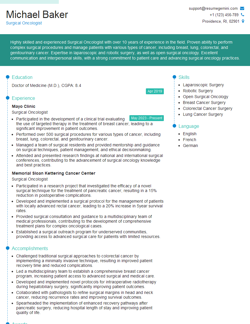
Mastering hepatectomy is a significant achievement that opens doors to advanced surgical practice and leadership roles. A strong understanding of this complex procedure is highly valued in the field. To showcase your expertise, it’s crucial to present your qualifications effectively. Creating an ATS-friendly resume is key to maximizing your job prospects. We highly recommend leveraging ResumeGemini to build a professional and impactful resume that highlights your skills and experience in hepatectomy. Examples of resumes tailored to hepatectomy specialists are available to help guide you through the process.
Explore more articles
Users Rating of Our Blogs
Share Your Experience
We value your feedback! Please rate our content and share your thoughts (optional).
What Readers Say About Our Blog
To the interviewgemini.com Webmaster.
Very helpful and content specific questions to help prepare me for my interview!
Thank you
To the interviewgemini.com Webmaster.
This was kind of a unique content I found around the specialized skills. Very helpful questions and good detailed answers.
Very Helpful blog, thank you Interviewgemini team.