Interviews are opportunities to demonstrate your expertise, and this guide is here to help you shine. Explore the essential Image-Guided Spinal Injections interview questions that employers frequently ask, paired with strategies for crafting responses that set you apart from the competition.
Questions Asked in Image-Guided Spinal Injections Interview
Q 1. Describe the different types of image guidance used in spinal injections.
Image guidance in spinal injections significantly improves accuracy and safety. The primary methods are fluoroscopy and computed tomography (CT) guidance. Fluoroscopy uses real-time X-ray imaging to visualize the needle’s trajectory, allowing for precise placement. CT guidance, on the other hand, provides cross-sectional images, offering a different perspective and enhancing visualization, particularly in complex anatomical situations. Ultrasound guidance is a newer, non-ionizing method gaining popularity for its real-time visualization and lack of radiation exposure. Each modality has its strengths and weaknesses, and the choice depends on the specific procedure, patient factors, and available resources. For instance, fluoroscopy is widely accessible and excellent for visualizing needle placement during epidural injections, while CT is particularly useful for navigating around bony structures in more challenging cases like facet joint injections. Ultrasound, while less widely available, is valuable for avoiding vascular structures and is particularly helpful in obese patients where bony landmarks may be less clear.
- Fluoroscopy: Real-time X-ray imaging.
- CT Guidance: Cross-sectional imaging using CT scans.
- Ultrasound Guidance: Real-time imaging using ultrasound waves.
Q 2. Explain the process of performing a fluoroscopy-guided epidural steroid injection.
A fluoroscopy-guided epidural steroid injection involves carefully placing a needle into the epidural space, the area surrounding the spinal cord. The procedure typically starts with prepping the injection site with antiseptic and administering local anesthesia. Then, under fluoroscopic guidance, a needle is carefully advanced towards the epidural space, the images on the fluoroscopy screen guiding the physician to avoid vital structures like nerves and blood vessels. The needle’s position is constantly monitored to ensure accurate placement. Once the needle is confirmed to be in the correct location, a contrast medium is often injected to confirm the position (this is called a test injection). If the contrast spreads appropriately in the epidural space, the medication (usually a steroid and local anesthetic) is injected. After the injection is complete, the needle is removed, and the site is dressed with a bandage. Post-procedure monitoring is essential to check for any immediate complications.
Think of it like navigating a ship—the fluoroscopy screen is your map, showing you the path to the desired location (epidural space) while guiding you around potential hazards (nerves, blood vessels).
Q 3. What are the contraindications for image-guided spinal injections?
Contraindications for image-guided spinal injections can be absolute or relative, meaning they may prohibit the procedure entirely or require careful consideration and potentially alternative approaches. Absolute contraindications include active local infection at the injection site, patient refusal, uncontrolled bleeding disorders (increased risk of hematoma), or severe spinal stenosis that may lead to cord compression during the procedure. Relative contraindications include conditions like uncontrolled hypertension (which could be exacerbated by stress of the procedure), severe kyphoscoliosis (making needle placement more challenging), or the presence of a spinal implant that may interfere with injection or visualization. Always consider the risks and benefits on a case-by-case basis. A thorough history and physical exam are critical in determining contraindications.
Q 4. How do you ensure patient safety during image-guided spinal injections?
Patient safety is paramount. This begins with a detailed explanation of the procedure, its risks, and benefits, ensuring informed consent. Strict adherence to sterile technique prevents infection. Continuous monitoring of vital signs (heart rate, blood pressure, oxygen saturation) is critical throughout the procedure. Careful needle placement under image guidance minimizes the risk of injury to nerves or blood vessels. The appropriate dose and type of medication are selected based on the patient’s condition and medical history. Post-procedure monitoring for signs of complications such as bleeding, infection, or neurological deficits is also essential. Having emergency medications and resuscitation equipment readily available is a crucial safety measure.
Q 5. What are the potential complications of image-guided spinal injections?
Potential complications are rare but serious and can include bleeding (hematoma), infection, nerve injury (leading to pain or neurological deficits), dural puncture (leakage of cerebrospinal fluid), and allergic reactions to the medication. Accidental intravascular injection of medication can also occur, leading to cardiovascular complications. Post-injection pain or discomfort is common, but typically mild and resolves within a few days. The incidence of these complications is greatly reduced by the use of image guidance and adherence to strict safety protocols.
Q 6. How do you manage complications during or after an image-guided spinal injection?
Management of complications depends on the specific complication. For example, a hematoma might require close monitoring, while a significant infection necessitates immediate antibiotic therapy. Nerve injury may require supportive care, pain management, and potentially physical therapy. A dural puncture typically involves bed rest and observation, sometimes with a blood patch procedure to seal the leak. Allergic reactions are treated with appropriate medications. In cases of accidental intravascular injection, supportive care and monitoring for cardiovascular complications are crucial. Prompt recognition, appropriate treatment, and close monitoring are vital for minimizing the severity and long-term consequences of any complication. Immediate consultation with a specialist (e.g., neurosurgeon or neurologist) might be necessary in serious cases.
Q 7. Describe your experience with different types of needles used in spinal injections.
The choice of needle depends on the target location and the specific procedure. We commonly use 22-gauge or 25-gauge needles for epidural injections, as these provide a balance between sufficient strength and reduced trauma. For facet joint injections, smaller-gauge needles are often used to minimize tissue damage. The length of the needle also varies according to patient anatomy. Different needle types, such as pencil-point or bevel-tip needles, exist, and the choice might be guided by the target location and the physician’s preference. All needles are sterile, disposable, and selected to minimize patient discomfort and potential complications. Choosing the right needle is an integral part of ensuring procedural success and patient comfort. It’s important that the needle be appropriately suited to the specific patient’s anatomy and the targeted region.
Q 8. What are the advantages and disadvantages of using fluoroscopy versus ultrasound guidance?
Choosing between fluoroscopy and ultrasound for image-guided spinal injections depends on the specific clinical scenario. Both modalities offer advantages and disadvantages.
- Fluoroscopy: Provides excellent visualization of bony anatomy, making it ideal for procedures near the spine, such as epidural injections or facet joint injections. It allows for precise needle placement and real-time assessment of contrast spread. However, it involves ionizing radiation, posing a risk to both the patient and the healthcare provider. The cost of equipment and maintenance is also higher.
- Ultrasound: Offers a real-time, radiation-free image of soft tissues, making it particularly useful for identifying nerves and ligaments. It’s often preferred for procedures targeting specific nerve roots or for patients who require repeated injections, minimizing cumulative radiation exposure. However, bone obscures visualization, limiting its utility in certain spinal locations. Operator skill and experience are crucial for optimal image interpretation and accurate needle placement.
For example, in a patient with lumbar spinal stenosis requiring an epidural steroid injection, fluoroscopy might be preferred for its clear visualization of the bony landmarks and the epidural space. In contrast, for a patient with cervical radiculopathy needing a selective nerve root block, ultrasound guidance might be a better option, offering clearer visualization of the nerve roots without radiation exposure. The ultimate choice often involves a shared decision-making process between the physician and patient, weighing the benefits and risks of each modality.
Q 9. How do you select the appropriate injection site for a specific spinal condition?
Selecting the appropriate injection site hinges on a precise diagnosis of the patient’s spinal condition. The goal is to target the source of pain or dysfunction as accurately as possible while minimizing risks to surrounding structures.
- History and Physical Examination: A thorough evaluation helps localize the pain and identify potential neurological deficits.
- Imaging Studies: X-rays, CT scans, or MRIs provide detailed anatomical information, helping pinpoint the affected area (e.g., specific disc level in a case of radiculopathy, or facet joint in facet joint pain).
- Specific Condition: Different spinal conditions require different injection sites. For instance, a patient with lumbar radiculopathy might need a selective nerve root block at the affected nerve root, while someone with spinal stenosis might benefit from an epidural injection at the appropriate lumbar level. A patient with sacroiliac joint pain would need injection into the sacroiliac joint itself.
For example, a patient presenting with right-sided L5 radiculopathy, confirmed by MRI showing a herniated disc at L4-L5, would necessitate a selective nerve root block targeting the right L5 nerve root. The injection site would be carefully chosen using fluoroscopy or ultrasound to avoid injuring the adjacent nerve roots or blood vessels.
Q 10. What are the key anatomical landmarks used for image-guided spinal injections?
Accurate image-guided spinal injections depend heavily on recognizing key anatomical landmarks. These landmarks provide a framework for needle placement, ensuring safe and effective delivery of medication.
- Spinous Processes: Palpable bony projections along the midline of the spine, used for counting vertebral levels.
- Transverse Processes: Bony projections extending laterally from each vertebra, aiding in lateral guidance.
- Intervertebral Spaces: The spaces between adjacent vertebrae, often targeted for epidural injections.
- Sacroiliac Joints: The joints between the sacrum and ilium, palpable in the posterior pelvis.
- Posterior Superior Iliac Spines (PSIS): Bony prominences easily palpable on the posterior iliac crests; frequently used as landmarks for sacroiliac joint injections.
- Pedicles and Laminae: The bony structures forming the vertebral arch; crucial for accurate needle placement in certain procedures, visible on fluoroscopy.
Think of these landmarks as a roadmap guiding us through the complex anatomy of the spine. Mastering them is essential for minimizing complications.
Q 11. Explain the principles of radiation safety during fluoroscopy-guided procedures.
Radiation safety is paramount during fluoroscopy-guided spinal injections. We adhere to the ALARA principle—As Low As Reasonably Achievable—to minimize radiation exposure to both the patient and healthcare personnel.
- Collimation: Restricting the X-ray beam to the area of interest reduces unnecessary radiation exposure.
- Pulse Fluoroscopy: Using pulsed fluoroscopy instead of continuous fluoroscopy reduces radiation dose without significantly compromising image quality.
- Image Intensifier: Positioning the image intensifier as close as possible to the patient reduces radiation scatter.
- Lead Shielding: Using lead aprons, thyroid shields, and other protective barriers for both the patient and the healthcare team significantly decreases radiation exposure.
- Time Optimization: Performing the procedure efficiently minimizes the duration of exposure.
- Distance: Maintaining a safe distance from the radiation source reduces exposure for personnel.
For example, we always collimate the x-ray beam to the smallest possible size encompassing the injection site, and we use pulse fluoroscopy whenever possible. Every member of the team wears appropriate lead shielding throughout the procedure.
Q 12. How do you interpret fluoroscopic images during spinal injections?
Interpreting fluoroscopic images during spinal injections requires a keen eye for anatomical detail and a thorough understanding of spinal anatomy. We look for several key features:
- Needle Position: Ensuring the needle is correctly positioned within the target structure (e.g., epidural space, facet joint, nerve root). We pay close attention to the needle’s relationship to the bony landmarks and surrounding structures.
- Contrast Spread: Observing how the contrast medium flows after injection helps confirm proper placement and identify any potential complications such as intravascular injection.
- Anatomical Structures: Identifying key anatomical structures such as the intervertebral discs, pedicles, and laminae, as well as the spinal cord and nerve roots.
- Complications: We look for signs of complications such as intravascular injection (contrast in the blood vessels), dural puncture (contrast leaking into the cerebrospinal fluid), or pneumothorax (air in the pleural space).
For instance, during an epidural injection, we would expect to see the contrast spread evenly within the epidural space, conforming to the shape of the vertebral column. Any extravasation into the surrounding tissues or blood vessels would be immediately apparent and necessitates a different approach.
Q 13. Describe your experience with different types of contrast media used in spinal injections.
Different types of contrast media are used in spinal injections, each with its own properties and advantages. The choice of contrast medium depends on the specific procedure and patient factors.
- Iodinated Contrast Media (e.g., Omnipaque, Isovue): These are commonly used and offer excellent radiopacity, allowing for clear visualization on fluoroscopy. They are water-soluble and generally well-tolerated.
- Non-ionic Contrast Media: These are less likely to cause adverse reactions compared to ionic contrast agents, and hence, are the preferred choice in many cases.
The choice between different types of iodinated contrast media often comes down to osmolality; lower osmolality agents are associated with reduced risk of adverse effects. I am always careful to review the patient’s medical history for allergies or previous adverse reactions to contrast media before selecting the appropriate agent. This ensures patient safety and prevents complications.
Q 14. How do you assess the success of an image-guided spinal injection?
Assessing the success of an image-guided spinal injection involves a multi-faceted approach combining immediate post-procedure assessment and follow-up.
- Immediate Post-Procedure Assessment: This involves evaluating the patient’s pain level and neurological status. Absence of significant bleeding or other complications at the injection site is also crucial.
- Fluoroscopic Confirmation: Careful review of the fluoroscopic images ensures the contrast medium was accurately placed in the target area and that there was no evidence of complications during the procedure.
- Clinical Follow-up: Post-procedure follow-up appointments are essential to evaluate the effectiveness of the injection. This involves reassessing the patient’s pain levels, range of motion, and neurological status. If there’s no significant improvement, we’ll re-evaluate the diagnosis and explore alternative treatment options.
For example, a successful epidural injection for lumbar radiculopathy would result in a significant reduction in leg pain within days to weeks, improved mobility, and absence of neurological deficits. Failure to see such improvement prompts a careful reassessment of the diagnosis, procedure technique, and alternative treatment approaches.
Q 15. What are the common post-procedure instructions for patients undergoing spinal injections?
Post-procedure instructions for patients who have undergone spinal injections are crucial for a safe and successful recovery. These instructions aim to minimize complications and promote healing. They typically include:
- Rest: Patients are advised to rest for a period of time, usually several hours, following the procedure. The exact duration depends on the type of injection and the patient’s individual response. This allows the injection site to settle and reduces the risk of bleeding or infection.
- Hydration: Drinking plenty of fluids helps to flush out any potential toxins or medications introduced during the procedure.
- Pain Management: Patients are provided with instructions on appropriate pain management, which may include over-the-counter medications like acetaminophen or ibuprofen, as prescribed by their physician. They are also educated on when to contact their doctor regarding increased pain.
- Activity Modification: Patients are usually advised to avoid strenuous activities, heavy lifting, and prolonged standing or sitting for several days following the procedure. Gradual return to normal activities is recommended, tailored to the patient’s condition and the physician’s recommendation. For instance, a patient might start with short walks and progressively increase their activity level.
- Infection Monitoring: Patients are taught how to monitor the injection site for signs of infection, such as increased pain, redness, swelling, or fever, and instructed to report any such symptoms immediately to their healthcare provider.
- Follow-up Appointment: A follow-up appointment is scheduled to assess the effectiveness of the injection and to monitor the patient’s progress.
For example, I always provide my patients with a detailed written instruction sheet along with a verbal explanation to ensure clarity and understanding. I also encourage them to ask questions to address any concerns they may have.
Career Expert Tips:
- Ace those interviews! Prepare effectively by reviewing the Top 50 Most Common Interview Questions on ResumeGemini.
- Navigate your job search with confidence! Explore a wide range of Career Tips on ResumeGemini. Learn about common challenges and recommendations to overcome them.
- Craft the perfect resume! Master the Art of Resume Writing with ResumeGemini’s guide. Showcase your unique qualifications and achievements effectively.
- Don’t miss out on holiday savings! Build your dream resume with ResumeGemini’s ATS optimized templates.
Q 16. How do you handle adverse events or unexpected outcomes during a procedure?
Handling adverse events during a spinal injection requires immediate action and a calm, decisive approach. My training emphasizes preparedness and rapid response. Adverse events, though rare with proper technique and precautions, can include bleeding, infection, nerve damage, or allergic reactions.
My immediate actions would involve:
- Assessment: Quickly assess the patient’s condition, focusing on vital signs (heart rate, blood pressure, respiration), level of consciousness, and the specific symptoms observed.
- Stabilization: Prioritize stabilizing the patient’s vital signs. This might involve administering oxygen, fluids, or medication as appropriate.
- Notification: Immediately notify the appropriate medical personnel, including the anesthesiologist and the hospital’s response team, depending on the severity of the event.
- Intervention: Implement appropriate interventions based on the specific adverse event. This may include administering medications to counteract allergic reactions, managing bleeding, or providing supportive care.
- Documentation: Meticulous documentation of the adverse event, including the time of occurrence, the symptoms observed, the interventions undertaken, and the patient’s response, is crucial for both medical and legal reasons.
For example, if a patient experiences a sudden drop in blood pressure, I would immediately initiate fluid resuscitation and monitor their vital signs while contacting the anesthesiologist. A detailed record of the event, including the patient’s response to treatment, would be added to their medical record. Post-procedure monitoring and communication with the patient’s primary care physician are also essential.
Q 17. What are the different types of spinal injections and their indications?
Spinal injections are categorized based on the target area and the intended therapeutic effect. Different injections address various pain conditions. Some common types include:
- Epidural Steroid Injections (ESI): These injections deliver corticosteroids into the epidural space, the area surrounding the spinal cord. ESIs are often used to treat radiculopathy (nerve root pain) caused by conditions like spinal stenosis or herniated discs. They reduce inflammation and pain.
- Facet Joint Injections: These injections target the facet joints, small joints in the spine that can become inflamed and painful. They are often used to diagnose and treat facet joint syndrome, a common source of back pain.
- Selective Nerve Root Blocks: These injections target specific nerve roots that are contributing to pain. They are often guided by fluoroscopy or CT/MRI to ensure accurate placement.
- Medial Branch Blocks: These injections target specific nerves that supply the facet joints, providing more targeted pain relief. They are often used to assess the contribution of a specific facet joint to a patient’s pain before considering more invasive treatments.
- Transforaminal Epidural Steroid Injections (TFESI): These injections deliver corticosteroids through the intervertebral foramen, a small opening between the vertebrae, aiming to reach the nerve roots directly. They are often employed in cases where radiculopathy is severe or doesn’t respond well to other interventions.
The choice of injection depends on the patient’s specific diagnosis, symptoms, and overall health. For example, a patient with sciatica (pain radiating down the leg) might benefit from an ESI or a selective nerve root block, while a patient with localized back pain might receive a facet joint injection. A thorough clinical evaluation is essential before determining the most appropriate injection type.
Q 18. Describe your experience with managing patients with complex anatomy or comorbidities.
Managing patients with complex anatomy or comorbidities presents unique challenges but is an integral part of my practice. These patients often require a more individualized approach, considering their specific anatomical variations and medical history.
My approach involves:
- Detailed Pre-Procedure Assessment: A comprehensive review of the patient’s medical history, including imaging studies (X-rays, CT, MRI), is crucial to identify any anatomical variations or comorbidities that could impact the procedure. This might include conditions like scoliosis, previous spine surgery, or cardiovascular disease.
- Adjusted Technique: I adapt my injection technique to accommodate the patient’s specific anatomy. For example, if a patient has severe scoliosis, I might need to adjust the needle trajectory to avoid vital structures. Similarly, a patient with osteoporosis might require a modified approach to minimize the risk of bone fracture.
- Careful Medication Selection: Comorbidities such as kidney disease or cardiac conditions may influence the choice of medications used during and after the procedure. I might need to consult with other specialists to ensure medication safety and efficacy.
- Enhanced Monitoring: Close monitoring of the patient’s vital signs during and after the procedure is crucial, particularly for patients with comorbidities. This allows for early detection and management of any complications.
- Collaboration: Collaboration with other medical specialists, such as cardiologists or nephrologists, is essential to ensure optimal patient care.
For example, I recently treated a patient with severe scoliosis and a history of heart failure who required an ESI. I carefully reviewed the patient’s CT scan to plan the needle trajectory, adjusted the medication dosage considering the cardiac condition, and closely monitored their vital signs throughout the procedure. The successful outcome reinforced the importance of a tailored approach for complex patients.
Q 19. What is your approach to pain management beyond spinal injections?
Pain management is a holistic endeavor, and spinal injections are just one tool in the arsenal. My approach goes beyond injections, encompassing a multi-modal strategy tailored to the individual patient.
This may include:
- Pharmacological Management: This includes not just pain medications but also medications to address underlying conditions contributing to pain, such as antidepressants for neuropathic pain or muscle relaxants for muscle spasms.
- Physical Therapy: Physical therapy plays a vital role in strengthening muscles, improving flexibility, and restoring functional movement. It helps patients regain independence and reduce reliance on pain medications.
- Lifestyle Modifications: This includes recommendations for weight management, posture correction, ergonomic adjustments at work or home, and regular exercise. Addressing lifestyle factors can significantly reduce pain and improve overall well-being.
- Cognitive Behavioral Therapy (CBT): CBT helps patients develop coping mechanisms for managing chronic pain, addressing emotional and psychological factors that can exacerbate pain perception.
- Interventional Procedures (Beyond Injections): In cases where conservative treatments fail, more advanced interventions, such as radiofrequency ablation or spinal cord stimulation, might be considered.
For instance, a patient with chronic back pain might receive a combination of ESI, physical therapy, and CBT to address both the physical pain and the psychological impact of chronic pain. My goal is to empower patients to take an active role in their pain management and improve their quality of life.
Q 20. How do you stay updated on the latest advancements in image-guided spinal injection techniques?
Staying updated on the latest advancements is paramount in this rapidly evolving field. My approach to continuous professional development involves several strategies:
- Professional Societies and Publications: I am an active member of several professional organizations, including the American Society of Interventional Pain Physicians (ASIPP) and the International Spinal Injection Society (ISIS). I regularly read peer-reviewed journals and attend their conferences and workshops to stay abreast of the latest research and techniques.
- Continuing Medical Education (CME): I actively participate in CME courses and workshops focused on image-guided spinal injections, advanced imaging techniques, and new treatment modalities.
- Collaboration with Colleagues: I engage in regular discussions and case reviews with colleagues specializing in pain management and interventional radiology. This collaborative exchange of knowledge and experience is invaluable.
- Online Resources: I utilize reputable online resources and databases, such as PubMed, to access the most current research and guidelines in the field.
- Mentorship and Peer Review: I actively seek mentorship from experienced practitioners and participate in peer review of research articles and clinical practices. This strengthens my knowledge and critical thinking skills.
By consistently engaging in these activities, I can ensure that my practice incorporates the most current and evidence-based techniques to provide the best possible care for my patients.
Q 21. Describe your experience with using advanced imaging techniques like CT or MRI guidance.
Advanced imaging techniques, like CT and MRI guidance, are indispensable for precise and safe spinal injections. They allow for visualization of the intricate anatomy of the spine, minimizing the risk of complications and enhancing accuracy.
My experience with these techniques includes:
- Fluoroscopy: Fluoroscopy, a type of real-time X-ray imaging, is often used for basic guidance, providing a dynamic view of needle placement during the procedure. It helps to ensure the needle reaches the intended target area, reducing the risk of complications such as nerve damage.
- CT Guidance: CT guidance provides high-resolution cross-sectional images of the spine, allowing for extremely precise targeting of the injection. This is particularly helpful in patients with complex anatomy or when injecting into small, difficult-to-access areas.
- MRI Guidance: MRI guidance offers excellent soft tissue contrast, providing detailed images of the spinal cord, nerves, and surrounding tissues. This is invaluable in cases where precise needle placement is critical, such as in selective nerve root blocks or when avoiding sensitive structures.
- Image Fusion: In certain cases, I utilize image fusion techniques that combine images from different modalities (e.g., CT and fluoroscopy) to achieve even greater accuracy in needle placement.
The choice of imaging modality depends on the specific procedure, the patient’s anatomy, and the availability of resources. For instance, I might use fluoroscopy for a basic ESI, but I might opt for CT guidance for a more complex procedure such as a transforaminal epidural injection. My goal is to always utilize the most appropriate imaging technique to maximize the safety and effectiveness of the procedure.
Q 22. How do you document procedures and patient outcomes for image-guided spinal injections?
Comprehensive documentation is crucial for image-guided spinal injections. We meticulously record every step, ensuring a clear, auditable trail for both medical and legal purposes. This begins with obtaining informed consent, detailing the procedure, risks, and alternatives discussed with the patient. During the procedure, I document the patient’s vital signs (heart rate, blood pressure, oxygen saturation) before, during, and after the injection. The type and amount of medication administered, the injection site (verified by imaging), any complications encountered, and the patient’s response are all meticulously noted. Post-procedure, we document the patient’s recovery, any pain relief achieved, and any adverse events. We use a standardized electronic health record (EHR) system for easy access and sharing of information. Imaging data, including fluoroscopy images or ultrasound scans, are integrated into the record. This ensures continuity of care and aids in future treatment decisions. For example, a specific note might read: “08:00 AM: Patient positioned prone, L3-L4 intervertebral level identified fluoroscopically. 1 ml of 0.5% bupivacaine with 40mg methylprednisolone injected. Patient reported 50% pain reduction immediately post-injection.”
Q 23. How do you communicate with patients and their families about risks and benefits?
Open and honest communication is paramount. I start by explaining the procedure in simple, understandable terms, avoiding medical jargon. I use visual aids like diagrams or videos to illustrate the process and address the patient’s concerns. We discuss the potential benefits – such as pain relief and improved mobility – alongside the risks, including infection, bleeding, nerve damage, and medication side effects. I answer all their questions patiently and encourage them to express any anxieties. If needed, I involve family members in the discussion to ensure everyone is well-informed. I emphasize that the decision to undergo the injection is theirs, and I respect their choice regardless. For instance, if a patient expresses fear of needles, I’ll address this specifically, discussing techniques to minimize discomfort and perhaps offering relaxation strategies. I strive for shared decision-making, where the patient feels empowered and actively participates in their care.
Q 24. Explain your understanding of the legal and ethical considerations related to spinal injections.
Legal and ethical considerations are central to my practice. Obtaining informed consent, ensuring patient autonomy, and maintaining confidentiality are non-negotiable. This includes providing clear and accurate information about the procedure, its potential benefits and risks, and alternative treatment options. I adhere to all relevant regulations and guidelines, maintaining detailed records that demonstrate adherence to best practices. I’m particularly mindful of avoiding conflicts of interest and ensuring that my recommendations are solely based on the patient’s best interests. Any deviations from standard protocols are carefully documented and justified. For example, if a patient has a specific religious or cultural belief affecting their treatment preferences, we discuss this openly and explore options that respect their values while maintaining safety and efficacy. The legal implications of medical malpractice, particularly related to negligence or incorrect technique, are always at the forefront of my mind, reinforcing the importance of meticulous practice and comprehensive documentation.
Q 25. What is your experience with managing difficult patients or those with significant anxiety?
Managing difficult or anxious patients requires patience, empathy, and strong communication skills. I actively listen to their concerns, validate their feelings, and address their anxieties. I often use a collaborative approach, involving the patient in decision-making and allowing them to express their preferences. In cases of significant anxiety, I may offer relaxation techniques, such as deep breathing exercises, or consider pre-procedural medication to alleviate their stress. I may also involve a psychologist or psychiatrist if necessary. For example, if a patient is verbally aggressive due to chronic pain, I maintain a calm and professional demeanor, focusing on reassuring them and addressing their pain concerns systematically. Building rapport and trust are key – it’s about understanding the patient’s perspective and working together to achieve the best possible outcome.
Q 26. How do you maintain sterility and asepsis during image-guided spinal injections?
Maintaining sterility and asepsis is paramount to prevent infection. We follow strict protocols, adhering to the principles of surgical asepsis. This begins with thorough hand hygiene, using an appropriate antiseptic solution. The injection site is prepared with a sterile drape and antiseptic solution, using a concentric circle approach to avoid contamination. We use sterile gloves, needles, and syringes. All equipment is carefully inspected for any signs of damage or contamination before use. The fluoroscopy or ultrasound equipment is also disinfected according to manufacturer guidelines. Throughout the procedure, we maintain a sterile field, ensuring that no non-sterile items come into contact with the injection site or sterile instruments. Any breaches in sterility are immediately addressed, and the procedure may need to be halted and restarted if necessary. This meticulous approach is essential to minimize the risk of infection, which can have severe consequences for the patient.
Q 27. Describe your experience with different types of medication used in spinal injections.
My experience encompasses a wide range of medications used in spinal injections, chosen based on the patient’s specific condition and needs. These include local anesthetics like lidocaine and bupivacaine to numb the area and reduce pain during the procedure. Corticosteroids, such as methylprednisolone, are often used to reduce inflammation and pain in conditions like spinal stenosis. Epidural analgesics, such as morphine or fentanyl, can be used for severe pain management. In some cases, a combination of medications may be employed to achieve optimal therapeutic effects. The choice of medication and dosage is carefully considered, taking into account the patient’s medical history, allergies, and potential drug interactions. For example, a patient with a history of cardiac problems might require a different medication profile than a patient with no known allergies. The selection process balances therapeutic effectiveness with minimizing potential adverse events.
Q 28. How do you troubleshoot equipment malfunctions during a procedure?
Troubleshooting equipment malfunctions during a procedure requires a calm, methodical approach. The first step is to assess the nature of the problem and its potential impact on patient safety. If the issue is minor, such as a malfunctioning display on the fluoroscopy unit, we may attempt to rectify it using established troubleshooting steps (e.g., checking connections, restarting the system). However, if the malfunction poses a risk to the patient or compromises the integrity of the procedure (e.g., loss of image quality, failure of the injection system), the procedure is immediately halted. Patient safety is the priority. The malfunction is reported, and we follow the established protocol, which may involve contacting biomedical engineering or using backup equipment. The incident is carefully documented, and corrective actions are implemented to prevent similar situations in the future. A thorough post-incident review is conducted to learn from any mistakes and refine our procedures.
Key Topics to Learn for Image-Guided Spinal Injections Interview
- Anatomy and Physiology: Thorough understanding of spinal anatomy, including vertebral levels, nerve roots, and surrounding structures. Focus on relevant anatomical variations and their implications for injection procedures.
- Imaging Modalities: Fluoroscopy, ultrasound, and CT-guided techniques. Compare and contrast their advantages, limitations, and appropriate applications in different clinical scenarios. Understand image interpretation and how to identify relevant anatomical landmarks.
- Injection Techniques: Master the practical aspects of various injection procedures, including epidural, facet joint, and selective nerve root blocks. Understand the steps involved, from patient preparation to post-procedure care. Practice explaining your procedural approach clearly and concisely.
- Needle Selection and Placement: Discuss the rationale behind choosing different needle sizes and types for various injections. Explain the principles of proper needle insertion and advancement, including strategies for minimizing complications.
- Patient Selection and Contraindications: Understand the criteria for selecting appropriate candidates for image-guided spinal injections and recognizing contraindications that might prevent a procedure or necessitate modifications.
- Complications and Management: Be prepared to discuss potential complications, such as bleeding, infection, nerve damage, and their management strategies. Demonstrate your understanding of risk mitigation techniques.
- Pain Management Principles: Explain the rationale behind using image-guided spinal injections in the context of overall pain management strategies. Discuss the integration of these injections with other therapeutic modalities.
- Legal and Ethical Considerations: Understand the legal and ethical aspects of obtaining informed consent, documenting procedures accurately, and adhering to relevant guidelines and regulations.
Next Steps
Mastering Image-Guided Spinal Injections significantly enhances your career prospects, opening doors to specialized roles and advanced opportunities in pain management and interventional radiology. To maximize your job search success, create an ATS-friendly resume that highlights your skills and experience effectively. ResumeGemini is a trusted resource that can help you build a professional and impactful resume. Examples of resumes tailored specifically to Image-Guided Spinal Injections are available to guide you through the process. Invest the time to showcase your expertise—it will pay off in your career advancement.

Medical Director Resume Sample

Medical Physicist Resume Sample

Anesthesiologist Resume Sample
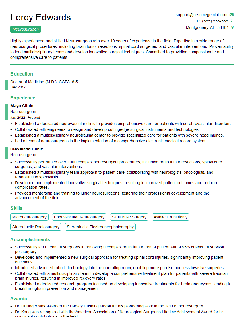
Neurosurgeon Resume Sample
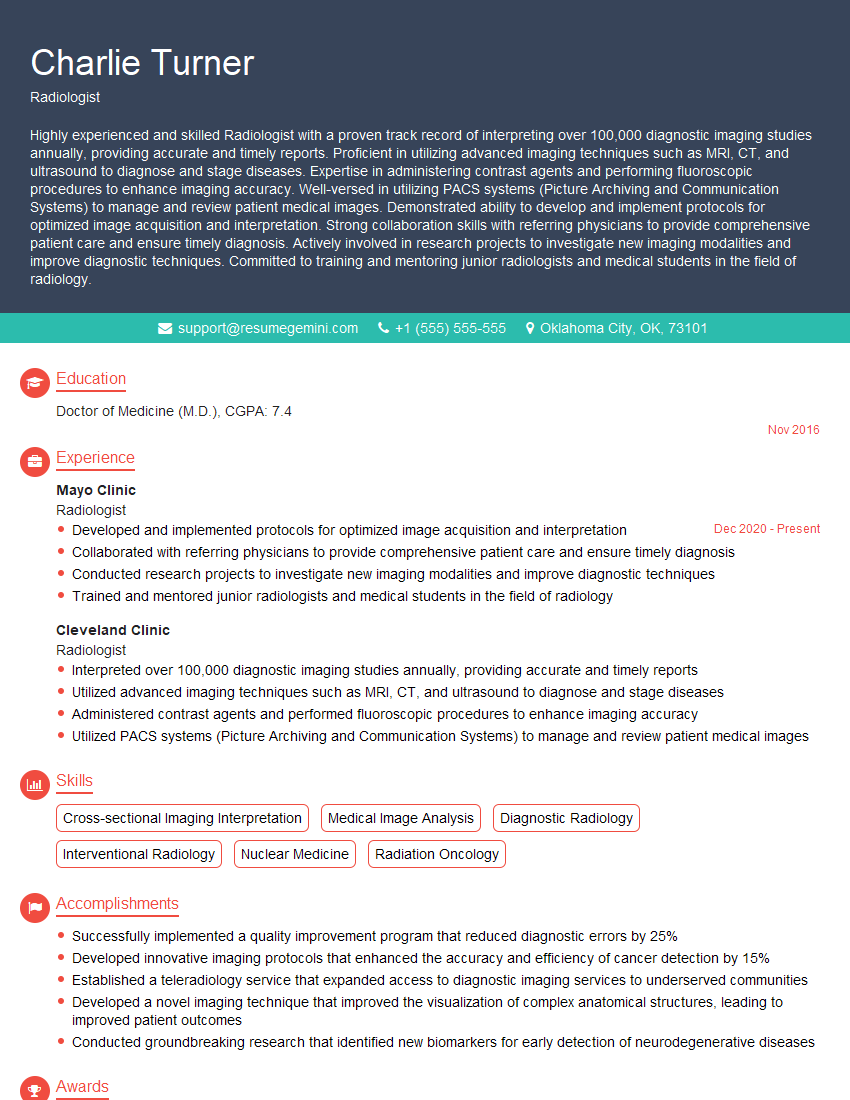
Radiologist Resume Sample
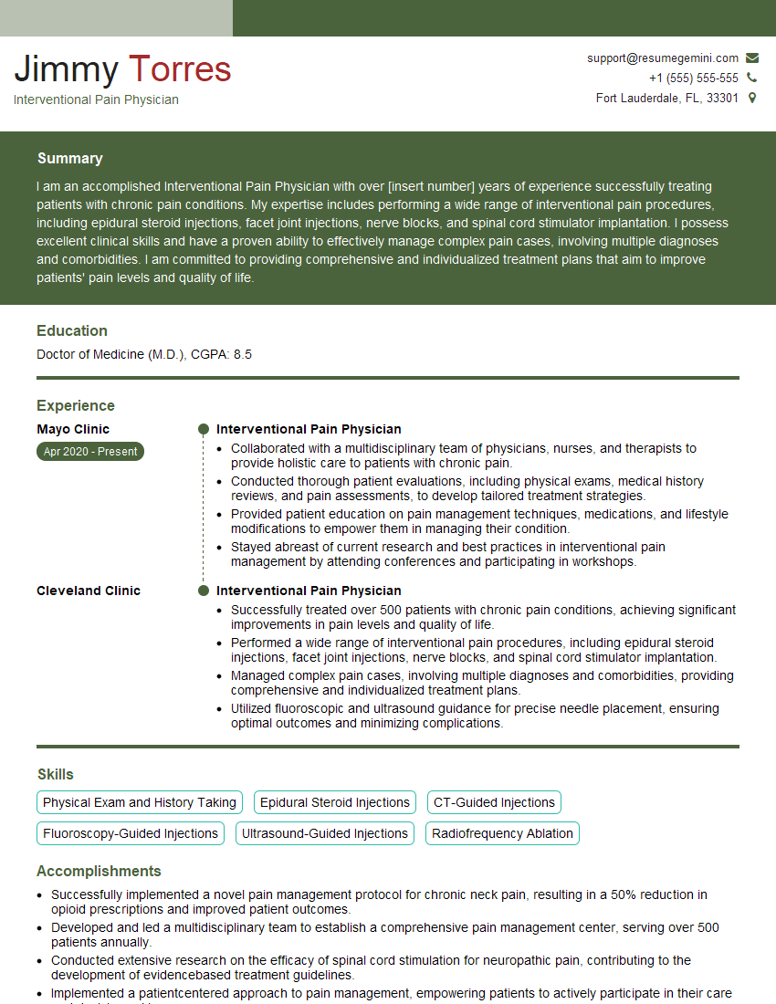
Interventional Pain Physician Resume Sample
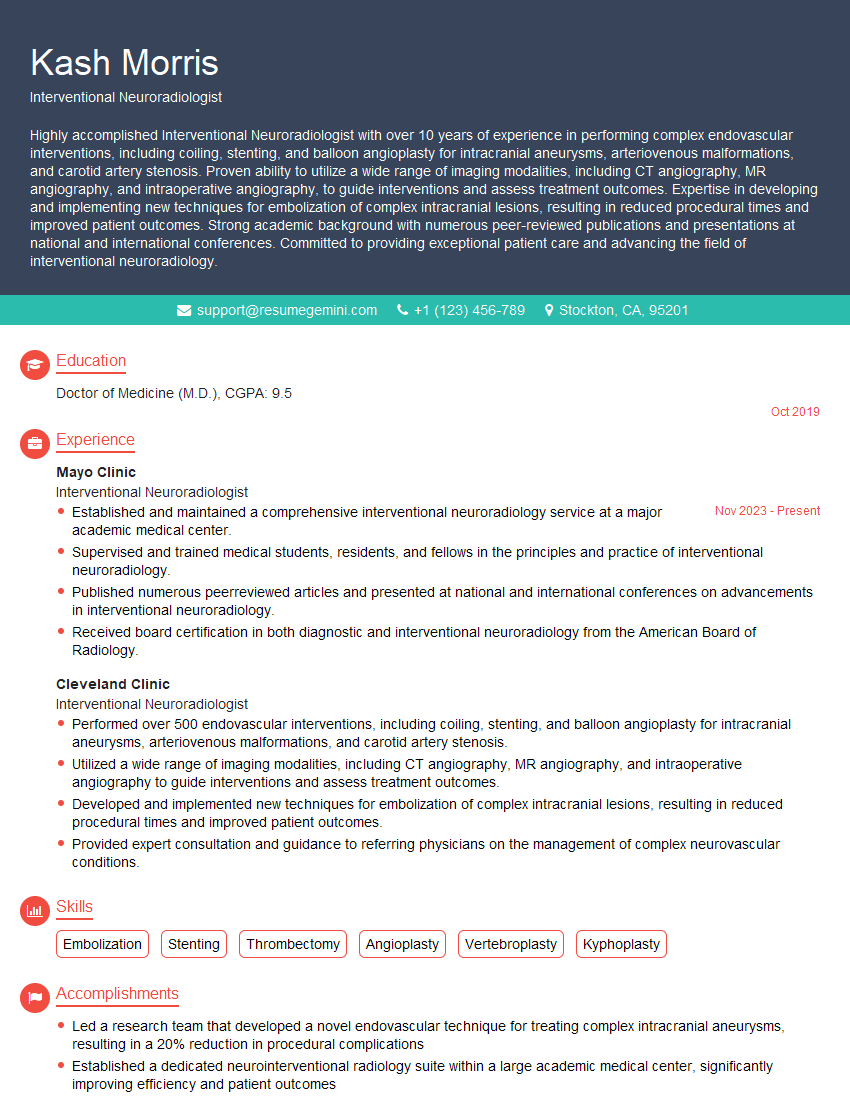
Interventional Neuroradiologist Resume Sample
Certified Registered Nurse Anesthetist (CRNA) Resume Sample

Chief Medical Officer Resume Sample

Radiology Technologist Resume Sample

Medical Imaging Technician Resume Sample
Explore more articles
Users Rating of Our Blogs
Share Your Experience
We value your feedback! Please rate our content and share your thoughts (optional).
What Readers Say About Our Blog
To the interviewgemini.com Webmaster.
Very helpful and content specific questions to help prepare me for my interview!
Thank you
To the interviewgemini.com Webmaster.
This was kind of a unique content I found around the specialized skills. Very helpful questions and good detailed answers.
Very Helpful blog, thank you Interviewgemini team.