Feeling uncertain about what to expect in your upcoming interview? We’ve got you covered! This blog highlights the most important Musculoskeletal Medicine interview questions and provides actionable advice to help you stand out as the ideal candidate. Let’s pave the way for your success.
Questions Asked in Musculoskeletal Medicine Interview
Q 1. Explain the difference between osteoarthritis and rheumatoid arthritis.
Osteoarthritis (OA) and rheumatoid arthritis (RA) are both forms of arthritis, causing joint pain and inflammation, but they differ significantly in their underlying causes and mechanisms. OA is a degenerative joint disease, essentially a ‘wear and tear’ condition. Cartilage, the cushioning between bones, gradually breaks down over time, leading to bone-on-bone friction, pain, and stiffness. It’s most common in weight-bearing joints like knees and hips. Think of it like the gradual wear on the tires of a car from continuous use.
RA, on the other hand, is an autoimmune disease. The body’s immune system mistakenly attacks the synovium, the lining of the joints. This inflammation leads to swelling, pain, and eventual joint damage. Unlike OA, RA can affect many joints simultaneously, often symmetrically (both hands or both feet equally). Imagine it as a car with a faulty internal mechanism causing widespread damage, rather than just wear and tear on the tires. The key difference lies in the underlying cause: degenerative changes in OA versus immune system dysfunction in RA.
- Osteoarthritis (OA): Degenerative, affects specific joints, cartilage breakdown.
- Rheumatoid Arthritis (RA): Autoimmune, systemic (affects multiple joints), synovial inflammation.
Q 2. Describe the pathophysiology of rotator cuff tears.
Rotator cuff tears involve damage to one or more of the four muscles and their tendons that surround the shoulder joint. These muscles – supraspinatus, infraspinatus, teres minor, and subscapularis – help with shoulder movement and stability. The pathophysiology is complex but generally involves overuse, trauma, or a combination of both. Repetitive overhead movements, such as in throwing sports or painting, can gradually weaken the tendons, making them susceptible to tears. Sudden, forceful movements, like a fall or a direct blow to the shoulder, can cause acute tears. Age-related degeneration also plays a significant role; as we age, the tendons become less elastic and more prone to injury.
The tear itself can be partial (only some of the tendon fibers are torn) or full-thickness (the tendon is completely severed). This damage leads to pain, weakness, and instability in the shoulder. The process can also be accompanied by inflammation, further hindering function. For example, a baseball pitcher might develop a rotator cuff tear due to the repetitive overhead throwing motion over years, while a construction worker might suffer an acute tear from a fall.
Q 3. What are the common causes of low back pain?
Low back pain is a remarkably common complaint, with a wide range of potential causes. Many cases are musculoskeletal in nature, meaning they originate from the structures of the back itself. These include:
- Muscle strains and sprains: Overexertion, poor posture, or sudden movements can injure the muscles and ligaments of the back.
- Disc herniation: The soft, gel-like center of an intervertebral disc can bulge or rupture, pressing on nerves and causing pain that can radiate down the leg (sciatica).
- Spinal stenosis: Narrowing of the spinal canal puts pressure on the spinal cord and nerves.
- Facet joint syndrome: Inflammation or degeneration of the small joints connecting the vertebrae.
Beyond musculoskeletal issues, low back pain can be caused by other conditions, including:
- Osteoarthritis and other arthritic conditions
- Infections
- Tumors
- Fibromyalgia
- Stress and psychological factors
Pinpointing the exact cause often requires a thorough medical evaluation, including a detailed history and physical examination.
Q 4. Discuss the diagnostic imaging techniques used in musculoskeletal evaluations.
Diagnostic imaging plays a crucial role in evaluating musculoskeletal problems. Various techniques provide different perspectives and information:
- Plain radiography (X-rays): A fundamental initial step, X-rays visualize bone structures, revealing fractures, dislocations, arthritis, and bone spurs. They are relatively inexpensive and readily available.
- Ultrasound: Uses sound waves to create images of soft tissues, such as muscles, tendons, and ligaments. Useful for assessing rotator cuff tears, tendonitis, and bursitis.
- Computed tomography (CT): Provides detailed cross-sectional images of bone and soft tissue. Excellent for visualizing complex fractures, spinal stenosis, and tumors.
- Magnetic resonance imaging (MRI): Uses magnetic fields and radio waves to create high-resolution images of all tissues, including bone, cartilage, ligaments, muscles, and nerves. Provides superior visualization of soft tissue injuries, like meniscus tears or ligament sprains, and is often the preferred imaging modality for assessing many musculoskeletal conditions.
- Bone scintigraphy (bone scan): Uses radioactive tracers to detect areas of increased bone metabolism, which can indicate stress fractures, infections, or tumors.
The choice of imaging technique depends on the specific clinical suspicion and the information needed. For instance, an X-ray is often sufficient for a suspected fracture, whereas an MRI is usually necessary for evaluating a suspected ligament tear.
Q 5. Outline the treatment options for carpal tunnel syndrome.
Carpal tunnel syndrome (CTS) occurs when the median nerve, which runs through the carpal tunnel in the wrist, becomes compressed. This compression causes pain, numbness, tingling, and weakness in the hand and fingers. Treatment options range from conservative to surgical:
- Conservative management: This is usually the first line of treatment and includes:
- Wrist splinting: Keeping the wrist in a neutral position at night reduces pressure on the median nerve.
- Nonsteroidal anti-inflammatory drugs (NSAIDs): Reduce pain and inflammation.
- Corticosteroid injections: Injected into the carpal tunnel, these medications can reduce inflammation and provide temporary relief.
- Physical therapy: Exercises and stretches to improve wrist mobility and strength.
- Surgical intervention: If conservative measures fail to provide adequate relief, surgical decompression of the carpal tunnel may be considered. This involves cutting a ligament to relieve pressure on the median nerve.
The decision to pursue surgery depends on the severity of symptoms, the duration of symptoms, and the response to conservative treatment. Many patients experience significant improvement with conservative management, while others may require surgery for long-term relief.
Q 6. Explain the principles of fracture healing.
Fracture healing is a complex process involving several stages. The body’s remarkable ability to repair broken bones depends on a cascade of cellular and molecular events:
- Hematoma formation: Immediately following a fracture, bleeding occurs, forming a blood clot (hematoma) at the fracture site.
- Inflammation: Inflammatory cells migrate to the area, initiating the healing process and removing debris.
- Callus formation: Specialized cells (osteoblasts) begin to form a soft callus of cartilage and fibrous tissue, bridging the fracture gap.
- Ossification: The soft callus is gradually replaced by bone, a process called ossification. This forms a hard callus, providing initial stability to the fracture.
- Remodeling: The hard callus is gradually remodeled, reshaping the bone to its original form. This stage can last for months or even years.
Several factors influence fracture healing, including the type of fracture, the patient’s age and overall health, and the presence of any underlying medical conditions. Adequate immobilization and proper nutrition are crucial for optimal healing. For instance, a simple, non-displaced fracture heals relatively quickly, while a complex, comminuted (shattered) fracture may require surgery and a longer healing time.
Q 7. What are the indications for surgical intervention in ACL injuries?
The anterior cruciate ligament (ACL) is a crucial knee ligament providing stability. ACL injuries, often occurring during sports activities involving sudden twisting or hyperextension, can cause significant instability. Surgical intervention (ACL reconstruction) is often indicated when:
- Significant instability: The knee gives way frequently, making activities like walking, running, and jumping difficult and potentially dangerous.
- Failure of conservative management: Physical therapy and bracing haven’t restored sufficient stability and function.
- High activity level: Individuals involved in high-impact sports are more likely to require surgery to return to their previous activity level.
- Young age: Younger patients have a greater likelihood of long-term complications if their knee remains unstable.
ACL reconstruction usually involves replacing the torn ligament with a graft from a tendon elsewhere in the body. The surgery aims to restore knee stability and allow for a return to normal activity. The decision to proceed with surgery is made on a case-by-case basis, considering the patient’s age, activity level, and the severity of their injury and symptoms.
Q 8. Describe the different types of arthritis and their distinguishing features.
Arthritis is an umbrella term encompassing numerous conditions characterized by joint inflammation, pain, and stiffness. Different types of arthritis have distinct underlying causes and clinical presentations.
- Osteoarthritis (OA): This is the most common type, often called ‘wear-and-tear’ arthritis. It involves the gradual breakdown of cartilage, the cushioning material between bones. This leads to bone-on-bone friction, causing pain, stiffness, and reduced range of motion. It’s particularly prevalent in weight-bearing joints like knees and hips. Imagine the cartilage as a sponge; in OA, it gradually loses its ability to absorb shock.
- Rheumatoid Arthritis (RA): This is an autoimmune disease where the body’s immune system mistakenly attacks the synovium, the lining of the joints. This leads to inflammation, swelling, pain, and potentially joint destruction. Unlike OA, RA can affect multiple joints symmetrically, often the hands and feet. Think of it as the immune system mistakenly attacking its own tissues.
- Gout: This is a type of inflammatory arthritis caused by a buildup of uric acid crystals in the joints. This leads to sudden, severe attacks of pain, swelling, redness, and tenderness, often in the big toe. Imagine tiny shards of glass causing intense inflammation.
- Psoriatic Arthritis (PsA): This inflammatory arthritis occurs in people with psoriasis, a skin condition. It can affect joints in a variety of ways, sometimes asymmetrically, and can cause stiffness and pain. It’s closely related to the immune system’s response to the skin condition.
Distinguishing features are crucial for diagnosis and treatment. For example, OA typically affects weight-bearing joints and improves with rest, while RA often presents with symmetrical joint involvement and morning stiffness lasting longer than 30 minutes. Gout is characterized by its sudden, intense attacks, and PsA is frequently linked with skin manifestations of psoriasis.
Q 9. Discuss the role of physical therapy in musculoskeletal rehabilitation.
Physical therapy plays a vital role in musculoskeletal rehabilitation, focusing on restoring function, reducing pain, and improving overall quality of life. It’s a cornerstone of treatment following injury, surgery (like hip or knee replacement), or chronic conditions like arthritis.
- Manual Therapy: Techniques like mobilization, manipulation, and massage are used to address joint stiffness, muscle spasms, and improve range of motion. Think of a skilled therapist gently mobilizing a stiff knee joint.
- Therapeutic Exercise: This encompasses a range of exercises, from strengthening and stretching to proprioceptive and balance training, aimed at improving muscle strength, flexibility, and coordination. Imagine practicing specific exercises to regain strength after a fracture.
- Modalities: These include modalities like ultrasound, electrical stimulation, heat, and cold therapy, to manage pain and inflammation. For example, ultrasound can be used to promote tissue healing.
- Patient Education: Physical therapists educate patients about their condition, proper body mechanics, and home exercise programs to maximize long-term outcomes. This empowerment is key to sustainable recovery.
For example, a patient recovering from a knee replacement might undergo physical therapy focusing on range-of-motion exercises, strengthening exercises for the quadriceps and hamstrings, and gait training to safely regain mobility. The tailored approach ensures the patient is able to fully participate in daily activities without pain or limitations.
Q 10. How do you assess for nerve root compression?
Assessing for nerve root compression, often referred to as radiculopathy, involves a combination of history-taking, physical examination, and sometimes imaging studies.
- History: This includes details about the onset, location, character, and radiation of pain, any numbness or tingling, and any associated weakness or reflexes.
- Physical Examination: Key elements include:
- Neurological exam: Assessing muscle strength, reflexes (deep tendon reflexes), sensation (light touch, pinprick), and dermatomes (areas of skin supplied by a specific nerve root).
- Specific tests: These may include straight leg raise (SLR) test and various other maneuvers to provoke symptoms indicative of nerve root compression.
- Imaging: Imaging studies like MRI or CT myelogram can provide visual confirmation of nerve root compression, identifying the exact location and severity of compression.
For instance, a patient complaining of pain radiating down their leg from their lower back might undergo a physical exam. A positive SLR test (pain reproduced with leg elevation) along with weakness in specific muscles innervated by the suspected compressed nerve root and altered reflexes, all support the diagnosis of nerve root compression. An MRI would then confirm the location and extent of the compression (e.g., disc herniation).
Q 11. What are the common complications of total hip replacement?
Total hip replacement (THR), or arthroplasty, is a highly successful procedure, but complications can occur. These complications can be early (occurring within the first few weeks) or late (developing months or years after surgery).
- Infection: A serious complication that can lead to implant failure and necessitate revision surgery. Prophylactic antibiotics are given before and during surgery to minimize risk.
- Dislocation: The hip joint can dislocate, requiring reduction (putting the joint back in place). Post-operative precautions are crucial to prevent this.
- Leg length discrepancy: A difference in leg length can occur, requiring adjustments to shoe lifts.
- Deep vein thrombosis (DVT) and pulmonary embolism (PE): Blood clots can form in the legs, potentially traveling to the lungs, causing a life-threatening pulmonary embolism. Prophylactic measures, including blood thinners, are utilized to prevent this.
- Periprosthetic fracture: A fracture around the implant can occur, particularly with trauma. This may need further surgical intervention.
- Loosening of the implant: Over time, the implant might loosen, requiring revision surgery.
- Nerve injury: Rare but potentially debilitating, resulting in weakness, numbness, or altered sensation.
Minimizing these complications requires meticulous surgical technique, appropriate post-operative care, including physical therapy and adherence to patient instructions on weight-bearing and activity levels.
Q 12. Explain the concept of Wolff’s Law in bone remodeling.
Wolff’s Law states that bone adapts to the loads placed upon it. Essentially, bone tissue remodels itself to become stronger in response to stress and weaker in response to lack of stress.
This means that bones subjected to greater stress, such as weight-bearing exercise, become denser and stronger, while bones that are not subjected to sufficient stress become thinner and weaker. Think of a weightlifter’s dense bones compared to those of a sedentary individual. The weightlifter’s bones are constantly stressed, leading to increased bone density.
Conversely, prolonged immobilization, like after a fracture, can lead to bone loss due to decreased stress. This illustrates the importance of weight-bearing exercise for maintaining bone health and preventing osteoporosis. The body constantly remodels bone, removing old bone tissue and forming new bone, always striving for an optimal structural adaptation.
Q 13. Describe the different types of fractures.
Fractures are classified in several ways, based on various factors.
- By the type of break:
- Complete fracture: The bone is broken completely through.
- Incomplete fracture: The bone is cracked but not broken all the way through (e.g., greenstick fracture in children).
- Comminuted fracture: The bone is broken into multiple pieces.
- Transverse fracture: The fracture line runs across the bone at a right angle.
- Oblique fracture: The fracture line runs at an angle to the bone’s long axis.
- Spiral fracture: The fracture line spirals around the bone, often caused by twisting forces.
- Segmental fracture: The bone is broken in two places, creating a floating segment.
- Avulsion fracture: A fragment of bone is pulled away from the main bone by a tendon or ligament.
- By the location of the fracture: The location indicates the specific bone and area affected.
- By displacement: This refers to how much the bone fragments are separated or misaligned.
- By the presence of an open wound: Open fractures are accompanied by a break in the skin, increasing the risk of infection.
Understanding the type of fracture is crucial for determining the appropriate treatment plan. For example, a simple transverse fracture might be treated with casting, while a complex comminuted fracture might require surgical intervention.
Q 14. What are the risk factors for osteoporosis?
Osteoporosis is a disease characterized by low bone mass and deterioration of bone tissue, leading to increased fragility and risk of fractures. Several risk factors increase the likelihood of developing osteoporosis.
- Age: Bone density naturally decreases with age, making older adults, particularly postmenopausal women, more susceptible.
- Sex: Women are at significantly higher risk than men, especially after menopause due to declining estrogen levels.
- Genetics: Family history of osteoporosis increases risk.
- Race: Caucasian and Asian women are at higher risk.
- Body size: Smaller and thinner individuals tend to have a higher risk.
- Hormonal factors: Low estrogen levels (menopause), low testosterone, and certain endocrine disorders can contribute to bone loss.
- Lifestyle factors:
- Lack of physical activity: Weight-bearing exercises are essential for maintaining bone density.
- Inadequate calcium and vitamin D intake: These nutrients are vital for bone health.
- Smoking: Smoking significantly increases the risk of osteoporosis.
- Excessive alcohol consumption: Alcohol interferes with calcium absorption and bone metabolism.
- Certain medications: Some medications, like corticosteroids, can increase the risk of osteoporosis.
- Medical conditions: Several conditions, such as rheumatoid arthritis, hyperthyroidism, and celiac disease, increase the risk.
Identifying these risk factors is crucial for preventive strategies, including lifestyle modifications, medication (like bisphosphonates), and regular bone density screenings.
Q 15. Discuss the management of osteoporosis.
Osteoporosis management is a multifaceted approach focusing on preventing fractures and improving bone health. It involves lifestyle modifications, medication, and regular monitoring.
- Lifestyle Modifications: This is crucial and includes a diet rich in calcium and vitamin D, regular weight-bearing exercise (like walking or weight training) to stimulate bone growth, and avoiding smoking and excessive alcohol consumption, both of which weaken bones. Think of your bones like a building – you need the right materials (calcium and vitamin D) and regular strengthening (exercise) to keep it sturdy.
- Medications: Several medications are available to slow bone loss or even increase bone density. Bisphosphonates are a common choice, working by inhibiting bone breakdown. Other options include denosumab, a monoclonal antibody that also slows bone resorption, and teriparatide, a parathyroid hormone analog that stimulates bone formation. The choice of medication depends on individual factors like age, risk factors, and other health conditions. For example, a postmenopausal woman with a history of fractures might benefit from bisphosphonates, while someone with a higher risk of fracture might need a more potent option like denosumab.
- Regular Monitoring: Bone density scans (DEXA scans) are used to track bone health and the effectiveness of treatment. Regular check-ups with a doctor or specialist are essential to monitor progress and adjust the management plan as needed. This allows for early detection of any changes and timely intervention.
Managing osteoporosis effectively is a long-term commitment that requires active participation from the patient and a collaborative approach from the healthcare team.
Career Expert Tips:
- Ace those interviews! Prepare effectively by reviewing the Top 50 Most Common Interview Questions on ResumeGemini.
- Navigate your job search with confidence! Explore a wide range of Career Tips on ResumeGemini. Learn about common challenges and recommendations to overcome them.
- Craft the perfect resume! Master the Art of Resume Writing with ResumeGemini’s guide. Showcase your unique qualifications and achievements effectively.
- Don’t miss out on holiday savings! Build your dream resume with ResumeGemini’s ATS optimized templates.
Q 16. How do you differentiate between a sprain and a strain?
Both sprains and strains are musculoskeletal injuries, but they affect different tissues. A sprain involves an injury to a ligament (the tissue that connects bones), while a strain involves an injury to a muscle or tendon (the tissue that connects muscle to bone).
- Sprain: Often caused by a sudden twisting or forceful movement beyond the joint’s normal range of motion. Think of a basketball player twisting their ankle – that’s a classic sprain. Symptoms include pain, swelling, bruising, and limited range of motion. Severity varies from mild (minor stretching) to severe (complete tear).
- Strain: Typically results from overuse, overstretching, or forceful muscle contraction. Imagine a weightlifter lifting too heavy a weight – they might strain a muscle in their back or arm. Symptoms are similar to sprains, including pain, swelling, and limited movement, but bruising is less common.
The key difference lies in the affected tissue: ligaments in sprains and muscles/tendons in strains. Diagnosis often involves a physical examination and sometimes imaging (like an X-ray or MRI) to rule out fractures or other injuries.
Q 17. What are the signs and symptoms of compartment syndrome?
Compartment syndrome is a serious condition that occurs when increased pressure within a confined muscle compartment compromises blood supply to the tissues. This can lead to muscle and nerve damage if not treated promptly.
Signs and Symptoms: The classic presentation includes the 6 Ps:
- Pain: Severe, disproportionate to the injury, and often unrelieved by analgesics (pain medications).
- Paresthesia: Numbness or tingling in the affected area.
- Pallor: Pale skin color.
- Paralysis: Weakness or inability to move the affected limb.
- Pulselessness: Absence of pulse in the affected extremity.
- Poikilothermia: Coolness or coldness to the touch.
These symptoms can develop gradually or rapidly. Early recognition is vital because delayed treatment can lead to permanent disability. If you suspect compartment syndrome, immediate medical attention is crucial.
Q 18. Explain the process of joint aspiration.
Joint aspiration, also known as arthrocentesis, is a procedure used to remove fluid from a joint. It’s a minimally invasive technique used for both diagnostic and therapeutic purposes. Think of it as a small sample for investigation.
Process:
- Preparation: The skin over the joint is cleaned and disinfected.
- Local Anesthesia: A local anesthetic is injected to numb the area.
- Needle Insertion: A sterile needle is inserted into the joint cavity under sterile conditions using ultrasound guidance for optimal accuracy and safety.
- Fluid Aspiration: Fluid is withdrawn using a syringe. The amount of fluid removed depends on the condition being treated.
- Analysis: The aspirated fluid is sent for laboratory analysis to identify the cause of the joint swelling (e.g., infection, inflammation, arthritis).
- Closure: A small bandage is applied to the insertion site.
Joint aspiration allows for accurate diagnosis and can provide symptomatic relief by reducing pressure and inflammation within the joint.
Q 19. Describe the various types of injections used in musculoskeletal pain management.
Various injections are used in musculoskeletal pain management, each with its own mechanism of action and indications.
- Corticosteroid Injections: These are potent anti-inflammatory medications that reduce swelling and pain in the joint or surrounding tissues. They are often used for conditions like bursitis, tendinitis, and arthritis.
- Hyaluronic Acid Injections: These are used to lubricate and cushion the joint, often employed in the management of osteoarthritis. They provide joint viscosity to improve joint function and reduce pain.
- Platelet-Rich Plasma (PRP) Injections: This therapy uses a concentrate of the patient’s own platelets to promote healing and reduce inflammation. This is considered a regenerative medicine approach, particularly useful in tendinopathies and other musculoskeletal injuries.
- Stem Cell Injections: Another regenerative approach that aims to stimulate tissue repair by using stem cells, either from the patient or a donor. They show promise for treating various musculoskeletal conditions but are still under investigation for optimal use and efficacy.
- Local Anesthetic Injections: These injections provide pain relief and can also be combined with corticosteroids to enhance the effect.
The choice of injection depends on the specific condition, the patient’s overall health, and other factors. Always discuss the benefits and risks with your healthcare provider before proceeding.
Q 20. What are the indications for using corticosteroid injections?
Corticosteroid injections are indicated for a variety of musculoskeletal conditions where inflammation plays a significant role. They provide powerful anti-inflammatory effects, reducing pain and swelling.
- Bursitis: Inflammation of the bursae (fluid-filled sacs that cushion joints).
- Tendinitis: Inflammation of the tendons (tissue connecting muscles to bones).
- Arthritis: Various types of arthritis, especially those with significant inflammation, can benefit from corticosteroid injections for pain management and improved mobility. Examples include rheumatoid arthritis and osteoarthritis flares.
- Epicondylitis: Inflammation of the tendons around the elbow (e.g., tennis elbow, golfer’s elbow).
- Spinal Stenosis: In certain cases, corticosteroid injections may be used to reduce inflammation and pain in the spine.
However, it’s crucial to consider potential side effects such as infection, joint thinning with repeated use, and systemic effects if large doses are given. These injections are most effective in the short term, often providing relief for several weeks, but they are not a cure for underlying conditions.
Q 21. Explain the use of NSAIDs in musculoskeletal conditions.
Nonsteroidal anti-inflammatory drugs (NSAIDs) are a widely used class of medications to manage pain and inflammation in musculoskeletal conditions. They work by inhibiting the production of prostaglandins, chemicals that contribute to pain and inflammation.
Use in Musculoskeletal Conditions:
- Osteoarthritis: NSAIDs help reduce pain and stiffness.
- Rheumatoid Arthritis: While not a primary treatment, NSAIDs can offer symptomatic relief.
- Soft Tissue Injuries: Sprains, strains, and other soft tissue injuries can benefit from NSAIDs to reduce pain and inflammation.
- Back Pain: NSAIDs can help manage pain associated with back pain, particularly acute episodes.
- Post-surgical Pain: NSAIDs can help control pain after musculoskeletal surgery.
However, NSAIDs have potential side effects, including gastrointestinal upset, kidney problems, and increased risk of cardiovascular events. Long-term use should be carefully monitored and only undertaken under a healthcare professional’s guidance.
Q 22. Discuss the role of ergonomics in preventing musculoskeletal injuries.
Ergonomics is the science of designing the workplace to fit the worker, minimizing the risk of musculoskeletal injuries (MSIs). It focuses on adapting tasks, tools, and the environment to reduce physical strain on the body.
Effective ergonomics involves analyzing workspaces for potential hazards. This includes assessing posture, repetitive movements, force exertion, and vibration exposure. For example, a poorly designed workstation might require a worker to reach excessively, leading to shoulder pain and potential rotator cuff injuries. Proper ergonomic design, in this case, might involve adjusting the desk height, chair, and monitor position to maintain a neutral posture, thus minimizing strain.
- Posture optimization: Maintaining a neutral spine and avoiding prolonged static postures are crucial. Think of your spine as a stack of building blocks; proper alignment prevents misalignment and stress.
- Repetitive movement reduction: Identifying and modifying tasks that involve repetitive motions is key. For instance, an assembly line worker might benefit from workstation modifications that reduce the frequency of repetitive wrist movements, lowering the risk of carpal tunnel syndrome.
- Force reduction: Utilizing tools and equipment that reduce the force needed for tasks is vital. A lever mechanism can drastically reduce the muscular effort required to lift heavy objects, reducing the risk of back injuries.
- Vibration control: Minimizing exposure to vibrations, especially hand-arm vibrations, prevents long-term damage. This is particularly important in industries using power tools.
Implementing ergonomic principles not only reduces the risk of MSIs but also increases worker productivity and job satisfaction by promoting comfort and reducing pain.
Q 23. What are the common causes of tendonitis?
Tendonitis, or tendinopathy, is an inflammation or irritation of a tendon. Several factors can contribute to its development:
- Overuse: Repetitive movements, like typing or playing sports, are a major culprit. Imagine constantly bending a paperclip – eventually, it’ll weaken and break. Similarly, overuse strains tendons.
- Acute injuries: A sudden, forceful strain or trauma can directly damage the tendon. A fall that forcefully twists your wrist might result in wrist tendonitis.
- Underlying conditions: Diseases like rheumatoid arthritis or diabetes can increase the risk of tendonitis.
- Poor posture: Maintaining poor posture can put excessive strain on certain tendons over time, such as neck and shoulder tendonitis from prolonged forward head posture.
- Improper training techniques: In athletes, incorrect training methods, sudden increases in training intensity, or inadequate warm-up routines can lead to tendon injuries.
Common locations for tendonitis include the shoulder (rotator cuff tendonitis), elbow (tennis or golfer’s elbow), wrist (de Quervain’s tenosynovitis), and knee (patellar tendonitis).
Q 24. Describe the assessment of gait abnormalities.
Gait analysis involves observing and evaluating how a person walks. Assessment can be both qualitative and quantitative.
Qualitative assessment involves visual observation, noting things like:
- Stance phase: The time the foot is in contact with the ground. A shortened stance phase on one side might indicate weakness on that side.
- Swing phase: The time the foot is off the ground. An abnormally long swing phase could suggest issues with the hip flexors.
- Step length and width: Variations can indicate neurological or musculoskeletal problems.
- Cadence: The number of steps per minute. Changes might point to pain or fatigue.
- Posture: Observing the spine alignment, head position, and pelvic tilt during walking.
- Arm swing: Assymetry can be indicative of neurological or musculoskeletal problems
Quantitative assessment involves using instruments such as motion capture systems, force plates, or electromyography to measure various parameters, providing more objective data.
The assessment helps identify causes of gait abnormalities, such as muscle weakness, joint pain, neurological disorders, or orthopedic conditions. For example, a limping gait could be caused by a hip injury, while a shuffling gait might be a sign of Parkinson’s disease. Accurate gait analysis is essential for diagnosis and creating a treatment plan.
Q 25. What are the different types of prosthetic devices?
Prosthetic devices replace missing limbs or body parts. Types vary depending on the site of amputation and individual needs:
- Upper limb prostheses: Range from simple cosmetic devices to sophisticated myoelectric prostheses controlled by muscle signals, allowing for more intricate movements.
- Lower limb prostheses: Include various designs depending on the level of amputation (e.g., transtibial, transfemoral). They’re designed to restore mobility and weight-bearing capabilities.
- Body-powered prostheses: These use the body’s own movements (e.g., shoulder movements) to control the prosthetic limb. They are generally more durable, but their functionality is limited compared to myoelectric devices.
- Myoelectric prostheses: Utilize sensors to detect electrical signals from muscles, providing more natural and controlled movements. These are often preferred for upper-limb prosthetics.
- Hybrid prostheses: Combining aspects of body-powered and myoelectric control systems.
- Targeted muscle reinnervation (TMR): A surgical procedure that reroutes nerves to different muscles allowing for better control of myoelectric prosthetics.
The selection of a prosthetic device is highly individualized, considering factors like the individual’s lifestyle, activity level, and the level of amputation.
Q 26. Explain the principles of biomechanics.
Biomechanics is the study of the structure and function of biological systems, particularly how forces interact with living organisms. It applies mechanical principles (like forces, moments, and energy) to understand movement and motion in living beings.
Key principles include:
- Levers: Bones act as levers, with muscles providing force and joints acting as fulcrums. Understanding lever systems is crucial for analyzing movement efficiency.
- Forces: Analyzing the forces acting on the body (e.g., gravity, muscle contractions, ground reaction forces) is vital for understanding joint loading and injury mechanisms.
- Torque (Moment): The rotational effect of a force, essential for understanding joint stability and movement control.
- Kinematics: The study of motion, focusing on displacement, velocity, and acceleration without considering the forces causing the motion. This helps analyze gait, range of motion, and movement patterns.
- Kinetics: The study of motion in relation to the forces causing it. This enables determining the forces generated by muscles or external loads and the stress on joints.
Biomechanical principles are used in various fields, including sports medicine, rehabilitation, ergonomics, and orthotics/prosthetics. For example, analyzing a runner’s gait using biomechanics can help identify inefficiencies that might lead to injury.
Q 27. Discuss the use of diagnostic ultrasound in musculoskeletal imaging.
Diagnostic ultrasound uses high-frequency sound waves to create images of soft tissues. In musculoskeletal imaging, it plays a crucial role due to its real-time imaging capabilities and its ability to differentiate between various soft tissue structures.
Its applications include:
- Assessing tendons: Identifying tendon tears, inflammation (tendonitis), and other pathologies. Ultrasound excels in visualizing tendon thickness, fiber orientation, and the presence of fluid.
- Evaluating muscles: Detecting muscle strains, tears, and hematomas. The visualization of muscle architecture helps determine the severity of injury.
- Examining ligaments: Identifying ligament injuries, such as sprains, tears, or instability. Ultrasound can help visualize ligament thickness and disruption.
- Assessing joints: Evaluating joint effusion (fluid buildup), synovitis (inflammation of the synovial membrane), and cartilage integrity. Though not as detailed as MRI for cartilage, it offers good real-time assessment.
- Guiding procedures: Used for guiding injections (e.g., corticosteroids into joints or tendons) to increase accuracy and reduce complications.
Compared to other imaging techniques like MRI or CT scans, ultrasound is relatively inexpensive, portable, and does not involve ionizing radiation. However, its imaging quality can be operator-dependent, and it may not visualize bone structures as effectively as other modalities.
Q 28. How would you manage a patient presenting with acute shoulder pain?
Managing a patient with acute shoulder pain requires a systematic approach:
- History and Physical Examination: This crucial first step involves detailed questioning about the onset, nature, location, and aggravating/relieving factors of the pain. The physical examination includes assessing range of motion, palpation for tenderness, and neurological testing to rule out nerve involvement. We also need to determine mechanisms of injury, like falls or lifting.
- Imaging: Depending on the initial assessment, imaging (X-ray, ultrasound, or MRI) may be necessary to exclude fractures, dislocations, or other serious injuries. For instance, a fall onto an outstretched hand warrants X-ray examination.
- Pain Management: Initial management often involves RICE (Rest, Ice, Compression, Elevation) to control inflammation and pain. Over-the-counter analgesics like ibuprofen or acetaminophen can provide pain relief. Depending on the severity, stronger analgesics or referral to a pain specialist might be needed.
- Referral to specialist: If the condition doesn’t improve after initial conservative management or if there’s suspicion of a more complex pathology (e.g., rotator cuff tear), referral to an orthopedic surgeon or physiatrist is indicated.
- Physical therapy: Once the acute phase subsides, physical therapy is often crucial for restoring range of motion, strengthening muscles, and improving function. Exercises will target range of motion, muscle strengthening, and posture corrections.
- Further interventions: In cases of significant pathology, further interventions like injections (e.g., corticosteroids) or surgery may be considered. Rotator cuff tears, for instance, might necessitate surgery in some instances.
The management strategy is tailored to the specific cause and severity of the shoulder pain. It’s vital to rule out serious conditions early on and to provide appropriate pain relief while focusing on restoring function and preventing future injuries.
Key Topics to Learn for Musculoskeletal Medicine Interview
- Musculoskeletal Anatomy & Physiology: Thorough understanding of bone structure, joint function, muscle groups, and relevant neurovascular supply. Consider the implications of anatomical variations.
- Imaging Interpretation: Proficiency in analyzing X-rays, CT scans, MRI, and ultrasound images to identify fractures, dislocations, arthritis, and other musculoskeletal pathologies. Practice interpreting images and correlating findings with clinical presentations.
- Common Musculoskeletal Conditions: Deep dive into the diagnosis, management, and prognosis of conditions such as osteoarthritis, rheumatoid arthritis, osteoporosis, low back pain, rotator cuff tears, and carpal tunnel syndrome. Understand the different treatment modalities available.
- Differential Diagnosis: Develop your skills in differentiating between various musculoskeletal conditions presenting with similar symptoms. Practice building a systematic approach to clinical decision-making.
- Non-Surgical and Surgical Management: Understand the principles of conservative management (physical therapy, medication, injections) and surgical interventions for various musculoskeletal problems. Be prepared to discuss indications, contraindications, and potential complications.
- Patient Communication & Empathy: Develop strong communication skills to effectively convey complex medical information to patients and their families, demonstrating empathy and understanding.
- Ethical and Legal Considerations: Familiarize yourself with ethical dilemmas and legal aspects relevant to musculoskeletal medicine, such as informed consent and patient confidentiality.
- Research & Evidence-Based Practice: Understand the importance of staying updated with the latest research and applying evidence-based approaches to patient care.
Next Steps
Mastering Musculoskeletal Medicine opens doors to a rewarding career with diverse opportunities for specialization and growth. A strong foundation in this field will significantly enhance your competitiveness in the job market. To maximize your chances of landing your dream role, creating an ATS-friendly resume is crucial. This ensures your application gets noticed by recruiters and hiring managers. We highly recommend using ResumeGemini to build a professional and impactful resume. ResumeGemini provides a user-friendly platform and offers examples of resumes tailored to Musculoskeletal Medicine to guide you through the process. Take advantage of these resources to present yourself effectively and confidently.
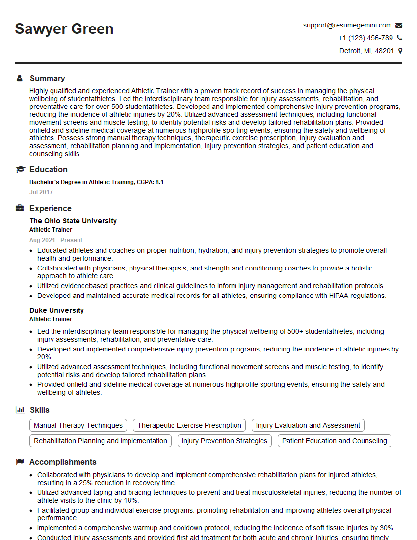
Athletic Trainer Resume Sample
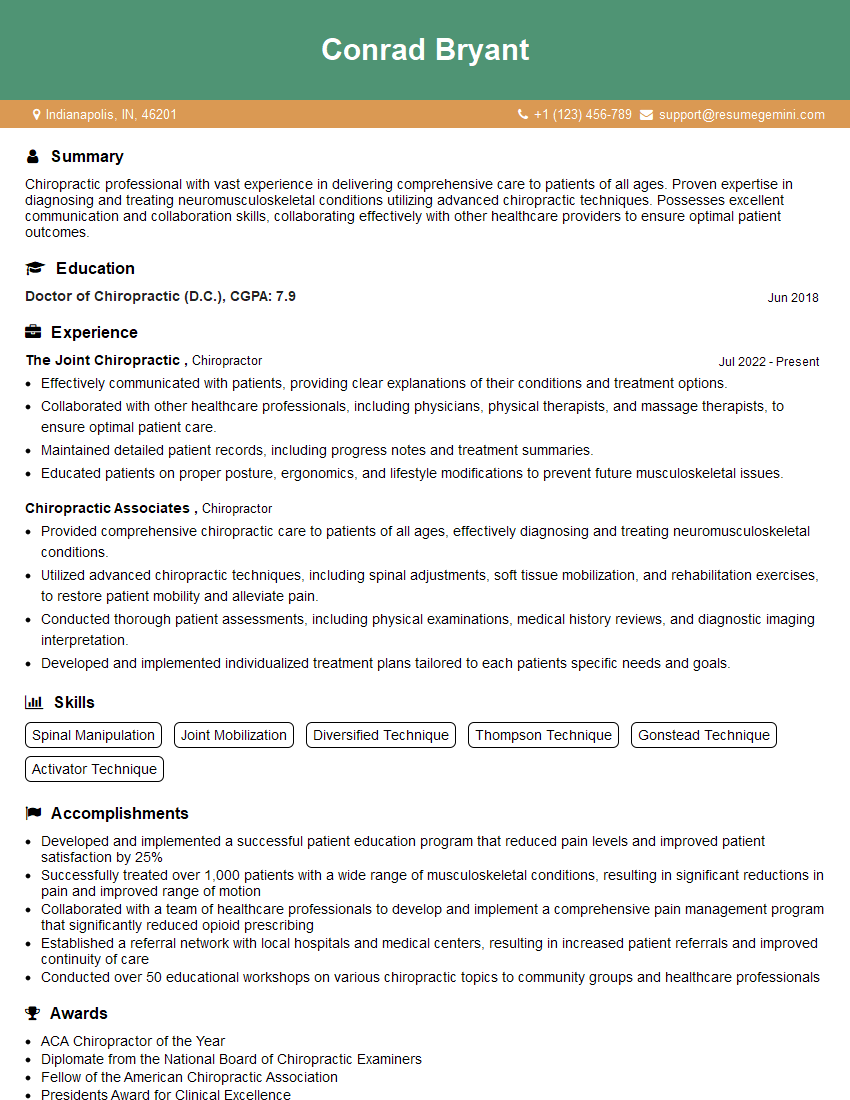
Chiropractor Resume Sample
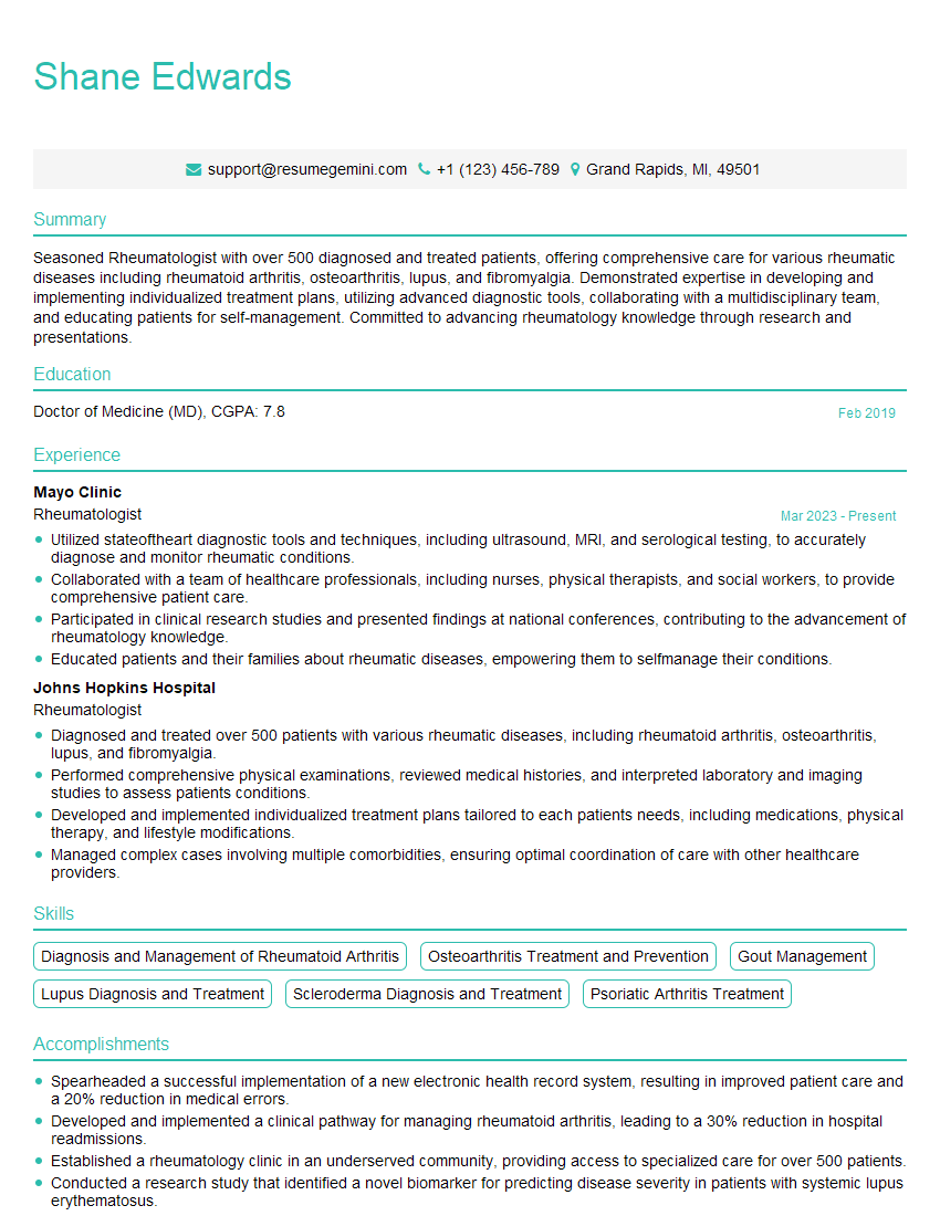
Rheumatologist Resume Sample
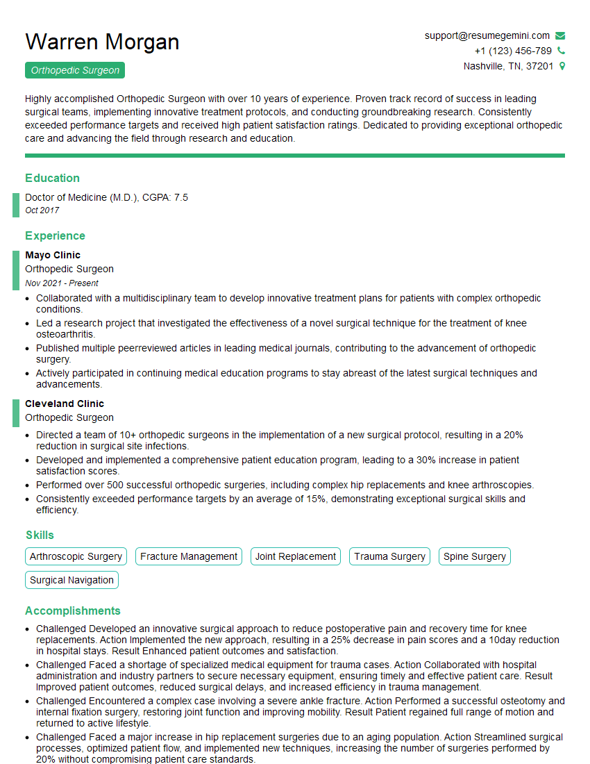
Orthopedic Surgeon Resume Sample
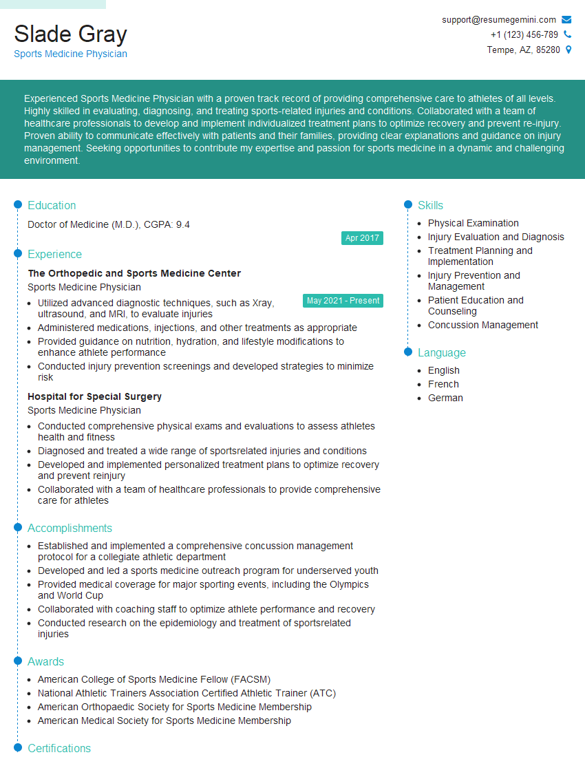
Sports Medicine Physician Resume Sample
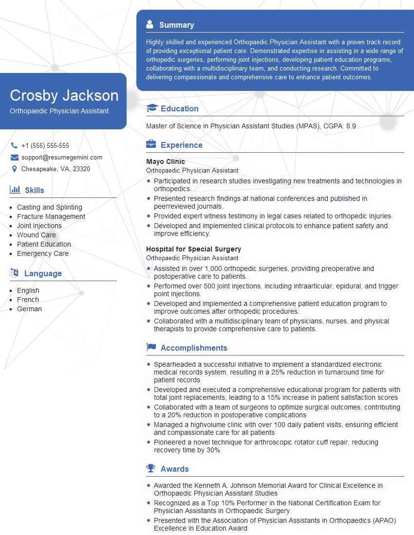
Orthopaedic Physician Assistant Resume Sample
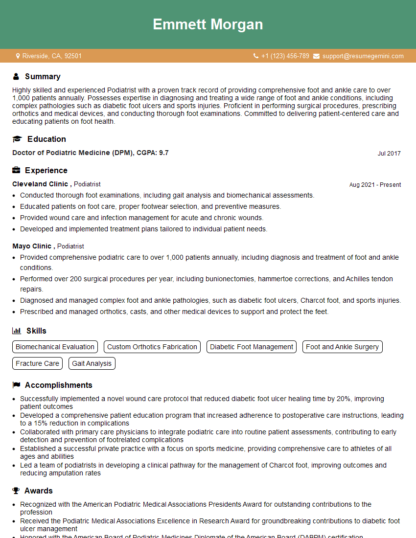
Podiatrist Resume Sample
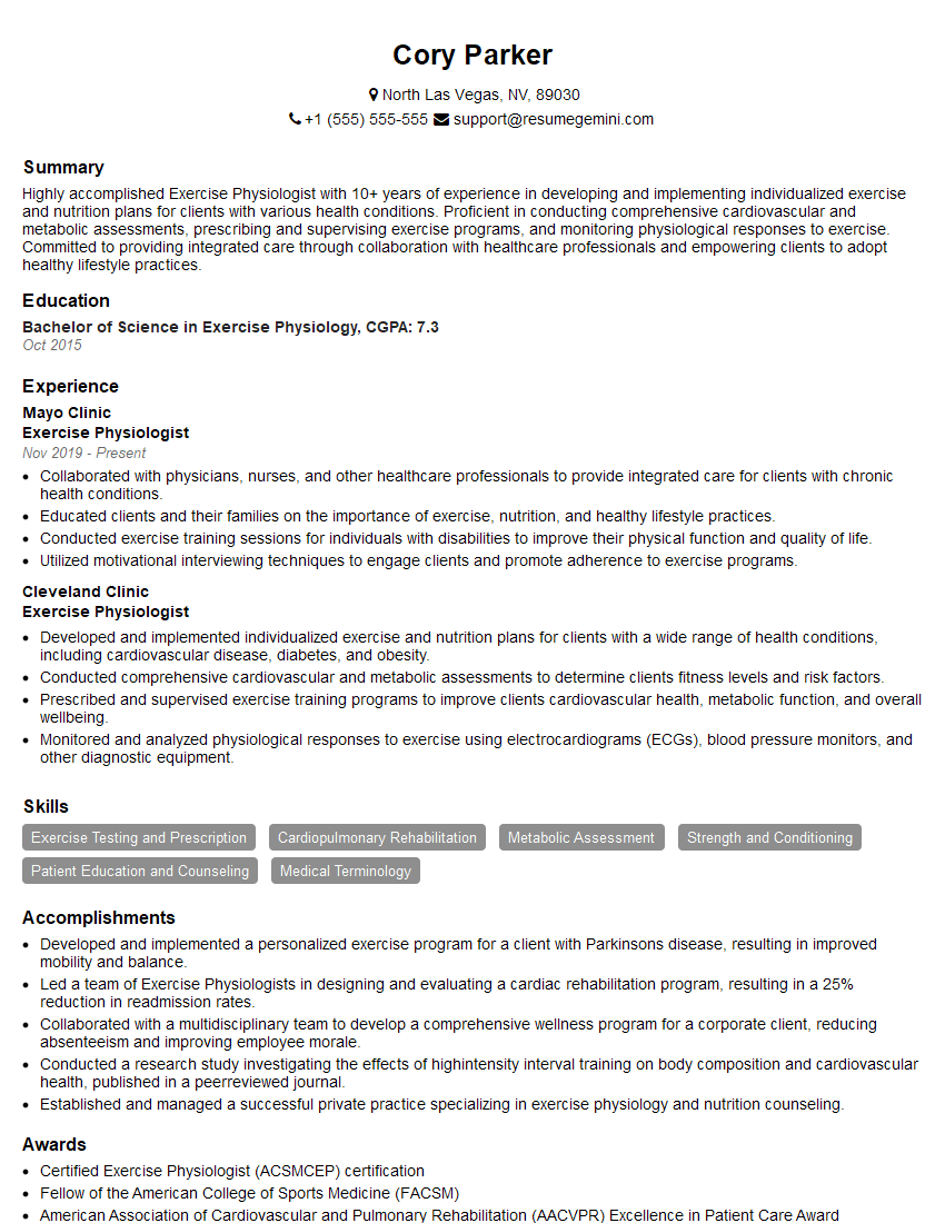
Exercise Physiologist Resume Sample
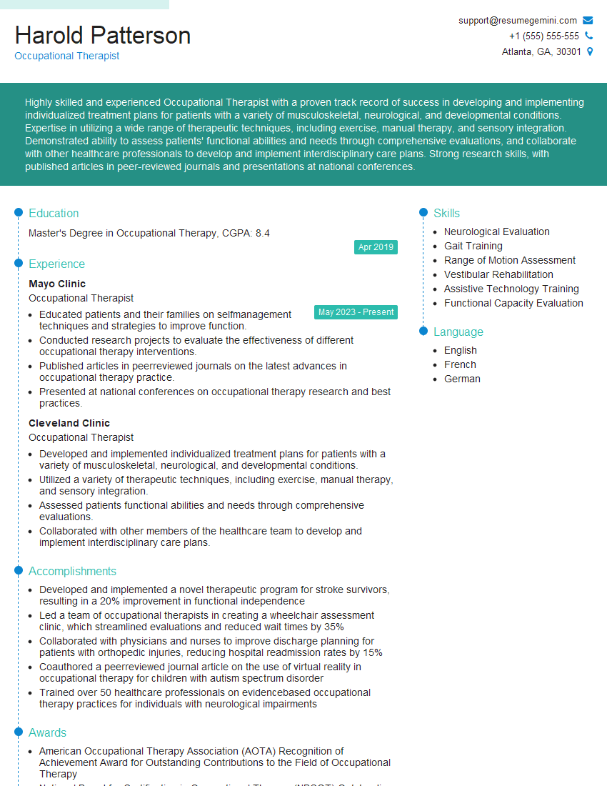
Occupational Therapist Resume Sample
Explore more articles
Users Rating of Our Blogs
Share Your Experience
We value your feedback! Please rate our content and share your thoughts (optional).
What Readers Say About Our Blog
To the interviewgemini.com Webmaster.
Very helpful and content specific questions to help prepare me for my interview!
Thank you
To the interviewgemini.com Webmaster.
This was kind of a unique content I found around the specialized skills. Very helpful questions and good detailed answers.
Very Helpful blog, thank you Interviewgemini team.