Preparation is the key to success in any interview. In this post, we’ll explore crucial Proctology interview questions and equip you with strategies to craft impactful answers. Whether you’re a beginner or a pro, these tips will elevate your preparation.
Questions Asked in Proctology Interview
Q 1. Describe your experience with hemorrhoid treatment techniques.
Hemorrhoid treatment varies greatly depending on the severity of the condition. Mild cases often respond well to conservative management, focusing on increasing fiber intake, drinking plenty of fluids, and using over-the-counter treatments like topical creams containing hydrocortisone or witch hazel. These help reduce inflammation and pain. For more advanced cases, I utilize a range of minimally invasive procedures. Rubber band ligation is a common technique where a small rubber band is placed around the base of the hemorrhoid, cutting off its blood supply and causing it to shrink and eventually fall off. Sclerotherapy involves injecting a solution into the hemorrhoid to reduce its size. Infrared coagulation uses heat to shrink the hemorrhoid. In severe cases requiring more extensive intervention, surgical hemorrhoidectomy might be necessary, which involves removing the hemorrhoid surgically. Post-operative care is crucial, focusing on pain management, hygiene, and dietary changes to promote healing.
For example, I recently treated a patient with grade II hemorrhoids who initially responded well to conservative measures. However, after several weeks, symptoms persisted, and we opted for rubber band ligation. The patient experienced minimal discomfort and had complete resolution of their symptoms within a few weeks. In another case, a patient with significant bleeding and prolapse required a hemorrhoidectomy, resulting in a successful outcome, but requiring a longer recovery period.
Q 2. Explain the different types of colonoscopy procedures and their indications.
Colonoscopy is a procedure that allows visualization of the entire colon. There are several types, primarily differentiated by the level of detail and the purpose of the examination. A standard colonoscopy uses a flexible endoscope to examine the entire large bowel, from the rectum to the cecum. This is the most common type and is used for screening for colorectal cancer, investigating symptoms like bleeding or changes in bowel habits, and removing polyps. A virtual colonoscopy (CT colonography) uses CT scanning to create three-dimensional images of the colon. It is less invasive than a traditional colonoscopy but may miss small polyps. A flexible sigmoidoscopy examines only the sigmoid colon and rectum. It’s often used for screening in lower-risk individuals or as a follow-up after a colonoscopy.
Indications for colonoscopy include screening for colorectal cancer in individuals over 50 (or earlier if there’s a family history), investigating gastrointestinal bleeding, diagnosing inflammatory bowel disease, evaluating changes in bowel habits such as persistent diarrhea or constipation, and removing polyps. The choice of procedure depends on the patient’s individual risk factors, symptoms, and medical history.
Q 3. What are the common causes and diagnostic approaches for anal fissures?
Anal fissures are tears in the lining of the anus, typically caused by passage of hard or large stools, leading to trauma. Chronic constipation is a major contributor. Other causes include childbirth, anal intercourse, and inflammatory bowel disease. Diagnosis is usually straightforward through a physical exam. I typically inspect the anus for the presence of a fissure, which often presents as a small tear or ulceration. Further investigations, such as anoscopy (using a short, rigid endoscope to examine the anal canal) might be necessary if the fissure is difficult to visualize or if there are concerns about other conditions. In chronic cases, additional investigations, such as excluding inflammatory bowel disease might be considered. Treatment initially focuses on conservative measures such as stool softeners, high-fiber diet, and warm sitz baths to promote healing and reduce pain. For fissures that don’t heal, topical medications like nitroglycerin or calcium channel blockers can be used to improve blood flow and healing. In some cases, surgical intervention, such as lateral internal sphincterotomy (a small incision in the anal sphincter muscle), might be necessary to reduce the pressure on the fissure and allow it to heal.
Q 4. Discuss your experience with managing colorectal cancer.
Managing colorectal cancer involves a multidisciplinary approach. Diagnosis begins with colonoscopy and biopsy to confirm the presence of cancer and determine its stage. Staging involves imaging studies such as CT scans, MRI, and PET scans to assess the extent of the cancer. Treatment options depend on the stage of the cancer. Early-stage cancers may be treated with surgery alone, typically a resection (removal of the affected portion of the colon or rectum). For more advanced cancers, surgery is often combined with chemotherapy and/or radiation therapy. Chemotherapy can be given before surgery (neoadjuvant) to shrink the tumor or after surgery (adjuvant) to reduce the risk of recurrence. Radiation therapy is typically used to shrink the tumor before surgery or to treat locally advanced cancers. Regular follow-up appointments, including colonoscopies, are crucial to monitor for recurrence. The approach is always personalized based on factors like tumor type, stage, patient’s overall health, and their preferences.
For example, I’ve worked with numerous patients, where a tailored approach involving surgery, chemotherapy and radiation delivered excellent outcomes, extending patient survival and improving quality of life. Palliative care becomes essential in advanced stages, focusing on pain management and improving the patient’s comfort.
Q 5. How do you approach the diagnosis and treatment of Crohn’s disease and ulcerative colitis?
Crohn’s disease and ulcerative colitis are both inflammatory bowel diseases (IBD), but they affect different parts of the gastrointestinal tract and have distinct characteristics. Crohn’s disease can affect any part of the digestive tract, from mouth to anus, and is characterized by transmural inflammation (inflammation that extends through all layers of the bowel wall). Ulcerative colitis, on the other hand, is limited to the colon and rectum and involves superficial inflammation of the mucosa (inner lining of the bowel). Diagnosis relies on a combination of clinical evaluation, imaging studies (such as endoscopy with biopsy and imaging studies like MRI or CT), and laboratory tests to assess inflammation markers like CRP and ESR. Treatment aims to induce remission and maintain long-term remission. This involves a combination of medications such as aminosalicylates, corticosteroids, immunomodulators, and biologics (anti-TNF agents). Surgery might be necessary in severe cases of both Crohn’s disease and ulcerative colitis, for complications like strictures, fistulas, perforation, or severe, unresponsive disease. The surgical approach for each disease is different given the differing anatomical distributions of disease.
Q 6. What are the surgical options for treating diverticulitis?
Diverticulitis is inflammation of diverticula, small pouches that can form in the colon wall. Treatment of diverticulitis depends on the severity of the disease. Mild cases often respond well to conservative management, which includes bowel rest (clear liquids), antibiotics to combat infection, and pain medication. In more severe cases, hospitalization may be required for intravenous antibiotics and fluid management. Surgical options are considered when conservative management fails, or in cases of complications such as abscess formation, perforation, fistula formation, or recurrent episodes of severe diverticulitis. Surgical options include: resection of the affected segment of the bowel (partial colectomy), often with creation of a colostomy, and minimally invasive approaches like laparoscopic surgery or robotic surgery that minimize invasiveness and accelerate recovery. The specific surgical technique chosen depends on several factors including the location and extent of the disease, and the presence of complications.
Q 7. Explain the procedure for a sigmoidoscopy and its significance.
A sigmoidoscopy is a procedure that examines the sigmoid colon and rectum using a flexible, lighted tube called a sigmoidoscope. The procedure begins with the patient lying on their side. A lubricated sigmoidoscope is gently inserted into the rectum and advanced into the sigmoid colon. Air is usually insufflated to expand the bowel, allowing for better visualization. The doctor can then examine the lining of the bowel for any abnormalities, such as polyps, inflammation, or bleeding. Biopsies can be taken if necessary. The procedure is typically well-tolerated, although some patients may experience mild discomfort or cramping. Preparation for a sigmoidoscopy usually involves bowel cleansing with laxatives to clear the bowel. Sigmoidoscopy is significant as it helps screen for colorectal cancer, detect and remove polyps, and diagnose other conditions such as inflammatory bowel disease, diverticulitis, and colorectal polyps. It is less extensive than a colonoscopy, examining only the distal portion of the large bowel, but it is useful in lower-risk patients or as a follow-up to other procedures.
Q 8. Describe your experience with managing post-surgical complications in colorectal surgery.
Managing post-surgical complications in colorectal surgery requires a vigilant and multi-faceted approach. Early identification and prompt intervention are key to minimizing morbidity and mortality. Common complications include infection (wound infection, intra-abdominal abscess), bleeding, anastomotic leaks (leakage at the surgical join), and bowel obstruction.
My approach involves meticulous surgical technique to minimize the risk of complications in the first place. Post-operatively, I closely monitor patients for signs and symptoms of complications, including fever, pain, changes in bowel habits, and abnormal lab values. Regular physical examinations and imaging studies (such as CT scans) are crucial. For instance, if a patient develops a fever and localized abdominal tenderness post-colectomy, I would suspect an abscess and order a CT scan to confirm and guide drainage.
Treatment strategies vary depending on the specific complication. Infections often require antibiotics and potentially surgical drainage. Bleeding may necessitate re-operation or interventional radiology procedures. Anastomotic leaks usually require surgical intervention, possibly including a diverting colostomy. Bowel obstruction might require medication to help the bowel relax, or surgical intervention to relieve the blockage. Proactive communication with the patient and their family is essential throughout the process, providing realistic expectations and addressing their concerns.
Q 9. How do you counsel patients about colorectal cancer screening?
Counseling patients about colorectal cancer screening is a critical aspect of preventative medicine. My approach involves a thorough discussion about their individual risk factors, including age, family history, personal medical history (e.g., inflammatory bowel disease), and ethnicity. I explain the different screening methods available, their benefits, risks, and limitations. These methods include colonoscopy (the gold standard), sigmoidoscopy, stool tests (fecal occult blood test, FIT, and Cologuard), and CT colonography.
I tailor my recommendations to each patient’s unique circumstances. For example, a patient with a strong family history of colorectal cancer might be recommended for earlier and more frequent screening than someone with no significant risk factors. I emphasize the importance of regular screening, explaining that early detection significantly improves the chances of successful treatment. I also address common patient concerns such as discomfort during the procedure and the importance of bowel preparation. I find visual aids like diagrams and pamphlets helpful in conveying this information effectively. Ultimately, the goal is to empower patients to make informed decisions about their health and participate actively in their care.
Q 10. What is your approach to managing fecal incontinence?
Managing fecal incontinence is a challenging but manageable condition. My approach involves a thorough evaluation to determine the underlying cause, which can range from anal sphincter dysfunction to neurological disorders. The evaluation includes a detailed history, physical examination (including a digital rectal examination and assessment of anal sphincter tone), and diagnostic tests such as anorectal manometry (measuring pressure in the anal canal) and endoanal ultrasound.
Treatment strategies are tailored to the cause and severity. Conservative measures may include dietary modifications (increasing fiber intake, avoiding certain foods), pelvic floor muscle exercises (to strengthen the anal sphincter), and biofeedback. Pharmacological interventions, such as antidiarrheal medications, may be used to manage symptoms. For more severe cases, surgical options such as sphincter repair, artificial bowel sphincter, or sacral nerve stimulation may be considered. In some cases, ostomy might be a temporary or permanent solution. The goal is to improve quality of life by reducing fecal incontinence episodes and improving continence.
Q 11. What are the indications for a colectomy?
A colectomy, the surgical removal of all or part of the colon, is indicated in a variety of conditions. The decision to proceed with a colectomy is made based on a careful consideration of the patient’s overall health, the severity of their condition, and the potential benefits and risks of surgery.
- Colorectal Cancer: This is the most common indication, involving the removal of the cancerous section of the colon along with a margin of healthy tissue.
- Inflammatory Bowel Disease (IBD): In cases of severe ulcerative colitis or Crohn’s disease that are refractory to medical management, a colectomy may be necessary to control symptoms and prevent complications.
- Diverticular Disease: Severe diverticulitis (inflammation of diverticula, small pouches in the colon) with complications such as abscesses or perforation may necessitate a colectomy.
- Ischemic Colitis: When a section of the colon experiences a lack of blood flow, resulting in tissue death, a colectomy is often required to remove the affected area.
- Polyps: Large or multiple polyps in the colon, which can be precancerous, might require a colectomy.
- Trauma: Severe injuries to the colon may necessitate a colectomy.
The type of colectomy performed (e.g., partial colectomy, total colectomy) depends on the specific condition and the location of the affected area. Post-operative management includes pain control, bowel management, and monitoring for complications.
Q 12. Explain the differences between inflammatory bowel disease (IBD) and irritable bowel syndrome (IBS).
Inflammatory bowel disease (IBD) and irritable bowel syndrome (IBS) are both chronic gastrointestinal disorders that cause abdominal pain and altered bowel habits, but they are distinctly different conditions.
IBD (Crohn’s disease and ulcerative colitis) is characterized by chronic inflammation of the digestive tract. This inflammation can affect any part of the gastrointestinal tract, from the mouth to the anus, but typically involves the terminal ileum and colon. IBD involves an autoimmune response leading to inflammation and damage of the bowel wall. This often results in symptoms such as diarrhea (often bloody in ulcerative colitis), abdominal pain, weight loss, and fever. Diagnosis involves colonoscopy with biopsy to confirm the presence of inflammation. Treatment often involves medication to suppress inflammation (corticosteroids, immunomodulators, biologics), and sometimes surgery.
IBS, on the other hand, is a functional gastrointestinal disorder. This means that there is no detectable inflammation or structural damage to the bowel wall. IBS is believed to be related to altered gut motility, visceral hypersensitivity, and changes in the gut microbiome. Symptoms can vary widely but typically include abdominal pain or discomfort, bloating, and changes in bowel habits (constipation, diarrhea, or both). Diagnosis is based primarily on clinical symptoms, as imaging tests and endoscopy are typically normal. Treatment focuses on managing symptoms through dietary changes, stress management techniques, and medication to alleviate specific symptoms (e.g., antispasmodics for pain, antidiarrheals or laxatives for bowel habit problems).
Q 13. Describe your experience with the use of minimally invasive surgical techniques in colorectal surgery.
Minimally invasive surgical techniques, such as laparoscopy and robotic surgery, have revolutionized colorectal surgery. My experience with these techniques is extensive. I’ve performed numerous laparoscopic colectomies, low anterior resections, and other colorectal procedures using these minimally invasive approaches.
The benefits of minimally invasive surgery include smaller incisions, reduced pain, shorter hospital stays, faster recovery times, and less scarring compared to open surgery. Laparoscopic surgery involves inserting small incisions in the abdomen and using a laparoscope (a thin, lighted tube with a camera) and specialized instruments to perform the surgery. Robotic surgery offers even greater precision and dexterity, with the surgeon controlling robotic arms that perform the operation. While minimally invasive surgery is not always suitable for every patient, it’s often preferred for appropriately selected cases, offering significant advantages. I carefully assess each patient’s individual condition to determine the most appropriate surgical approach. For example, a patient with a complex colorectal cancer requiring a large resection might be a better candidate for an open procedure, while a patient with uncomplicated diverticulitis might be an ideal candidate for laparoscopic surgery.
Q 14. How do you manage anal fistulas?
Anal fistulas are abnormal connections between the anal canal and the skin around the anus. Management of anal fistulas requires a tailored approach based on the fistula’s characteristics (location, tract complexity, and presence of infection).
My approach starts with a thorough examination, including a careful evaluation of the external opening and internal opening of the fistula. Imaging studies, such as endoanal ultrasound or MRI, are often helpful in determining the complexity of the fistula tract. For simple fistulas, conservative treatment with antibiotics to address any infection might be considered.
However, most fistulas require surgical intervention. The choice of surgical technique depends on the fistula’s characteristics. Options include fistulotomy (laying open the fistula tract), seton placement (inserting a drain to allow for gradual healing), advancement flaps, and fibrin glue. The goal of surgery is to completely excise the fistula tract and promote healing while minimizing damage to the anal sphincter to preserve continence. Postoperative care focuses on pain management, wound care, and monitoring for infection or complications. The choice of the best procedure is determined based on a combination of the fistula anatomy, its relationship to the anal sphincter muscles and the patient’s overall health.
Q 15. What are the common complications associated with colorectal surgery?
Colorectal surgery, while highly successful, carries potential complications. These can be broadly categorized into early (occurring within the first 30 days post-surgery) and late (occurring after 30 days) complications. Early complications frequently include infections (wound infections, peritonitis, pneumonia), bleeding, and anastomotic leaks (leakage at the surgical join). Late complications are often more subtle and can include strictures (narrowing of the bowel), fistulas (abnormal connections between bowel and other organs), and recurrent disease.
- Infections: These are managed with antibiotics and, in severe cases, may require surgical drainage. For instance, a wound infection might require debridement (removal of infected tissue) and further antibiotic treatment.
- Bleeding: This can range from minor oozing to life-threatening hemorrhage and requires careful monitoring and, if severe, potentially a return to the operating room.
- Anastomotic Leaks: A leak at the surgical join is a serious complication, potentially leading to peritonitis (infection of the abdominal cavity). Treatment involves drainage, antibiotics, and sometimes further surgery.
- Strictures: These narrowings can cause bowel obstructions and may require dilation or surgical revision.
Minimizing complications requires meticulous surgical technique, appropriate patient selection, and robust postoperative care, including vigilant monitoring for signs of infection or bleeding.
Career Expert Tips:
- Ace those interviews! Prepare effectively by reviewing the Top 50 Most Common Interview Questions on ResumeGemini.
- Navigate your job search with confidence! Explore a wide range of Career Tips on ResumeGemini. Learn about common challenges and recommendations to overcome them.
- Craft the perfect resume! Master the Art of Resume Writing with ResumeGemini’s guide. Showcase your unique qualifications and achievements effectively.
- Don’t miss out on holiday savings! Build your dream resume with ResumeGemini’s ATS optimized templates.
Q 16. Explain the role of imaging (CT, MRI) in diagnosing colorectal conditions.
Imaging plays a crucial role in diagnosing colorectal conditions. CT (Computed Tomography) scans provide detailed cross-sectional images of the abdomen and pelvis, allowing for the assessment of bowel wall thickness, the presence of masses, and the extent of any spread to lymph nodes or distant organs. CT colonography, a virtual colonoscopy, is a non-invasive alternative to traditional colonoscopy, although it’s less sensitive for detecting small polyps.
MRI (Magnetic Resonance Imaging) offers superior soft tissue contrast compared to CT, making it particularly valuable for evaluating the local extent of colorectal cancer, particularly its invasion into surrounding structures like the bladder or uterus. It also plays a key role in assessing for recurrence after treatment.
Example: A patient presenting with rectal bleeding and a palpable mass might undergo both a CT scan to assess for distant metastasis and an MRI to determine the exact size and depth of invasion of the rectal cancer.
Q 17. How do you interpret a colonoscopy report?
Interpreting a colonoscopy report requires a systematic approach. The report typically includes a description of bowel preparation quality, the extent of colon visualization, findings during the examination (polyps, inflammation, masses, etc.), and any biopsies taken. A clear description of the location, size, and morphology (shape and appearance) of any abnormalities is provided. The report will also often include recommendations for further management.
Example: A report might state: “Colonoscopy revealed a well-differentiated 1.5 cm polyp in the ascending colon, which was removed via snare polypectomy. Biopsy revealed a tubular adenoma without high-grade dysplasia. No other significant findings were noted. Follow-up colonoscopy recommended in 5-10 years.”
Understanding the terminology used in the report (e.g., adenoma, dysplasia, ulcerative colitis) is crucial for accurate interpretation. I would look for any signs of malignancy, the need for further investigation (e.g., biopsies), and any specific recommendations.
Q 18. What are the various types of bowel obstructions and their management strategies?
Bowel obstructions can be broadly classified as mechanical or non-mechanical (functional). Mechanical obstructions are caused by a physical blockage within the bowel lumen (the inside of the bowel), such as a tumor, adhesions (scar tissue), or impacted stool (fecal impaction). Functional obstructions, on the other hand, result from a problem with the motility (movement) of the bowel, often due to paralytic ileus (a temporary paralysis of the bowel) after surgery or from conditions like inflammatory bowel disease.
- Mechanical Obstructions: Management depends on the cause and severity and might involve non-surgical methods (like bowel rest, nasogastric suction, or enemas to relieve impaction) or surgery to remove the obstruction. For example, a large tumor causing a blockage would usually require surgical resection.
- Functional Obstructions: These often resolve with conservative management. Treatment includes bowel rest, intravenous fluids, and sometimes medications to stimulate bowel motility. In cases of paralytic ileus, addressing the underlying cause is critical; for example, relieving post-operative pain and bowel inflammation are important.
Diagnosis usually involves clinical assessment, abdominal X-rays, and possibly CT scans to identify the location and cause of the obstruction. Prompt management is crucial to prevent complications such as bowel perforation and peritonitis.
Q 19. Describe your understanding of the TNM staging system for colorectal cancer.
The TNM staging system is the most widely used system for classifying colorectal cancer. It provides a standardized method of describing the extent of the disease based on three key factors:
- T (Tumor): Describes the size and extent of the primary tumor. T1 indicates a small tumor confined to the bowel wall, while T4 indicates a very large tumor invading adjacent organs.
- N (Nodes): Refers to the involvement of regional lymph nodes. N0 means no lymph node involvement, while N2 indicates significant lymph node metastasis.
- M (Metastasis): Indicates the presence of distant metastases (spread to other parts of the body). M0 means no distant metastasis, while M1 signifies the presence of distant metastases.
These three factors are combined to create a complete TNM stage, which then determines the prognosis and influences treatment decisions. For example, a stage II colorectal cancer (typically T3-T4, N0, M0) would have a different treatment strategy than a stage IV (any T, any N, M1) cancer, which would usually involve systemic chemotherapy.
Q 20. Explain the process of bowel preparation before colonoscopy.
Bowel preparation before colonoscopy is essential to ensure a clear view of the colon’s lining. The goal is to completely clear the bowel of stool, allowing for optimal visualization of polyps, tumors, or other abnormalities. The process typically involves a combination of dietary restrictions and bowel cleansing agents.
Dietary Restrictions: Patients are typically advised to avoid foods high in fiber and red meat for several days leading up to the procedure, which may also require low-residue foods, like white bread, broth, etc. This reduces stool bulk.
Bowel Cleansing Agents: These agents, such as polyethylene glycol (PEG)-based solutions, are taken orally on the day before the procedure. These solutions draw water into the colon, promoting bowel movements and effectively clearing the colon. The choice and dosage are determined based on the patient’s individual needs and the specific recommendations by the physician. Instructions are always given on the appropriate preparation based on factors such as patient age, health conditions, and individual tolerances.
Adequate bowel preparation is crucial for the success of the colonoscopy, as inadequate preparation can hinder visualization and lead to the need for repeat procedures.
Q 21. Discuss your experience with the management of postoperative pain.
Postoperative pain management after colorectal surgery is a crucial aspect of patient care. It aims to minimize pain, promote early mobilization, and accelerate recovery. A multimodal approach, combining various pain relief techniques, is often most effective.
My approach involves a combination of strategies, starting with preemptive analgesia (pain medication given before surgery to prevent pain). This is often supplemented by regional anesthesia techniques (such as an epidural) and opioid analgesics (like morphine or fentanyl) in the immediate post-operative period. Once the patient is stable, we switch to less potent analgesics (such as NSAIDs or acetaminophen) combined with adjuvant therapies, like nerve blocks or physiotherapy.
Example: A patient recovering from a colectomy may receive an epidural catheter for the first 24-48 hours, followed by a combination of oral oxycodone and acetaminophen. Regular assessment of pain levels and adjustment of medication are essential. Patient education on pain management techniques (such as relaxation exercises and deep breathing) is also included in the care plan. Careful monitoring and managing any potential side effects of pain medication is critical to ensure patient safety and comfort.
Q 22. What are your thoughts on the use of robotic surgery in proctology?
Robotic surgery is revolutionizing proctology, offering several advantages over traditional laparoscopic or open procedures. The enhanced dexterity and precision of robotic instruments allow for minimally invasive approaches to complex colorectal procedures, leading to smaller incisions, reduced pain, shorter hospital stays, and faster recovery times. For example, robotic-assisted rectal resections are now routinely performed, offering significant improvements in patient outcomes compared to open surgery. The 3D high-definition visualization provided by the robotic system enhances surgical precision, particularly in intricate anatomical areas like the pelvis. However, the high cost of equipment and the need for specialized training remain significant barriers to widespread adoption.
It’s crucial to remember that robotic surgery isn’t a panacea. Careful patient selection remains paramount; the benefits might not outweigh the risks in all cases. The decision to use robotic surgery should be made on a case-by-case basis, taking into account the patient’s overall health, the complexity of the procedure, and the surgeon’s experience with the robotic platform.
Q 23. Describe your familiarity with different types of bowel anastomosis techniques.
Bowel anastomosis, the surgical connection of two segments of bowel, employs various techniques. The choice depends on factors such as location, bowel condition, and surgeon preference. Common techniques include:
- End-to-end anastomosis: The simplest type, where the cut ends of both bowel segments are directly joined.
- Side-to-side anastomosis: A side-to-side connection is created between the two bowel segments. This is often used when one segment is significantly shorter than the other.
- End-to-side anastomosis: One end of one bowel segment is connected to the side of another.
Specific suture techniques vary, but generally involve careful apposition of the bowel mucosa, submucosa, muscularis, and serosa layers to ensure a secure and leak-proof connection. Stapling devices are frequently used to expedite the process and improve precision, minimizing the risk of anastomotic leaks, a serious complication that can lead to infection or peritonitis.
Choosing the optimal technique requires a thorough understanding of colorectal anatomy, surgical principles, and potential complications. For example, in cases of compromised bowel perfusion, a side-to-side anastomosis might be preferred to minimize tension on the suture line.
Q 24. Explain the importance of proper wound care after colorectal surgery.
Proper wound care after colorectal surgery is critical to prevent infection and promote healing. This involves several key steps:
- Meticulous wound cleansing: Gentle cleaning with soap and water or a prescribed antiseptic solution, avoiding harsh scrubbing.
- Dressing changes: Regular changes of the dressing, as directed by the surgeon, to maintain a clean and dry wound environment.
- Monitoring for signs of infection: Close observation for redness, swelling, increased pain, pus, or fever, requiring prompt medical attention if any signs appear.
- Pain management: Effective pain control to encourage movement and deep breathing, promoting better circulation and healing.
- Nutritional support: Adequate nutrition is essential for tissue repair and overall recovery.
In cases of complex wounds or compromised tissue, specialized wound care techniques may be required, potentially involving negative pressure wound therapy or other advanced modalities. The goal is to create an optimal healing environment, minimizing the risk of complications and improving patient outcomes.
Think of it like tending a garden: proper care, including regular tending and protection from harmful elements, is essential for healthy growth. Similarly, appropriate wound care ensures the surgical site heals properly.
Q 25. How do you manage complications related to ostomy care?
Managing ostomy complications requires vigilance and proactive care. Common issues include:
- Skin irritation: Proper skin barrier protection with appropriate ostomy appliances is crucial. Frequent changes and careful attention to peri-stoma skin hygiene can prevent irritation and breakdown.
- Obstruction: Constipation or blockage of the ostomy can cause abdominal pain and distension. Dietary modifications, stool softeners, and possibly manual disimpaction may be needed.
- Infection: Signs of infection (redness, swelling, pus) require prompt treatment with antibiotics. Maintaining meticulous hygiene is key to prevention.
- Prolapse: The protrusion of the stoma may require surgical intervention.
- Hernia: The development of a hernia around the stoma requires surgical repair.
Patient education is key. I work closely with ostomy nurses and dieticians to provide patients and their families with comprehensive support and guidance, empowering them to manage their ostomy effectively and maintain their quality of life. Regular follow-up appointments allow for early detection and management of any potential issues.
Q 26. What are the latest advancements in the field of Proctology?
Proctology is constantly evolving. Some of the latest advancements include:
- Minimally invasive surgical techniques: Robotic surgery and advanced laparoscopic approaches continue to improve outcomes and reduce morbidity.
- Improved imaging modalities: Advanced imaging techniques like magnetic resonance imaging (MRI) and computed tomography (CT) colonography enhance the diagnosis and staging of colorectal diseases.
- Targeted therapies for colorectal cancer: Development of novel chemotherapeutic agents and targeted therapies is improving cancer treatment efficacy and survival rates.
- Advances in ostomy care: New ostomy appliances and technologies are improving comfort and reducing complications for patients with ostomies.
- Biologic therapies for inflammatory bowel disease: Novel biologics and targeted therapies are revolutionizing the management of inflammatory bowel diseases, improving symptoms and preventing disease progression.
These advancements continuously improve patient care, leading to better outcomes and an improved quality of life for individuals with proctologic conditions.
Q 27. Discuss your experience with colorectal pathology and biopsy interpretation.
My experience with colorectal pathology and biopsy interpretation encompasses a broad range of conditions. I’m proficient in interpreting biopsies for various diseases, including inflammatory bowel disease (ulcerative colitis, Crohn’s disease), colorectal polyps (adenomas, hyperplastic polyps), and colorectal cancer. I utilize a systematic approach, assessing histological features such as architecture, cellular morphology, and immunohistochemical stains to reach an accurate diagnosis.
For example, in evaluating a colorectal polyp biopsy, I would carefully assess the architectural features for signs of dysplasia (abnormal cell growth), the degree of nuclear atypia, and the presence of any invasive features. Immunohistochemical stains can be helpful in differentiating between different types of polyps and in identifying specific molecular markers associated with colorectal cancer risk. Accurate interpretation of biopsies is crucial for guiding appropriate clinical management.
Correlating the biopsy findings with the patient’s clinical presentation and imaging studies allows for a comprehensive evaluation, leading to effective treatment planning. This integrated approach ensures that patients receive the most appropriate care, whether it’s endoscopic surveillance, surgical intervention, or medical management.
Q 28. How do you communicate complex medical information to patients and their families?
Communicating complex medical information effectively is paramount. I believe in a patient-centered approach, using clear, concise language, avoiding medical jargon whenever possible. I begin by assessing the patient’s understanding of the condition and tailoring my explanation accordingly. I use analogies and visual aids, such as diagrams or models, to illustrate complex concepts. I encourage questions and answer them patiently, addressing concerns and clarifying misunderstandings.
I always involve the family in discussions, ensuring they understand the diagnosis, treatment plan, and potential risks and benefits. I provide written summaries of the discussion and encourage patients to keep a list of questions for our next meeting. I view effective communication not just as conveying information but also as building trust and creating a collaborative partnership between me and my patient. This approach ensures informed decision-making and promotes patient empowerment.
For example, explaining the risks of a surgical procedure involves breaking down complex statistical data into easily digestible terms and presenting realistic scenarios to help patients grasp the potential outcomes. This approach allows patients to actively participate in making decisions that best serve their needs.
Key Topics to Learn for Your Proctology Interview
Preparing for a proctology interview requires a comprehensive understanding of the field. Focus on mastering these key areas to showcase your expertise and confidence.
- Anorectal Anatomy and Physiology: Deep understanding of the anatomical structures and their physiological functions is crucial. Be prepared to discuss variations and clinical significance.
- Diagnostic Procedures: Demonstrate knowledge of various diagnostic techniques used in proctology, including anoscopy, sigmoidoscopy, and colonoscopy. Discuss their indications, contraindications, and interpretation of findings.
- Hemorrhoids and Anal Fissures: Master the diagnosis, management, and treatment options for these common conditions. Be ready to discuss both conservative and surgical approaches.
- Inflammatory Bowel Disease (IBD): Understand the various types of IBD, their presentation, and the role of proctology in their management. This includes recognizing complications and appropriate referrals.
- Colorectal Cancer Screening and Prevention: Discuss current screening guidelines, risk factors, and the importance of early detection. Be familiar with various screening modalities and their interpretation.
- Functional Bowel Disorders: Develop an understanding of conditions like constipation, fecal incontinence, and irritable bowel syndrome (IBS). Be prepared to discuss diagnostic approaches and management strategies.
- Pelvic Floor Dysfunction: Familiarize yourself with the anatomy and physiology of the pelvic floor and its role in bowel function. Understand common disorders and treatment approaches.
- Surgical Techniques: While depth depends on the specific role, a general understanding of common surgical procedures in proctology is beneficial. Focus on principles and indications rather than intricate details.
- Patient Communication and Counseling: Highlight your ability to effectively communicate complex medical information to patients and their families. Discuss your approach to sensitive topics and building rapport.
Next Steps: Launch Your Proctology Career
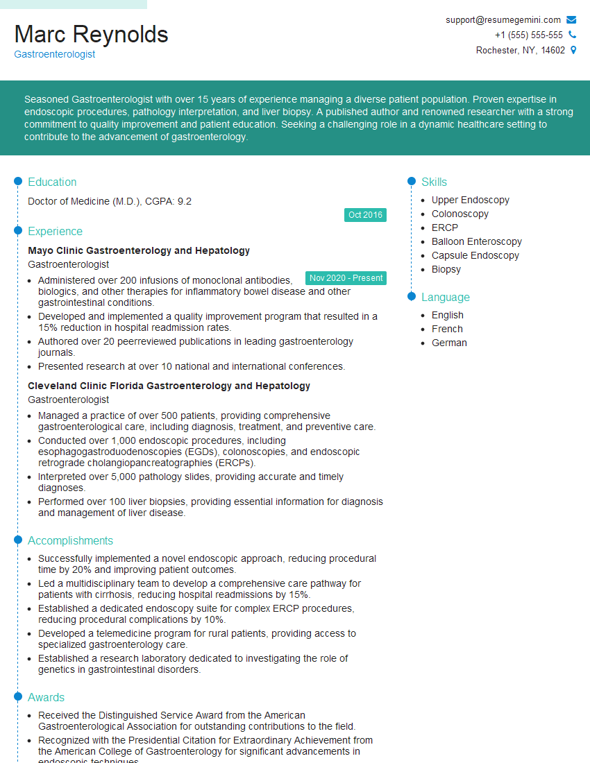
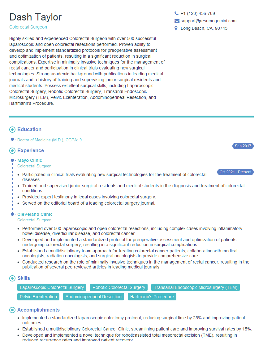
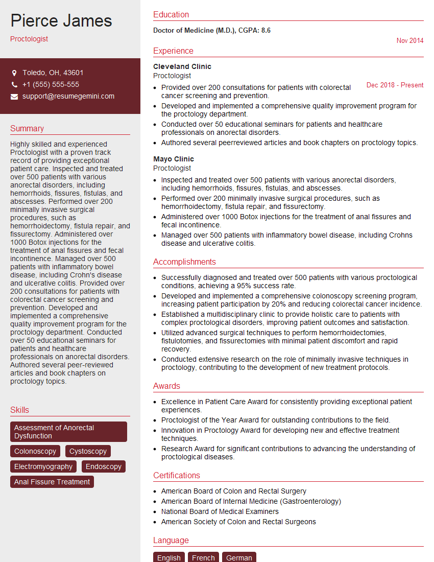
Mastering these key areas will significantly enhance your interview performance and demonstrate your readiness for a successful career in proctology. A strong resume is the first step in landing your dream job. Creating an ATS-friendly resume is crucial to ensuring your application is seen by recruiters. We recommend using ResumeGemini to build a professional and impactful resume that highlights your skills and experience. ResumeGemini provides examples of resumes tailored specifically for Proctology professionals to help guide you.
Explore more articles
Users Rating of Our Blogs
Share Your Experience
We value your feedback! Please rate our content and share your thoughts (optional).
What Readers Say About Our Blog
To the interviewgemini.com Webmaster.
Very helpful and content specific questions to help prepare me for my interview!
Thank you
To the interviewgemini.com Webmaster.
This was kind of a unique content I found around the specialized skills. Very helpful questions and good detailed answers.
Very Helpful blog, thank you Interviewgemini team.